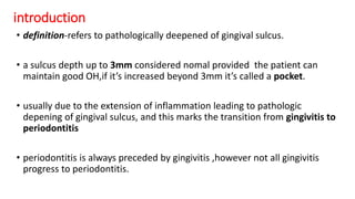
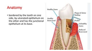
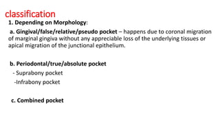
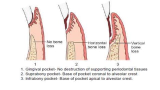
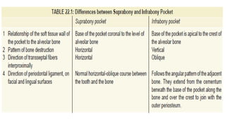
3. introduction
• definition-refers to pathologically deepened of gingival sulcus.
• a sulcus depth up to 3mm considered nomal provided the patient can
maintain good OH,if it’s increased beyond 3mm it’s called a pocket.
• usually due to the extension of inflammation leading to pathologic
depening of gingival sulcus, and this marks the transition from gingivitis to
periodontitis
• periodontitis is always preceded by gingivitis ,however not all gingivitis
progress to periodontitis.
4. Anatomy
• bordered by the tooth on one
side, by ulcerated epithelium on
the other and has the junctional
epithelium at its base.
5. classification
1. Depending on Morphology:
a. Gingival/false/relative/pseudo pocket – happens due to coronal migration
of marginal gingiva without any appreciable loss of the underlying tissues or
apical migration of the junctional epithelium.
b. Periodontal/true/absolute pocket
- Suprabony pocket
-Infrabony pocket
c. Combined pocket
6.
7.
8. 2.Depending on the number of surfaces involved
a. Simple - involve one tooth surface.
b. Compound - involves two or more tooth surfaces. The base of the
pockets is in direct communication with the gingival margin along each of
the involved surface.
c. Complex/Spiral- originates on one tooth surface and twists around the
tooth to involve one or more additional surfaces. The only communication
with gingival margin is at surface where the pocket originates.
9.
10. 3. Depending on disease activity:
a. Active pocket
b. Inactive pocket
4. Depending on the nature of soft tissue wall:
a. Edematous
b. Fibrotic
5. Depending on the lateral wall of the pocket:
a. Suprabony: consist of soft tissue alone
b. Infrabony: consist of both soft tissue and bone. The alveolar bone
becomes a part of the pocket wall.
11. clinical features
• Symptoms
1. Localized pain or a sensation of pressure after eating,which gradually
diminishes.
2. Radiating pain deep in the bone.
3. A foul taste in localized areas.
4. A gnawing feeling or feeling of itching in the gingiva.
5. Urge to dig with pointed instrument into the gingiva.
6. A tendency to suck material from the interproximal spaces.
7. Sensitivity to heat and cold and toothache in theabsence of caries.
12. Signs
1. Enlarged, bluish red thickened marginal gingiva with a rolled edge separated from the
tooth surface.
2. Bluish-red vertical zone extending from the gingival margin to the alveolar mucosa.
3. A break in the faciolingual continuity of the interdental gingiva.
4. Shiny, discolored and puffy gingiva associated with the exposed root surfaces.
5. Gingival bleeding and suppuration from the gingival margin.
6. Extrusion, mobility, diastema and migration of teeth.
14. Pathogenesis
• Pocket formation starts as an inflammatory change in the connective tissue wall
of the gingival sulcus as a response to supragingival and subgingival plaque.
• The cellular and fluid inflammatory exudate causes degeneration of the
surrounding connective tissue, including the gingival fibers.
• Just apical to the junctional epithelium, collagen fibers are destroyed and the
area is occupied by inflammatory cells and edema
• As a result of inflammation, PMNs invade the coronal end of the junctional
epithelium in increasing numbers
• When the relative volume of PMNs reaches approximately 60% or more of the
junctional epithelium, the tissue loses cohesiveness and detaches from the tooth
surface.
• Thus, the coronal portion of the junctional epithelium detaches from the root as
the apical portion migrates, thereby resulting in its apical shift.
15.
16. • Two mechanisms associated with collagen loss:
• 1. Collagenases and other enzymes secreted by cells in inflamed tissue, such as
fibroblasts and macrophages become extracellular and destroy collagen.
• 2. Fibroblasts phagocytize collagen fibers by extending cytoplasmic processes to the
ligament–cementum interface and degrading the inserted collagen fibrils and the fibrils
of the cementum matrix.
• As a consequence of the loss of collagen, the apical cells of the junctional epithelium
proliferate along the root and extend fingerlike projections that are two or three cells
in thickness.
17. HISTOLOGY
• The connective tissue is edematous and densely infiltrated
with plasma cells, lymphocytes, and PMNs.
• The blood vessels are increased in number, dilated, and
engorged, particularly in the subepithelial connective tissue
layer.
• The connective tissue exhibits varying degrees of
degeneration.
• The connective tissue shows proliferation of the
endothelial cells, with newly formed capillaries, fibroblasts,
and collagen fibers .
• The junctional epithelium at the base of the pocket is
usually much shorter than that of a normal sulcus.
• The corono-apical length of the junctional epithelium is
reduced to 50 to 100 µm.
• The cells may be well formed and in good condition, or they
may exhibit light to marked degeneration.
21. Clinical;
• Exploration of the gingival margin with a periodontal probe
• Inserted parallel to the vertical axis of the tooth
• The area with the deepest reading is used
• Readings more than 3 mm are considered pathologic
22. Radiographically;
• GP or calibrated silver points can be used with a radiograph to
determine the level of attachment in pockets