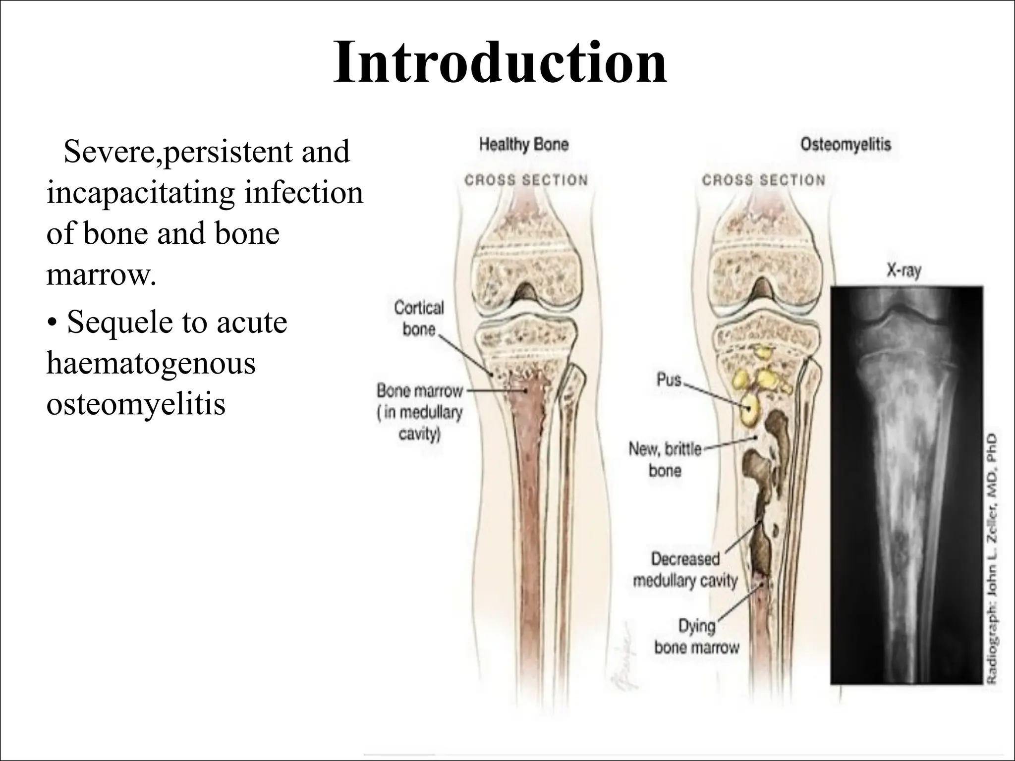
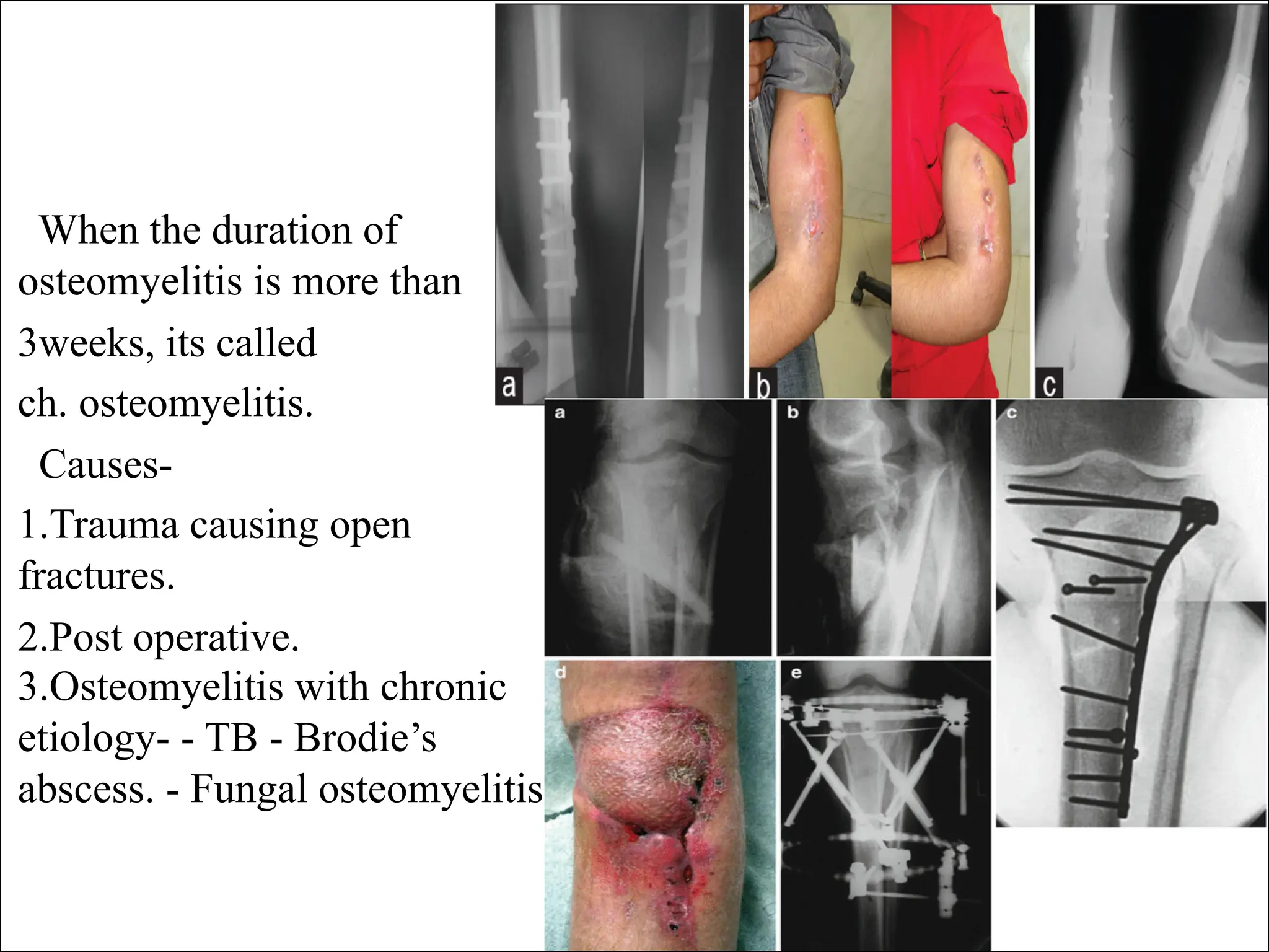
Chronic osteomyelitis
Presented by
Dr. Karrar Kareem Al-Jubory
Orthopedic surgeon
FICMS, CABMS (ORTHO)
Introduction
Severe,persistent and
incapacitating infection
of bone and bone
marrow.
• Sequele to acute
haematogenous
osteomyelitis
Introduction
Definition:
An inflammation of the bone
caused by an infecting
organism.
The infection may be limited
to a single portion of the bone
or may involve the marrow,
cortex, periosteum, and the
surrounding soft tissue.
Classification
Duration of symptoms
(acute, subacute, and
chronic)
Mechanism of infection
exogenous or
hematogenous.
Based on the host response to
the disease.
pyogenic or nonpyogenic
When the duration of
osteomyelitis is more than
3weeks, its called
ch. osteomyelitis.
Causes-
1.Trauma causing open
fractures.
2.Post operative.
3.Osteomyelitis with chronic
etiology- - TB - Brodie’s
abscess. - Fungal osteomyelitis
Chronic osteomyelitis is
difficult to eradicate
completely.
Systemic symptoms may
subside, but one or more foci
in the bone may contain
purulent material, infected
granulation tissue, or a
sequestrum
Pathology
Necrosis >> stage of new bone
formation >> involucrum. >>
with sequestrum inside, with a
persistent discharging sinus.
>> pus from bone escapes
through multiple hole in
involucrum
The hallmark of chronic
osteomyelitis is infected dead
bone within a compromised
soft-tissue envelope.
The infected foci within the
bone are surrounded by
sclerotic, relatively avascular
bone covered by a thickened
periosteum and scarred muscle
and subcutaneous tissue.
This avascular envelope of
scar tissue leaves systemic
antibiotics essentially
ineffective.
Secondary infections are
common, and sinus track
cultures usually do not
correlate with cultures obtained
at bone biopsy.
Multiple organisms may grow
from cultures taken from sinus
tracks and from open biopsy
specimens of surrounding soft
tissue and bone.
Clinically
Pain, swelling.
Discharging sinus.
Bone thickening.
Deformity.
Joint stiffness.
Shortening of limb,
Pathological fracture.
Sinus track
malignancy.
Cierny and Mader Staging System
I Medullary >> Endosteal disease
II Superficial >> Cortical surface
infected because of coverage defect
III Localized >> Cortical sequestrum
that can be excised without
compromising stability
IV Diffuse >> Features of I, II, and
III plus mechanical instability before
or after débridementl sequestrum
Differential diagnosis and diagnosis
1.TB osteomyelitis- watery
discharge. - previous h/o TB,
sinus with undermined margin
with blue colour.
2. Ewing's sarcoma- A primary
malignant tumor of bone,
usually arising as a central
tumor in long bone. (biopsy)
3. Soft tissue chronic infection.
(X-ray)
Diagnosis :
Clinical, Laboratory, and
Imaging studies.
The “gold standard” is to
obtain a biopsy specimen for
histological and
microbiological evaluation of
the infected bone
Diagnosis
Clinical examination - Integrity of the skin and soft tissue -areas
of tenderness -assess bone stability - neurovascular status of the
limb.
Laboratory investigations Blood counts ESR CRP
Imaging studies
Plain radiographs
Cortical destruction
Periosteal reaction
Sequestrum deformity
Sclerotic bone
Sinography
CT Scan >> Cortical bone and
surrounding soft tissues and is
especially useful in identifying
sequestra.
MRI
The extent of the pathological
insult by showing the margins
of bone and soft-tissue edema.
Well-defined rim of high signal
intensity surrounding the focus
of active disease seen
(rim sign)
Treatment
Supportive treatment .
Antibiotics – to prevent
spread.
Surgery – sequestretomy +
saucerization [cannot be
eradicated without surgical
intervention]
Sinus tracks can be injected
with methylene blue 24 hours
before surgery to make them
easier to locate and excise.
After Treatment
6-week course of intravenous
antibiotics is given after
surgical débridement
The limb is splinted until the
wound has healed, and then it
is protected to prevent
pathological fracture
Methods described to eliminate dead
space
(1) Bone grafting with primary or secondary closure
(2) Antibiotic polymethyl methacrylate (PMMA) beads as a
temporary filler of the dead space before reconstruction
(3) Local muscle flaps and skin grafting with or without bone
grafting
(4) Microvascular transfer of muscle, myocutaneous, osseous,
and osteocutaneous flaps
(5) The use of bone transport (Ilizarov technique).
Complications
Joint stiffness.
Shortening.
Muscle contracture.
Pathological fracture.
Sinus track malignancy (sq. cell CA).
Amyloidosis.
THANK YOU

Chronic osteomy lllhhhhhhhhhhhhhhhhelitis (2).pdf

  • 1.
    Chronic osteomyelitis Presented by Dr.Karrar Kareem Al-Jubory Orthopedic surgeon FICMS, CABMS (ORTHO)
  • 2.
    Introduction Severe,persistent and incapacitating infection ofbone and bone marrow. • Sequele to acute haematogenous osteomyelitis
  • 3.
    Introduction Definition: An inflammation ofthe bone caused by an infecting organism. The infection may be limited to a single portion of the bone or may involve the marrow, cortex, periosteum, and the surrounding soft tissue.
  • 4.
    Classification Duration of symptoms (acute,subacute, and chronic) Mechanism of infection exogenous or hematogenous. Based on the host response to the disease. pyogenic or nonpyogenic
  • 5.
    When the durationof osteomyelitis is more than 3weeks, its called ch. osteomyelitis. Causes- 1.Trauma causing open fractures. 2.Post operative. 3.Osteomyelitis with chronic etiology- - TB - Brodie’s abscess. - Fungal osteomyelitis
  • 6.
    Chronic osteomyelitis is difficultto eradicate completely. Systemic symptoms may subside, but one or more foci in the bone may contain purulent material, infected granulation tissue, or a sequestrum
  • 8.
    Pathology Necrosis >> stageof new bone formation >> involucrum. >> with sequestrum inside, with a persistent discharging sinus. >> pus from bone escapes through multiple hole in involucrum
  • 9.
    The hallmark ofchronic osteomyelitis is infected dead bone within a compromised soft-tissue envelope. The infected foci within the bone are surrounded by sclerotic, relatively avascular bone covered by a thickened periosteum and scarred muscle and subcutaneous tissue. This avascular envelope of scar tissue leaves systemic antibiotics essentially ineffective. Secondary infections are common, and sinus track cultures usually do not correlate with cultures obtained at bone biopsy. Multiple organisms may grow from cultures taken from sinus tracks and from open biopsy specimens of surrounding soft tissue and bone.
  • 11.
    Clinically Pain, swelling. Discharging sinus. Bonethickening. Deformity. Joint stiffness. Shortening of limb, Pathological fracture. Sinus track malignancy.
  • 12.
    Cierny and MaderStaging System I Medullary >> Endosteal disease II Superficial >> Cortical surface infected because of coverage defect III Localized >> Cortical sequestrum that can be excised without compromising stability IV Diffuse >> Features of I, II, and III plus mechanical instability before or after débridementl sequestrum
  • 13.
    Differential diagnosis anddiagnosis 1.TB osteomyelitis- watery discharge. - previous h/o TB, sinus with undermined margin with blue colour. 2. Ewing's sarcoma- A primary malignant tumor of bone, usually arising as a central tumor in long bone. (biopsy) 3. Soft tissue chronic infection. (X-ray) Diagnosis : Clinical, Laboratory, and Imaging studies. The “gold standard” is to obtain a biopsy specimen for histological and microbiological evaluation of the infected bone
  • 14.
    Diagnosis Clinical examination -Integrity of the skin and soft tissue -areas of tenderness -assess bone stability - neurovascular status of the limb. Laboratory investigations Blood counts ESR CRP
  • 15.
    Imaging studies Plain radiographs Corticaldestruction Periosteal reaction Sequestrum deformity Sclerotic bone
  • 16.
    Sinography CT Scan >>Cortical bone and surrounding soft tissues and is especially useful in identifying sequestra.
  • 17.
    MRI The extent ofthe pathological insult by showing the margins of bone and soft-tissue edema. Well-defined rim of high signal intensity surrounding the focus of active disease seen (rim sign)
  • 18.
    Treatment Supportive treatment . Antibiotics– to prevent spread. Surgery – sequestretomy + saucerization [cannot be eradicated without surgical intervention] Sinus tracks can be injected with methylene blue 24 hours before surgery to make them easier to locate and excise.
  • 19.
    After Treatment 6-week courseof intravenous antibiotics is given after surgical débridement The limb is splinted until the wound has healed, and then it is protected to prevent pathological fracture
  • 20.
    Methods described toeliminate dead space (1) Bone grafting with primary or secondary closure (2) Antibiotic polymethyl methacrylate (PMMA) beads as a temporary filler of the dead space before reconstruction (3) Local muscle flaps and skin grafting with or without bone grafting (4) Microvascular transfer of muscle, myocutaneous, osseous, and osteocutaneous flaps (5) The use of bone transport (Ilizarov technique).
  • 22.
    Complications Joint stiffness. Shortening. Muscle contracture. Pathologicalfracture. Sinus track malignancy (sq. cell CA). Amyloidosis.
  • 23.