Downloaded 209 times


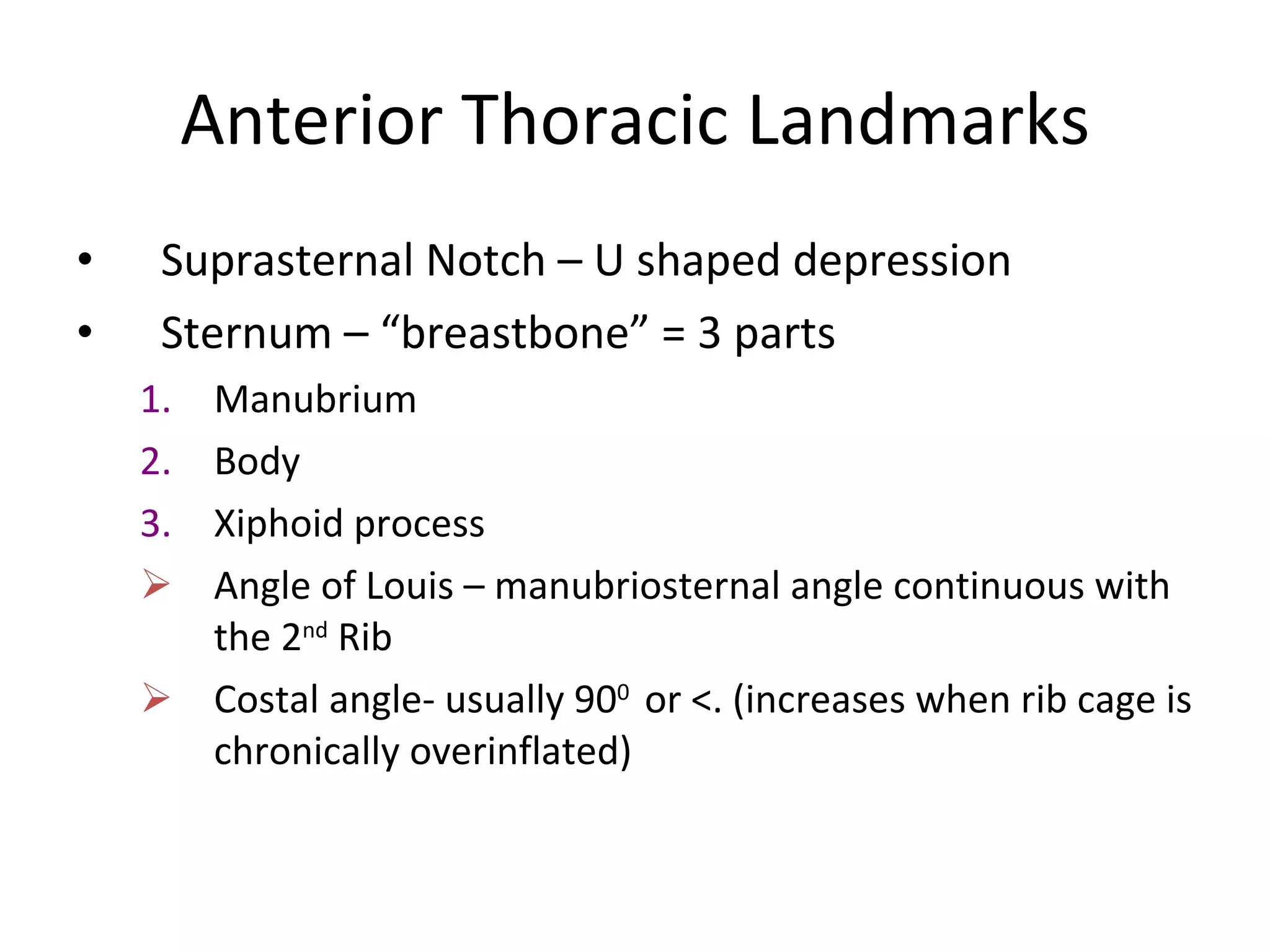


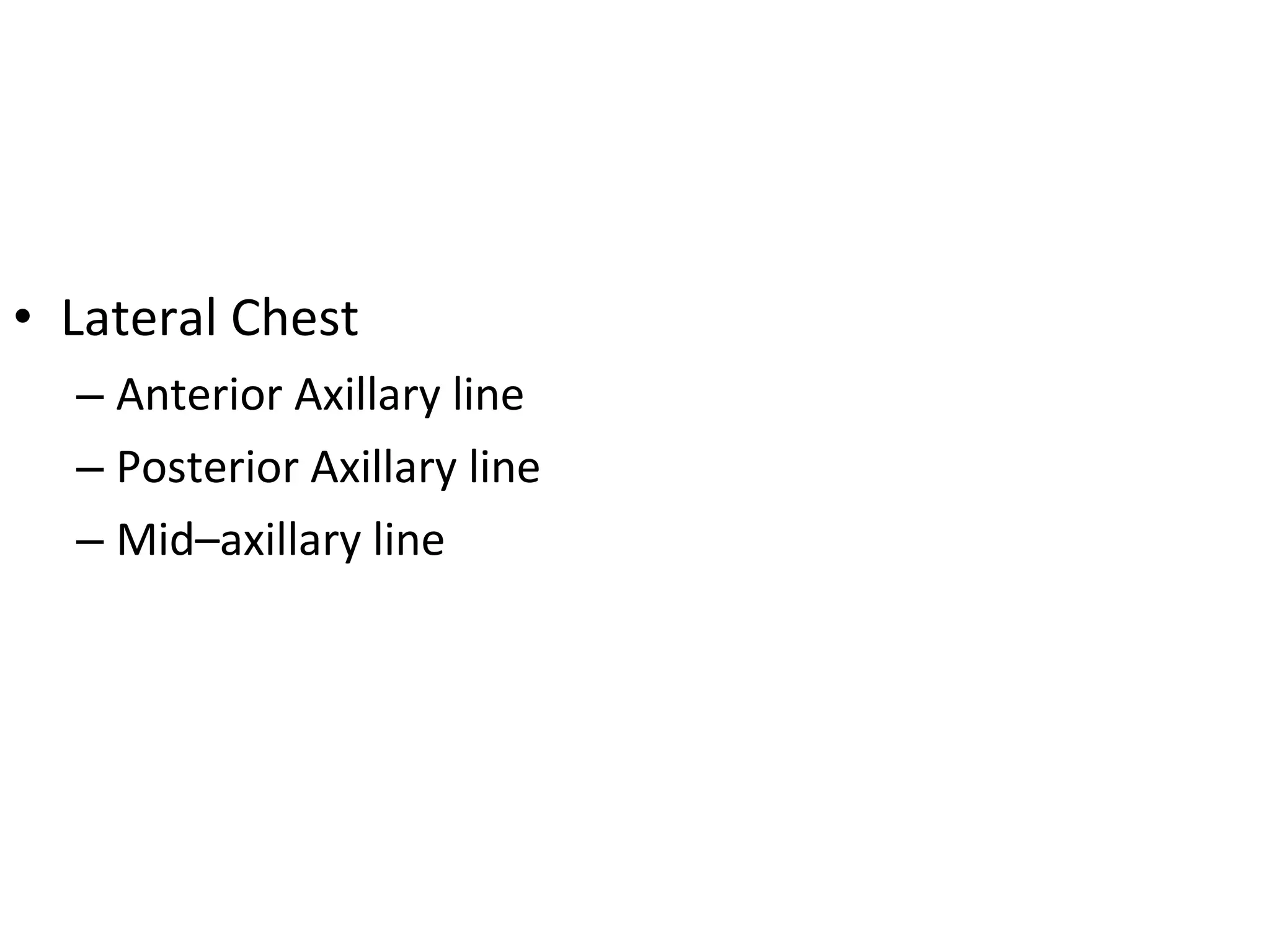


The document provides a detailed overview of the anatomy and physiology of the thorax and lungs. It describes the structures of the thoracic cage and mediastinum. It outlines the lobes and borders of the lungs, as well as the mechanics of respiration and gas exchange. It also discusses the examination techniques for inspecting, palpating, percussing, and auscultating the lungs, including normal and abnormal breath sound findings.


