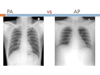
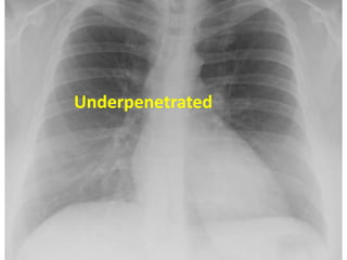
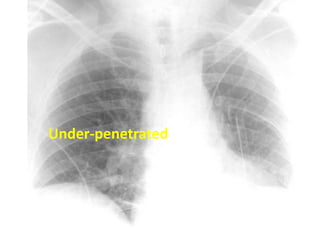
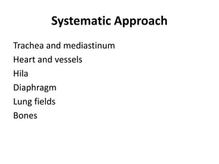
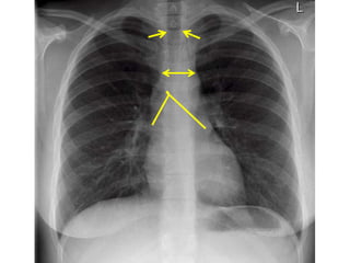
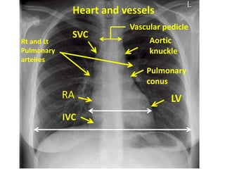
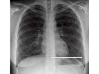
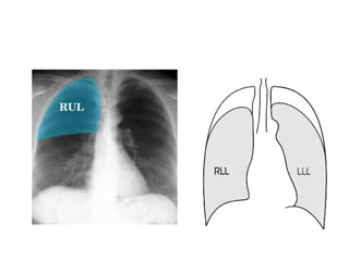
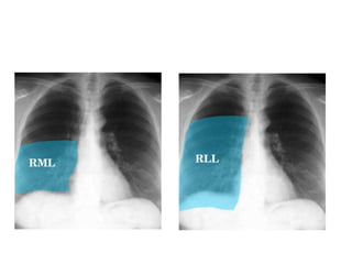
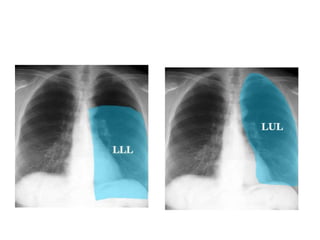
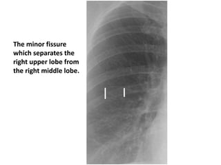
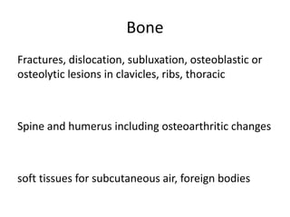
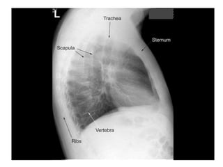
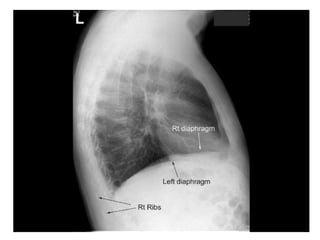
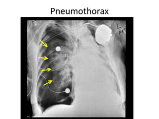
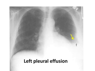
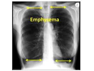
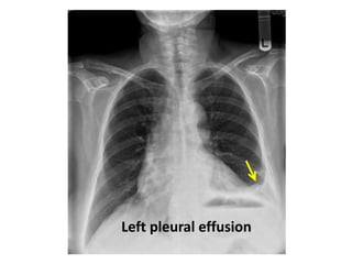
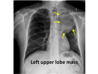
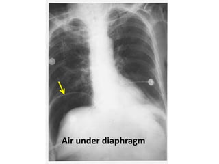
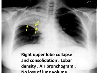
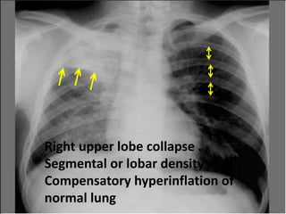
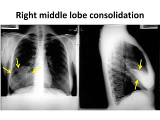
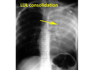
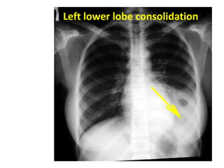
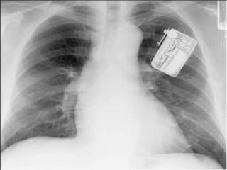
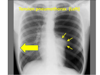
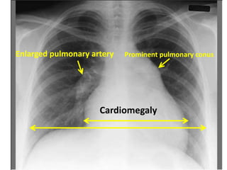
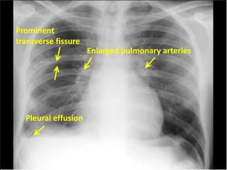
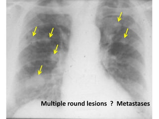
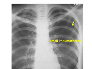
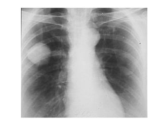
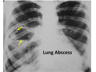
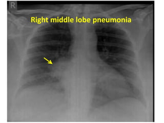
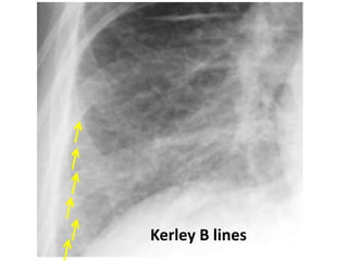
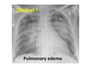
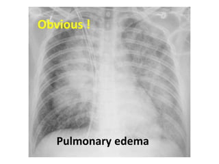
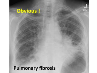
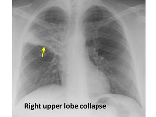
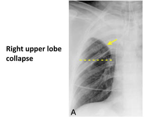
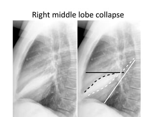
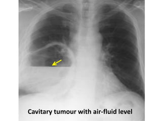
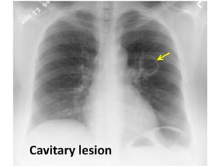
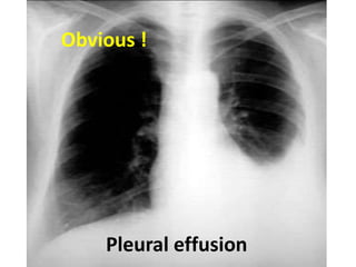
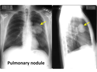
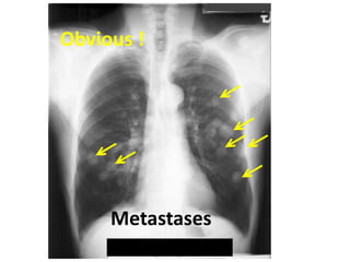
Chest X-RAY interpretation
Before you interpret … check for the
following :
Name
Date
Orientation : Left, right
Position : erect , supine , PA or AP
Inspiration, expiration
Rotation : central or tilted
Distance from the medial end of each clavicle to the
spinous process of the vertebra at the same level
Exposure : You must barely see the thoracic vertebrae
behind the heart
Identify both costophrenic angles and lung apices
Name
Date Orientation
Adequate
inspiration
9 ribs
posteriourly
Exposure
Adequate tissue
penetration
Adequate anatomical exposure
Gastric air bubble
Rotation
Underpenetrated
Under-penetrated
Systematic Approach
Trachea and mediastinum
Heart and vessels
Hila
Diaphragm
Lung fields
Bones
PA Film
Trachea and Mediastinum
Ensure trachea is visible
If deviated to one side :
Pushed away by pleural effusion or tension pneumothorax
Pulled towards collapse
The carina, (60 –100 degrees)
Angle of the carina increases left atrial enlargement, lymph
node enlargement and left upper lobe collapse (left main
bronchus is pulled up)
Check for a widened mediastinum :
Mass lesions (eg tumour, lymph nodes, aneurysm)
Heart and vessels
Heart size and heart borders
Check aorta : Widening, calcification
Check SVC, IVC, azygos vein widening
LV
Pulmonary
conus
Aortic
knuckle
RA
Rt and Lt
Pulmonary
arteires
SVC
IVC
Heart and vessels
Vascular pedicle
Hila
Note position and size bilaterally
Check for enlarged lymph nodes
Calcified nodes or nodules
Mass lesions
Pulmonary arteries (normally < 1.5cm )
Diaphragm
Right hemidiaphragm slightly higher than the
left
If right is much higher than the left : ? effusion,
lobar collapse, diaphragmatic paralysis
Haziness : consider lung infiltrate (eg
pneumonia) or effusion
Air under the diaphragm : perforated viscus
Lung fields
Parenchymal infiltrates :
1- Determine location :
Loss of heart borders or of the contour of the diaphragm
E.g. : Right middle lobe consolidation blurs the heart
border
The lingula blurs the left side border of the heart
2- Identify the pattern of infiltration :
Interstitial pattern → reticular
Alveolar → patchy or nodular pattern
Patchy infiltrates may be homogenous or heterogenous
Lung fields (cont’d)
Check also for :
Lobar collapse
Lobar consolidation : Look for air bronchograms
Pulmonary nodules
Apices
Effusions
Kerley B lines
Fissures : they become wide if there is fluid
collection
Costophrenic angle
The pleura : Thickening, loculations, calcifications
and pneumothorax
The minor fissure
which separates the
right upper lobe from
the right middle lobe.
Bone
Fractures, dislocation, subluxation, osteoblastic or
osteolytic lesions in clavicles, ribs, thoracic
Spine and humerus including osteoarthritic changes
soft tissues for subcutaneous air, foreign bodies
Lateral Film
Left Lateral
Right Lateral
Self Assessment
Right middle lobe infiltrates (or early consildation)
Hilar mass ( ? Enlarged ymph node)
Pneumothorax
Left pleural effusion
Emphysema
Left pleural effusion
Left upper lobe mass
Air under diaphragm
Right upper lobe collapse
and consolidation . Lobar
density . Air bronchogram .
RUL infiltration with air bronchogram +
atelectasis
Right upper lobe collapse .
Segmental or lobar density
Compensatory hyperinflation of
normal lung
Right middle lobe consolidation
LUL consolidation
Left lower lobe consolidation
pacemaker
Tension pneumothorax
Tension pneumothorax (Left)
Pulmonary hypertension
Prominent pulmonary conus
Enlarged pulmonary artery
Cardiomegaly
Pleural effusion
Enlarged pulmonary arteries
Pleural effusion
Prominent
transverse fissure
metastasis
Multiple round lesions ? Metastases
pneumothorax
Small Pneumothorax
Metastatic Ca colon
Infected hydatid cyst
Lung Abscess
Figure 7: Right middle lobe pneumonia
Right middle lobe pneumonia
Kerley B lines
Obvious !
Pulmonary edema
Obvious !
Pulmonary edema
Pulmonary fibrosis
Obvious !
Right upper lobe collapse
- - - - - - - -
Right upper lobe
collapse
Right middle lobe collapse
Cavitary tumour with air-fluid level
Cavitary lesion
Pleural effusion
Obvious !
Pulmonary nodule
Obvious !
Metastases
Hilar lymph node
Any suggestion????
Large Hiatus hernia
Any suggestion????
Bilateral numerous small dots
Miliary Tuberculosis

Chest xray interpretation made easy basics