SlideShare a Scribd company logo
1 of 35
Chara
Systematic Position
Occurrence of Chara
Chara is a fresh water, green alga found submerged in shallow water
ponds, tanks, lakes and slow running water. C. baltica is found growing is
brackish water and C. fragilis is found in hot springs. Chara is found
mostly in hard fresh water, rich in organic matter, calcium and deficient in
oxygen. Chara plants are often encrusted with calcium carbonate and hence
are commonly called stone wort. Chara often emits disagreeable onion like
odour due to presence of sulphur compounds. C. hatei grows trailing on the
soil C. nuda and C. grovesii are found on mountains, C. wallichii and C.
liydropitys are found in plains.
Thallus Structure of Chara
The thallus of Chara is branched, multicellular and macroscopic. The
thallus is normally 20-30 cm. in height but often may be up to 90 cm to l
m. Some species like C. hatei are small and may be 2-3 cm. long. The
plants in appearance resemble Equisetum hence Chara is commonly called
as aquatic horsetail. The thallus is mainly differentiated into rhizoids and
main axis
The rhizoids are white, thread like, multicellular,
uniseriate and branched structures. The rhizoids
arise from rhizoidal plates which are formed at the
base of main axis or from peripheral cells of lower
nodes. The rhizoids are characterized by presence
of oblique Septa. The tips of rhizoids possess
minute solid particles which function as statoliths.
The rhizoids show apical growth. Rhizoids help in
attachment of plant to substratum i.e., mud or sand,
in absorption
of minerals and in vegetative multiplication of
plants by forming bulbils and secondary
protonema.
Rhizoids:
The main axis is erect, long, branched and differentiated into nodes and
internodes. The internode consists of single, much elongated or oblong
cell. The inter-nodal cells in some species may be surrounded by one
celled thick layer called cortex and such species are called as corticate
species. T he species in which cortical layer is absent are called
ecorticate species (Fig. 3 A, B). The node consists of a pair of central
small cells surrounded by 6-20 peripheral cells (Fig. 3 C). The central
cells and peripheral cells arise from a single nodal initial cell.
Main Axis:
On nodes develop these following four types of appendages:
(i) Branches of limited growth.
The branches of limited growth arise in whorls of 6-20 from peripheral cells of
the nodes of main axis or on branches of unlimited growth. These are also
called branchlets, branches of first order, primary laterals or leaves. These
branches stop to grow after forming 5-15 nodes and hence are called branches
of limited growth. The stipulodes and reproductive structures are formed on the
node of these branches.
(ii) Branches of unlimited growth:
The branches – of unlimited growth arise from the axils of the branches of
limited growth hence these are also called auxiliary branches or long laterals.
These are differentiated into nodes and internodes. At nodes they bear primary
laterals and these branches look like the main axis. Their growth is also
unlimited like main axis.
(iii) Stipulodes:
The basal node of the branches of limited
growth develops short, oval, pointed single cell
outgrowths called stipulodes. In most of the species of
Chara e.g., C. burmanica, the number of stipulodes at
each node is twice the number of primary laterals,
such species are called as bi-stipulate. In some species
of Chara e.g., C. nuda and C. braunii, the number of
stipulodes at each node, is equal to number of primary
laterals at that node, such species are called
unitipulate. When stipulodes are present in one whorl
at each node the species are called as haplostephanous
and with two whorls on each node are called
diplostephanous (Fig. 4 A, B).
(iv) Cortex:
Many species of Chara e.g., C. aspera, C. inferma have inter-
nodal cells of main axis en-sheathed by cortex cells. Such species are
called corticated species. The cortex consists of vertically elongated
narrow cells. The internode up to half of its length by corticating
filaments developed from upper node called descending the lower half of
internode is covered by filaments developed from lower node
called filaments. The ascending and descending filaments meet at the
middle of internode. T species without cortex e.g., C. corallina are called
ecorticated species.
The main axis of Chara consists of mainly two types of cells:
(i) Nodal cells
(ii) Inter-nodal cells.
The nodal cells are smaller in size and isodiametric. The cells are dense
cytoplasmic, uninucleate with few small ellipsoidal chloroplasts. The central
vacuole is not developed instead many small vacuoles may be present. The
cytoplasm can be differentiated in outer exoplasm and inner endoplasm (Fig. 5
A). The inter-nodal cells are much elongated. The cytoplasm is present around a
large central vacuole. The cells are multinucleate and contain many discoid
chloroplasts. The cytoplasm is also differentiated into outer exoplasm and inner
endoplasm. The endoplasm shows streaming movements (Fig. 5 B). The cell
walls between the nodal cell and inter-nodal cells are porous to help in
cytoplasmic continuity between cells.
Cell Structure of Chara:
Reproduction in Chara takes place by vegetative and sexual methods. Asexual
reproduction is absent.
(i) Vegetative Reproduction in Chara:
Vegetative reproduction in Chara takes place by following methods:
(a) Bulbils:
The bulbils are spherical or oval tube-like structures which develop on rhizoids.
C. aspora or on lower nodes of main axis e.g., C. baltica. The bulbils on
detachment from plants germinate into new thallus (Fig. 6 A).
(b) Amylum Stars:
In some species of Chara e.g., C. stelligna, on the lower nodes of main axis
develop multicellular star shape aggregates of cells (Fig. 6 B). These cells are
full of amylum starch and hence are called Amylum stars. The amylum stars do
detachment from plants develops into new Chara thalli.
Reproduction in Chara
(c) Amorphous bulbils:
The amoiphous bulbils are group, many cells, irregular in shape which
develop on lower node main axis e.g., C. delicatula or on rhizoids e.g., C.
fragifera and C. baltica. The amorphous bulbils are perennating structures,
when the main plant dies under unfavorable conditions; these bulbils
survive and make Chara plants on return of favourable conditions.
(d) Secondary Protonema:
These are tubular or filamentous structure which develops from primary
protonema or the basal cells of the rhizoids. The secondary protonema like
primary protonema form Chara plants.
Sexual Reproduction in Chara:
The sexual reproduction in Chara is of highly advanced oogamous
type. The sex organs are macroscopic and complex in organization. The
male sex organs are called antheridium or globule and the female oogonium
or nucule. Most of the Chara species are homothallic i.e., the male and male
sex organs are borne on the same nodes, (Fig. 7) e.g., C. zeylanica. Some
species e.g., C. wallichii are heterothallic i.e., male and female sex organs
are borne on different plants. The sex organs arise on the branches of limited
growth or primary laterals, the nucule above the globule. The development
of globule and nucule takes place simultaneously but species globule
matures before nucule (Fig. 7 A, B).
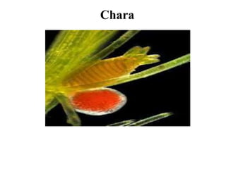
Globule:
The globule is large, spherical, red or yellow structure.
Development and Structure:
The early development of globule and nucule is similar. The pripheral
cell of the lower node of the primary lateral divides periclinally to form and an
inner cell. The outer cell functions as antheridial initial and the lower cell
again divides a periclinal division. Out of these three cells formed, the
lowermost functions as inter-nodal cell the middle forms basal node, the
uppermost functions as the antehridial initial (Fig. 8 A, B). The middle basal
node cell divides to make 5 peripheral cells. Out of these five peripheral cells,
the upper one develops into oogonium, two lateral ones form unicellular
bracteoles and two lower ones, one on either side of oogonium forms cortex or
remains non-functional (Fig. 8 C, D).
The antheridial initial divides by transverse division to make basal
pedicel cell and a terminal antheridial mother cell (Fig. 9 A, B). The pedicel
cell does not divide further and forms pedicel or stalk of mature antheridium.
The antheridial mother cell enlarges and divides by two successive vertical
division at right angle to each other to make a quadrant (Fig. 9 C-D). All
these four cells divide by a transverse wall to make eight cells or octant stage.
Each eel of the octant divides periclinally and forms two layers of eight cells
each. The cells of inner or outer layer divide periclinally to make three radial
layers of eight cells each. The outermost eight cells enlarge laterally to form a
curved plate of eight shield cells (Fig. 9 E, F). The cells of the middle layer
elongate towards centre to make eight rod-shaped manubrial cells (Fig. 9 F).
The cells of the inner layer function as eight primary capitulum cells.
Each primary capitulum cell divides to form six secondary capitulum
cells. Sometimes the secondary capitulum cells divide to make tertiary
capitulum cells. Each capitulum cell divides repeatedly to form 2-4 long,
multicellular, branched or un-branched antheridial Filaments or
sperinatogenous filaments (Fig. 9 G-H). The antheridial filament has up to 250
uninucleate cells. These cells function as sperm mother cell and each cell gives
rise to a single spirally coiled, uninucleate, bi-flagellated antherozoid (Fig. 9 J-
L). The mature globule thus is made up of 8 curved shield cells, 8 elongated
manubrial cells, 8 centrally located primary capitulum cells and 48 secondary
capitulum cells. The secondary capitulum cells give rise to many antheridial
filaments. Each sperm mother cell forms a single bi-flagellated antherozoid
(Fig. 9 K).
Liberation of Antherozoids:
At maturity the shield cells of antheridium separate from each other
exposing antheridial filaments in water. The sperm mother cell gelatinizes to
liberate the antherozoids.
Nucule:
The nucule of Chara is large, green, oval structure with short stalk. It
is borne at the node of the primary lateral. It lies just above the globule in
homothallic species. Development and Structure: The upper peripheral cell of
the basal node of the antheridium functions as the oogonial initial. The oogonial
initial divides by two transverse divisions to make three celled filament. It has
lower pedical cell, the middle nodal cell and the upper oogonial mother cell
(Fig. 10 A-C). The pedicell does not divide further and makes pedicel of the
oogonium. The middle nodal cell under many vertical divisions to make five
sheath cells or peripheral cell which surrounds the central cell (Fig. 10 D-F).
The central cell does not divide and functions as the node of the oogonium.
The oogonial mother cell elongates vertically and divides by
transverse division to make lower small stalk cell and an upper large
oogonium. The oogonium contains uninucleate ovum or egg. (Fig. 10 G, H).
The peripheral cells or five sheath initials elongate and divide by transverse
division to make two tiers of five cells each. The five upper tier cells from
coronary cells which form the corona of nucule. The five lower tier functions
as tube cells, the tube cells elongate and get spirally twisted in clockwise
directions on oogonium. The nucleus migrates on lower side and receptive spot
develops at the tip of oogonium. Large amount of starch and oil get deposited
in oogonium. The mature nucule is attached to the node by the pedicel cell.
The nucule is surrounded by five tube cells. The tips of tube cells from
corona at the top of nucule. The oogonial cell possesses a single large egg or
ovum. The nucule contains large amount of starch and
oil. The receptive spot is present at the upper part of nucule.
Fertilization:
When the oogonium is mature, the five tube cells get separated from each other
forming narrow slits between them. Antherozoids are chemotactically attracted
towards ovum. The antherozoids enter through these slits and penetrate gelatinized
wall of the oogonium. Many antherozoids enter oogonium but one of those fertilizes
the egg to make a diploid zygote. The zygote secretes a thick wall around itself to
make oospore.
Oospore:
The mature oospore is hard, oval, ellipsoid structure which may be
brown e.g., C. inferma, black e.g., C. corallina or golden brown e.g., C. flauda.
The oospore inside contains a diploid nucleus and many oil globules in
cytoplasm.
On maturity of oospore the inner walls of tube cells get thickened,
suberised and silicified. The oogonial as well as oospore walls become thick.
The oospore nucleus moves towards the apical region. In advanced stage the
outer walls of the envelope or sheath cells fall off and the inner parts remain
attached to mature oospore in form of ridges.
Germination of Oospore:
The oospore germinates when favourable conditions appear. The diploid nucleus present
in apical colourless region divides by meiosis forming four haploid daughter nuclei (Fig.
II A-B). At this stage a septum divides oospore into two unequal cells. The upper
smaller apical cell contains a single nucleus and the large basal cell contains three
nuclei. The three nuclei of basal cell degenerate gradually. The oospore apical cell
divides by longitudinal division to make a rhizoidal initial and protonemal
initial (Fig. 11 D). The rhizoidal initial shows positive geotropism and forms primary
rhizoid, the protonemal initial shows negative geotropism and forms primary
protonema. The primary protonema differentiates into nodes and internodes. The
peripheral cells of the basal node give rise to rhizoids and secondary protonema. The
peripheral cells of the upper nodes give rise to lateral branches (Fig. 11 E-I).
Life Cycle of Chara
The plant body of Chara is haploid. The vegetative reproduction takes
place by the formation of amylum stars, bulbils and secondary
protonema. Asexual reproduction is absent. The sexual reproduction
is advanced oogamous type. The male and female sex organs are
globule and nucule respectively. After fertilization a diploid spore is
formed. At the time of germination diploid oospore nucleus divides to
make hapoid nuclei and haploid Chara plant. Thus the life cycle of
Chara a predominantly haploid type (Fig. 12, 13).
chara.pptx
chara.pptx
chara.pptx

More Related Content

What's hot

Morphology, Structure and Reproduction of Equisetum.
Morphology, Structure and Reproduction of Equisetum. Morphology, Structure and Reproduction of Equisetum.
Morphology, Structure and Reproduction of Equisetum. Dhaneesh Ram
 
Marchantia
MarchantiaMarchantia
MarchantiaAkumpaul
 
General characters of pteridophytes
General characters of pteridophytesGeneral characters of pteridophytes
General characters of pteridophytesAliAbbasAslam
 
Xanthophyceae
XanthophyceaeXanthophyceae
XanthophyceaeNikkiM12
 
Pteris ppt
Pteris pptPteris ppt
Pteris pptmamtey1
 
Economic and Ecological Importance of Bryophytes.pptx
Economic and Ecological Importance of Bryophytes.pptxEconomic and Ecological Importance of Bryophytes.pptx
Economic and Ecological Importance of Bryophytes.pptxDilip Gavande
 
General characters and clasification of gymnosperm
General characters and clasification of gymnospermGeneral characters and clasification of gymnosperm
General characters and clasification of gymnospermgohil sanjay bhagvanji
 
Marsilea.pptx
Marsilea.pptxMarsilea.pptx
Marsilea.pptxjntuhcej
 
Types of stele and stelar evolution
Types of stele and stelar evolutionTypes of stele and stelar evolution
Types of stele and stelar evolutionRimiRoy6
 
Stelar evolution in Pteridophytes-BOTANY
Stelar evolution in Pteridophytes-BOTANYStelar evolution in Pteridophytes-BOTANY
Stelar evolution in Pteridophytes-BOTANYANJALIJAYAN6
 
Marsilea structure and reproduction
Marsilea structure and reproductionMarsilea structure and reproduction
Marsilea structure and reproductionMuhammad ArSlan
 

What's hot (20)

Morphology, Structure and Reproduction of Equisetum.
Morphology, Structure and Reproduction of Equisetum. Morphology, Structure and Reproduction of Equisetum.
Morphology, Structure and Reproduction of Equisetum.
 
Marchantia
MarchantiaMarchantia
Marchantia
 
Hydrodictyon
HydrodictyonHydrodictyon
Hydrodictyon
 
General characters of pteridophytes
General characters of pteridophytesGeneral characters of pteridophytes
General characters of pteridophytes
 
Oedogonium
OedogoniumOedogonium
Oedogonium
 
Xanthophyceae
XanthophyceaeXanthophyceae
Xanthophyceae
 
Pteris ppt
Pteris pptPteris ppt
Pteris ppt
 
Economic and Ecological Importance of Bryophytes.pptx
Economic and Ecological Importance of Bryophytes.pptxEconomic and Ecological Importance of Bryophytes.pptx
Economic and Ecological Importance of Bryophytes.pptx
 
Vaucheria ppt.pptx
Vaucheria ppt.pptxVaucheria ppt.pptx
Vaucheria ppt.pptx
 
Marchantia ppt
Marchantia pptMarchantia ppt
Marchantia ppt
 
General characters and clasification of gymnosperm
General characters and clasification of gymnospermGeneral characters and clasification of gymnosperm
General characters and clasification of gymnosperm
 
Marsilea.pptx
Marsilea.pptxMarsilea.pptx
Marsilea.pptx
 
Types of stele and stelar evolution
Types of stele and stelar evolutionTypes of stele and stelar evolution
Types of stele and stelar evolution
 
Volvox
VolvoxVolvox
Volvox
 
Telome Theory
Telome TheoryTelome Theory
Telome Theory
 
Stelar evolution in Pteridophytes-BOTANY
Stelar evolution in Pteridophytes-BOTANYStelar evolution in Pteridophytes-BOTANY
Stelar evolution in Pteridophytes-BOTANY
 
Batrachospermum.pptx
Batrachospermum.pptxBatrachospermum.pptx
Batrachospermum.pptx
 
Notothylas by p.n
Notothylas by p.nNotothylas by p.n
Notothylas by p.n
 
Marsilea structure and reproduction
Marsilea structure and reproductionMarsilea structure and reproduction
Marsilea structure and reproduction
 
5 2 -pinus
5   2 -pinus5   2 -pinus
5 2 -pinus
 

Similar to chara.pptx

Similar to chara.pptx (20)

CHARA.pptx
CHARA.pptxCHARA.pptx
CHARA.pptx
 
Vegetative characters of equisetum
Vegetative characters of equisetumVegetative characters of equisetum
Vegetative characters of equisetum
 
Marchantia jb
Marchantia jbMarchantia jb
Marchantia jb
 
Bryopsida – funaria
Bryopsida – funariaBryopsida – funaria
Bryopsida – funaria
 
Anatomy of dicot and monocot stem
Anatomy of dicot and monocot stemAnatomy of dicot and monocot stem
Anatomy of dicot and monocot stem
 
Algae thallus
Algae thallusAlgae thallus
Algae thallus
 
Type polytrichum
Type polytrichumType polytrichum
Type polytrichum
 
Taxus
TaxusTaxus
Taxus
 
35 plantstructure text
35  plantstructure text35  plantstructure text
35 plantstructure text
 
Funaria
FunariaFunaria
Funaria
 
General plant organization
General plant organizationGeneral plant organization
General plant organization
 
Life-cycle-of-Sphagnum.pdf
Life-cycle-of-Sphagnum.pdfLife-cycle-of-Sphagnum.pdf
Life-cycle-of-Sphagnum.pdf
 
8. Vegetative and sexual reproduction in funaria
8. Vegetative and sexual reproduction in funaria8. Vegetative and sexual reproduction in funaria
8. Vegetative and sexual reproduction in funaria
 
CYCAS-1.pdf
CYCAS-1.pdfCYCAS-1.pdf
CYCAS-1.pdf
 
ANTHOCEROS(bryophyta) Faizan botany department GPGC kohat
ANTHOCEROS(bryophyta) Faizan botany department GPGC kohatANTHOCEROS(bryophyta) Faizan botany department GPGC kohat
ANTHOCEROS(bryophyta) Faizan botany department GPGC kohat
 
Gnetum Botany Characters used for B>Sc and M.Sc.
Gnetum Botany Characters used for B>Sc and M.Sc.Gnetum Botany Characters used for B>Sc and M.Sc.
Gnetum Botany Characters used for B>Sc and M.Sc.
 
Aspergillus
AspergillusAspergillus
Aspergillus
 
Developement of Embryo
Developement of Embryo Developement of Embryo
Developement of Embryo
 
Plant Kingdom III
Plant Kingdom IIIPlant Kingdom III
Plant Kingdom III
 
structure and function in plants
structure and function in plantsstructure and function in plants
structure and function in plants
 

More from rnath286

Phaeophyceae.ppt
Phaeophyceae.pptPhaeophyceae.ppt
Phaeophyceae.pptrnath286
 
control of microbes.pptx
control of microbes.pptxcontrol of microbes.pptx
control of microbes.pptxrnath286
 
BIOINFO unit 1.pptx
BIOINFO unit 1.pptxBIOINFO unit 1.pptx
BIOINFO unit 1.pptxrnath286
 
cell membrane.pptx
cell membrane.pptxcell membrane.pptx
cell membrane.pptxrnath286
 
coleochaetea.pptx
coleochaetea.pptxcoleochaetea.pptx
coleochaetea.pptxrnath286
 
Volvox.pptx
Volvox.pptxVolvox.pptx
Volvox.pptxrnath286
 
algae classification.pptx
algae classification.pptxalgae classification.pptx
algae classification.pptxrnath286
 
virus.pptx
virus.pptxvirus.pptx
virus.pptxrnath286
 
Microbio.pptx
Microbio.pptxMicrobio.pptx
Microbio.pptxrnath286
 

More from rnath286 (9)

Phaeophyceae.ppt
Phaeophyceae.pptPhaeophyceae.ppt
Phaeophyceae.ppt
 
control of microbes.pptx
control of microbes.pptxcontrol of microbes.pptx
control of microbes.pptx
 
BIOINFO unit 1.pptx
BIOINFO unit 1.pptxBIOINFO unit 1.pptx
BIOINFO unit 1.pptx
 
cell membrane.pptx
cell membrane.pptxcell membrane.pptx
cell membrane.pptx
 
coleochaetea.pptx
coleochaetea.pptxcoleochaetea.pptx
coleochaetea.pptx
 
Volvox.pptx
Volvox.pptxVolvox.pptx
Volvox.pptx
 
algae classification.pptx
algae classification.pptxalgae classification.pptx
algae classification.pptx
 
virus.pptx
virus.pptxvirus.pptx
virus.pptx
 
Microbio.pptx
Microbio.pptxMicrobio.pptx
Microbio.pptx
 

Recently uploaded

Manassas R - Parkside Middle School 🌎🏫
Manassas R - Parkside Middle School 🌎🏫Manassas R - Parkside Middle School 🌎🏫
Manassas R - Parkside Middle School 🌎🏫qfactory1
 
Transposable elements in prokaryotes.ppt
Transposable elements in prokaryotes.pptTransposable elements in prokaryotes.ppt
Transposable elements in prokaryotes.pptArshadWarsi13
 
TOTAL CHOLESTEROL (lipid profile test).pptx
TOTAL CHOLESTEROL (lipid profile test).pptxTOTAL CHOLESTEROL (lipid profile test).pptx
TOTAL CHOLESTEROL (lipid profile test).pptxdharshini369nike
 
Best Call Girls In Sector 29 Gurgaon❤️8860477959 EscorTs Service In 24/7 Delh...
Best Call Girls In Sector 29 Gurgaon❤️8860477959 EscorTs Service In 24/7 Delh...Best Call Girls In Sector 29 Gurgaon❤️8860477959 EscorTs Service In 24/7 Delh...
Best Call Girls In Sector 29 Gurgaon❤️8860477959 EscorTs Service In 24/7 Delh...lizamodels9
 
‏‏VIRUS - 123455555555555555555555555555555555555555
‏‏VIRUS -  123455555555555555555555555555555555555555‏‏VIRUS -  123455555555555555555555555555555555555555
‏‏VIRUS - 123455555555555555555555555555555555555555kikilily0909
 
Vision and reflection on Mining Software Repositories research in 2024
Vision and reflection on Mining Software Repositories research in 2024Vision and reflection on Mining Software Repositories research in 2024
Vision and reflection on Mining Software Repositories research in 2024AyushiRastogi48
 
Solution chemistry, Moral and Normal solutions
Solution chemistry, Moral and Normal solutionsSolution chemistry, Moral and Normal solutions
Solution chemistry, Moral and Normal solutionsHajira Mahmood
 
Call Girls in Aiims Metro Delhi 💯Call Us 🔝9953322196🔝 💯Escort.
Call Girls in Aiims Metro Delhi 💯Call Us 🔝9953322196🔝 💯Escort.Call Girls in Aiims Metro Delhi 💯Call Us 🔝9953322196🔝 💯Escort.
Call Girls in Aiims Metro Delhi 💯Call Us 🔝9953322196🔝 💯Escort.aasikanpl
 
Evidences of Evolution General Biology 2
Evidences of Evolution General Biology 2Evidences of Evolution General Biology 2
Evidences of Evolution General Biology 2John Carlo Rollon
 
Harmful and Useful Microorganisms Presentation
Harmful and Useful Microorganisms PresentationHarmful and Useful Microorganisms Presentation
Harmful and Useful Microorganisms Presentationtahreemzahra82
 
Welcome to GFDL for Take Your Child To Work Day
Welcome to GFDL for Take Your Child To Work DayWelcome to GFDL for Take Your Child To Work Day
Welcome to GFDL for Take Your Child To Work DayZachary Labe
 
Call Girls in Hauz Khas Delhi 💯Call Us 🔝9953322196🔝 💯Escort.
Call Girls in Hauz Khas Delhi 💯Call Us 🔝9953322196🔝 💯Escort.Call Girls in Hauz Khas Delhi 💯Call Us 🔝9953322196🔝 💯Escort.
Call Girls in Hauz Khas Delhi 💯Call Us 🔝9953322196🔝 💯Escort.aasikanpl
 
Spermiogenesis or Spermateleosis or metamorphosis of spermatid
Spermiogenesis or Spermateleosis or metamorphosis of spermatidSpermiogenesis or Spermateleosis or metamorphosis of spermatid
Spermiogenesis or Spermateleosis or metamorphosis of spermatidSarthak Sekhar Mondal
 
insect anatomy and insect body wall and their physiology
insect anatomy and insect body wall and their  physiologyinsect anatomy and insect body wall and their  physiology
insect anatomy and insect body wall and their physiologyDrAnita Sharma
 
Dashanga agada a formulation of Agada tantra dealt in 3 Rd year bams agada tanta
Dashanga agada a formulation of Agada tantra dealt in 3 Rd year bams agada tantaDashanga agada a formulation of Agada tantra dealt in 3 Rd year bams agada tanta
Dashanga agada a formulation of Agada tantra dealt in 3 Rd year bams agada tantaPraksha3
 
Forest laws, Indian forest laws, why they are important
Forest laws, Indian forest laws, why they are importantForest laws, Indian forest laws, why they are important
Forest laws, Indian forest laws, why they are importantadityabhardwaj282
 
BIOETHICS IN RECOMBINANT DNA TECHNOLOGY.
BIOETHICS IN RECOMBINANT DNA TECHNOLOGY.BIOETHICS IN RECOMBINANT DNA TECHNOLOGY.
BIOETHICS IN RECOMBINANT DNA TECHNOLOGY.PraveenaKalaiselvan1
 
Call Us ≽ 9953322196 ≼ Call Girls In Lajpat Nagar (Delhi) |
Call Us ≽ 9953322196 ≼ Call Girls In Lajpat Nagar (Delhi) |Call Us ≽ 9953322196 ≼ Call Girls In Lajpat Nagar (Delhi) |
Call Us ≽ 9953322196 ≼ Call Girls In Lajpat Nagar (Delhi) |aasikanpl
 

Recently uploaded (20)

Hot Sexy call girls in Moti Nagar,🔝 9953056974 🔝 escort Service
Hot Sexy call girls in  Moti Nagar,🔝 9953056974 🔝 escort ServiceHot Sexy call girls in  Moti Nagar,🔝 9953056974 🔝 escort Service
Hot Sexy call girls in Moti Nagar,🔝 9953056974 🔝 escort Service
 
Manassas R - Parkside Middle School 🌎🏫
Manassas R - Parkside Middle School 🌎🏫Manassas R - Parkside Middle School 🌎🏫
Manassas R - Parkside Middle School 🌎🏫
 
Transposable elements in prokaryotes.ppt
Transposable elements in prokaryotes.pptTransposable elements in prokaryotes.ppt
Transposable elements in prokaryotes.ppt
 
TOTAL CHOLESTEROL (lipid profile test).pptx
TOTAL CHOLESTEROL (lipid profile test).pptxTOTAL CHOLESTEROL (lipid profile test).pptx
TOTAL CHOLESTEROL (lipid profile test).pptx
 
Best Call Girls In Sector 29 Gurgaon❤️8860477959 EscorTs Service In 24/7 Delh...
Best Call Girls In Sector 29 Gurgaon❤️8860477959 EscorTs Service In 24/7 Delh...Best Call Girls In Sector 29 Gurgaon❤️8860477959 EscorTs Service In 24/7 Delh...
Best Call Girls In Sector 29 Gurgaon❤️8860477959 EscorTs Service In 24/7 Delh...
 
Engler and Prantl system of classification in plant taxonomy
Engler and Prantl system of classification in plant taxonomyEngler and Prantl system of classification in plant taxonomy
Engler and Prantl system of classification in plant taxonomy
 
‏‏VIRUS - 123455555555555555555555555555555555555555
‏‏VIRUS -  123455555555555555555555555555555555555555‏‏VIRUS -  123455555555555555555555555555555555555555
‏‏VIRUS - 123455555555555555555555555555555555555555
 
Vision and reflection on Mining Software Repositories research in 2024
Vision and reflection on Mining Software Repositories research in 2024Vision and reflection on Mining Software Repositories research in 2024
Vision and reflection on Mining Software Repositories research in 2024
 
Solution chemistry, Moral and Normal solutions
Solution chemistry, Moral and Normal solutionsSolution chemistry, Moral and Normal solutions
Solution chemistry, Moral and Normal solutions
 
Call Girls in Aiims Metro Delhi 💯Call Us 🔝9953322196🔝 💯Escort.
Call Girls in Aiims Metro Delhi 💯Call Us 🔝9953322196🔝 💯Escort.Call Girls in Aiims Metro Delhi 💯Call Us 🔝9953322196🔝 💯Escort.
Call Girls in Aiims Metro Delhi 💯Call Us 🔝9953322196🔝 💯Escort.
 
Evidences of Evolution General Biology 2
Evidences of Evolution General Biology 2Evidences of Evolution General Biology 2
Evidences of Evolution General Biology 2
 
Harmful and Useful Microorganisms Presentation
Harmful and Useful Microorganisms PresentationHarmful and Useful Microorganisms Presentation
Harmful and Useful Microorganisms Presentation
 
Welcome to GFDL for Take Your Child To Work Day
Welcome to GFDL for Take Your Child To Work DayWelcome to GFDL for Take Your Child To Work Day
Welcome to GFDL for Take Your Child To Work Day
 
Call Girls in Hauz Khas Delhi 💯Call Us 🔝9953322196🔝 💯Escort.
Call Girls in Hauz Khas Delhi 💯Call Us 🔝9953322196🔝 💯Escort.Call Girls in Hauz Khas Delhi 💯Call Us 🔝9953322196🔝 💯Escort.
Call Girls in Hauz Khas Delhi 💯Call Us 🔝9953322196🔝 💯Escort.
 
Spermiogenesis or Spermateleosis or metamorphosis of spermatid
Spermiogenesis or Spermateleosis or metamorphosis of spermatidSpermiogenesis or Spermateleosis or metamorphosis of spermatid
Spermiogenesis or Spermateleosis or metamorphosis of spermatid
 
insect anatomy and insect body wall and their physiology
insect anatomy and insect body wall and their  physiologyinsect anatomy and insect body wall and their  physiology
insect anatomy and insect body wall and their physiology
 
Dashanga agada a formulation of Agada tantra dealt in 3 Rd year bams agada tanta
Dashanga agada a formulation of Agada tantra dealt in 3 Rd year bams agada tantaDashanga agada a formulation of Agada tantra dealt in 3 Rd year bams agada tanta
Dashanga agada a formulation of Agada tantra dealt in 3 Rd year bams agada tanta
 
Forest laws, Indian forest laws, why they are important
Forest laws, Indian forest laws, why they are importantForest laws, Indian forest laws, why they are important
Forest laws, Indian forest laws, why they are important
 
BIOETHICS IN RECOMBINANT DNA TECHNOLOGY.
BIOETHICS IN RECOMBINANT DNA TECHNOLOGY.BIOETHICS IN RECOMBINANT DNA TECHNOLOGY.
BIOETHICS IN RECOMBINANT DNA TECHNOLOGY.
 
Call Us ≽ 9953322196 ≼ Call Girls In Lajpat Nagar (Delhi) |
Call Us ≽ 9953322196 ≼ Call Girls In Lajpat Nagar (Delhi) |Call Us ≽ 9953322196 ≼ Call Girls In Lajpat Nagar (Delhi) |
Call Us ≽ 9953322196 ≼ Call Girls In Lajpat Nagar (Delhi) |
 

chara.pptx

  • 3. Occurrence of Chara Chara is a fresh water, green alga found submerged in shallow water ponds, tanks, lakes and slow running water. C. baltica is found growing is brackish water and C. fragilis is found in hot springs. Chara is found mostly in hard fresh water, rich in organic matter, calcium and deficient in oxygen. Chara plants are often encrusted with calcium carbonate and hence are commonly called stone wort. Chara often emits disagreeable onion like odour due to presence of sulphur compounds. C. hatei grows trailing on the soil C. nuda and C. grovesii are found on mountains, C. wallichii and C. liydropitys are found in plains.
  • 4. Thallus Structure of Chara The thallus of Chara is branched, multicellular and macroscopic. The thallus is normally 20-30 cm. in height but often may be up to 90 cm to l m. Some species like C. hatei are small and may be 2-3 cm. long. The plants in appearance resemble Equisetum hence Chara is commonly called as aquatic horsetail. The thallus is mainly differentiated into rhizoids and main axis
  • 5.
  • 6. The rhizoids are white, thread like, multicellular, uniseriate and branched structures. The rhizoids arise from rhizoidal plates which are formed at the base of main axis or from peripheral cells of lower nodes. The rhizoids are characterized by presence of oblique Septa. The tips of rhizoids possess minute solid particles which function as statoliths. The rhizoids show apical growth. Rhizoids help in attachment of plant to substratum i.e., mud or sand, in absorption of minerals and in vegetative multiplication of plants by forming bulbils and secondary protonema. Rhizoids:
  • 7. The main axis is erect, long, branched and differentiated into nodes and internodes. The internode consists of single, much elongated or oblong cell. The inter-nodal cells in some species may be surrounded by one celled thick layer called cortex and such species are called as corticate species. T he species in which cortical layer is absent are called ecorticate species (Fig. 3 A, B). The node consists of a pair of central small cells surrounded by 6-20 peripheral cells (Fig. 3 C). The central cells and peripheral cells arise from a single nodal initial cell. Main Axis:
  • 8.
  • 9. On nodes develop these following four types of appendages: (i) Branches of limited growth. The branches of limited growth arise in whorls of 6-20 from peripheral cells of the nodes of main axis or on branches of unlimited growth. These are also called branchlets, branches of first order, primary laterals or leaves. These branches stop to grow after forming 5-15 nodes and hence are called branches of limited growth. The stipulodes and reproductive structures are formed on the node of these branches. (ii) Branches of unlimited growth: The branches – of unlimited growth arise from the axils of the branches of limited growth hence these are also called auxiliary branches or long laterals. These are differentiated into nodes and internodes. At nodes they bear primary laterals and these branches look like the main axis. Their growth is also unlimited like main axis.
  • 10. (iii) Stipulodes: The basal node of the branches of limited growth develops short, oval, pointed single cell outgrowths called stipulodes. In most of the species of Chara e.g., C. burmanica, the number of stipulodes at each node is twice the number of primary laterals, such species are called as bi-stipulate. In some species of Chara e.g., C. nuda and C. braunii, the number of stipulodes at each node, is equal to number of primary laterals at that node, such species are called unitipulate. When stipulodes are present in one whorl at each node the species are called as haplostephanous and with two whorls on each node are called diplostephanous (Fig. 4 A, B).
  • 11. (iv) Cortex: Many species of Chara e.g., C. aspera, C. inferma have inter- nodal cells of main axis en-sheathed by cortex cells. Such species are called corticated species. The cortex consists of vertically elongated narrow cells. The internode up to half of its length by corticating filaments developed from upper node called descending the lower half of internode is covered by filaments developed from lower node called filaments. The ascending and descending filaments meet at the middle of internode. T species without cortex e.g., C. corallina are called ecorticated species.
  • 12. The main axis of Chara consists of mainly two types of cells: (i) Nodal cells (ii) Inter-nodal cells. The nodal cells are smaller in size and isodiametric. The cells are dense cytoplasmic, uninucleate with few small ellipsoidal chloroplasts. The central vacuole is not developed instead many small vacuoles may be present. The cytoplasm can be differentiated in outer exoplasm and inner endoplasm (Fig. 5 A). The inter-nodal cells are much elongated. The cytoplasm is present around a large central vacuole. The cells are multinucleate and contain many discoid chloroplasts. The cytoplasm is also differentiated into outer exoplasm and inner endoplasm. The endoplasm shows streaming movements (Fig. 5 B). The cell walls between the nodal cell and inter-nodal cells are porous to help in cytoplasmic continuity between cells. Cell Structure of Chara:
  • 13.
  • 14. Reproduction in Chara takes place by vegetative and sexual methods. Asexual reproduction is absent. (i) Vegetative Reproduction in Chara: Vegetative reproduction in Chara takes place by following methods: (a) Bulbils: The bulbils are spherical or oval tube-like structures which develop on rhizoids. C. aspora or on lower nodes of main axis e.g., C. baltica. The bulbils on detachment from plants germinate into new thallus (Fig. 6 A). (b) Amylum Stars: In some species of Chara e.g., C. stelligna, on the lower nodes of main axis develop multicellular star shape aggregates of cells (Fig. 6 B). These cells are full of amylum starch and hence are called Amylum stars. The amylum stars do detachment from plants develops into new Chara thalli. Reproduction in Chara
  • 15.
  • 16. (c) Amorphous bulbils: The amoiphous bulbils are group, many cells, irregular in shape which develop on lower node main axis e.g., C. delicatula or on rhizoids e.g., C. fragifera and C. baltica. The amorphous bulbils are perennating structures, when the main plant dies under unfavorable conditions; these bulbils survive and make Chara plants on return of favourable conditions. (d) Secondary Protonema: These are tubular or filamentous structure which develops from primary protonema or the basal cells of the rhizoids. The secondary protonema like primary protonema form Chara plants.
  • 17. Sexual Reproduction in Chara: The sexual reproduction in Chara is of highly advanced oogamous type. The sex organs are macroscopic and complex in organization. The male sex organs are called antheridium or globule and the female oogonium or nucule. Most of the Chara species are homothallic i.e., the male and male sex organs are borne on the same nodes, (Fig. 7) e.g., C. zeylanica. Some species e.g., C. wallichii are heterothallic i.e., male and female sex organs are borne on different plants. The sex organs arise on the branches of limited growth or primary laterals, the nucule above the globule. The development of globule and nucule takes place simultaneously but species globule matures before nucule (Fig. 7 A, B).
  • 18.
  • 19. Globule: The globule is large, spherical, red or yellow structure. Development and Structure: The early development of globule and nucule is similar. The pripheral cell of the lower node of the primary lateral divides periclinally to form and an inner cell. The outer cell functions as antheridial initial and the lower cell again divides a periclinal division. Out of these three cells formed, the lowermost functions as inter-nodal cell the middle forms basal node, the uppermost functions as the antehridial initial (Fig. 8 A, B). The middle basal node cell divides to make 5 peripheral cells. Out of these five peripheral cells, the upper one develops into oogonium, two lateral ones form unicellular bracteoles and two lower ones, one on either side of oogonium forms cortex or remains non-functional (Fig. 8 C, D).
  • 20.
  • 21. The antheridial initial divides by transverse division to make basal pedicel cell and a terminal antheridial mother cell (Fig. 9 A, B). The pedicel cell does not divide further and forms pedicel or stalk of mature antheridium. The antheridial mother cell enlarges and divides by two successive vertical division at right angle to each other to make a quadrant (Fig. 9 C-D). All these four cells divide by a transverse wall to make eight cells or octant stage. Each eel of the octant divides periclinally and forms two layers of eight cells each. The cells of inner or outer layer divide periclinally to make three radial layers of eight cells each. The outermost eight cells enlarge laterally to form a curved plate of eight shield cells (Fig. 9 E, F). The cells of the middle layer elongate towards centre to make eight rod-shaped manubrial cells (Fig. 9 F). The cells of the inner layer function as eight primary capitulum cells.
  • 22. Each primary capitulum cell divides to form six secondary capitulum cells. Sometimes the secondary capitulum cells divide to make tertiary capitulum cells. Each capitulum cell divides repeatedly to form 2-4 long, multicellular, branched or un-branched antheridial Filaments or sperinatogenous filaments (Fig. 9 G-H). The antheridial filament has up to 250 uninucleate cells. These cells function as sperm mother cell and each cell gives rise to a single spirally coiled, uninucleate, bi-flagellated antherozoid (Fig. 9 J- L). The mature globule thus is made up of 8 curved shield cells, 8 elongated manubrial cells, 8 centrally located primary capitulum cells and 48 secondary capitulum cells. The secondary capitulum cells give rise to many antheridial filaments. Each sperm mother cell forms a single bi-flagellated antherozoid (Fig. 9 K).
  • 23.
  • 24. Liberation of Antherozoids: At maturity the shield cells of antheridium separate from each other exposing antheridial filaments in water. The sperm mother cell gelatinizes to liberate the antherozoids.
  • 25. Nucule: The nucule of Chara is large, green, oval structure with short stalk. It is borne at the node of the primary lateral. It lies just above the globule in homothallic species. Development and Structure: The upper peripheral cell of the basal node of the antheridium functions as the oogonial initial. The oogonial initial divides by two transverse divisions to make three celled filament. It has lower pedical cell, the middle nodal cell and the upper oogonial mother cell (Fig. 10 A-C). The pedicell does not divide further and makes pedicel of the oogonium. The middle nodal cell under many vertical divisions to make five sheath cells or peripheral cell which surrounds the central cell (Fig. 10 D-F). The central cell does not divide and functions as the node of the oogonium.
  • 26.
  • 27. The oogonial mother cell elongates vertically and divides by transverse division to make lower small stalk cell and an upper large oogonium. The oogonium contains uninucleate ovum or egg. (Fig. 10 G, H). The peripheral cells or five sheath initials elongate and divide by transverse division to make two tiers of five cells each. The five upper tier cells from coronary cells which form the corona of nucule. The five lower tier functions as tube cells, the tube cells elongate and get spirally twisted in clockwise directions on oogonium. The nucleus migrates on lower side and receptive spot develops at the tip of oogonium. Large amount of starch and oil get deposited in oogonium. The mature nucule is attached to the node by the pedicel cell. The nucule is surrounded by five tube cells. The tips of tube cells from corona at the top of nucule. The oogonial cell possesses a single large egg or ovum. The nucule contains large amount of starch and oil. The receptive spot is present at the upper part of nucule.
  • 28. Fertilization: When the oogonium is mature, the five tube cells get separated from each other forming narrow slits between them. Antherozoids are chemotactically attracted towards ovum. The antherozoids enter through these slits and penetrate gelatinized wall of the oogonium. Many antherozoids enter oogonium but one of those fertilizes the egg to make a diploid zygote. The zygote secretes a thick wall around itself to make oospore.
  • 29. Oospore: The mature oospore is hard, oval, ellipsoid structure which may be brown e.g., C. inferma, black e.g., C. corallina or golden brown e.g., C. flauda. The oospore inside contains a diploid nucleus and many oil globules in cytoplasm. On maturity of oospore the inner walls of tube cells get thickened, suberised and silicified. The oogonial as well as oospore walls become thick. The oospore nucleus moves towards the apical region. In advanced stage the outer walls of the envelope or sheath cells fall off and the inner parts remain attached to mature oospore in form of ridges.
  • 30. Germination of Oospore: The oospore germinates when favourable conditions appear. The diploid nucleus present in apical colourless region divides by meiosis forming four haploid daughter nuclei (Fig. II A-B). At this stage a septum divides oospore into two unequal cells. The upper smaller apical cell contains a single nucleus and the large basal cell contains three nuclei. The three nuclei of basal cell degenerate gradually. The oospore apical cell divides by longitudinal division to make a rhizoidal initial and protonemal initial (Fig. 11 D). The rhizoidal initial shows positive geotropism and forms primary rhizoid, the protonemal initial shows negative geotropism and forms primary protonema. The primary protonema differentiates into nodes and internodes. The peripheral cells of the basal node give rise to rhizoids and secondary protonema. The peripheral cells of the upper nodes give rise to lateral branches (Fig. 11 E-I).
  • 31.
  • 32. Life Cycle of Chara The plant body of Chara is haploid. The vegetative reproduction takes place by the formation of amylum stars, bulbils and secondary protonema. Asexual reproduction is absent. The sexual reproduction is advanced oogamous type. The male and female sex organs are globule and nucule respectively. After fertilization a diploid spore is formed. At the time of germination diploid oospore nucleus divides to make hapoid nuclei and haploid Chara plant. Thus the life cycle of Chara a predominantly haploid type (Fig. 12, 13).