Chapter 6

 A Tour of the Cell


PowerPoint® Lecture Presentations for

        Biology
       Eighth Edition
Neil Campbell and Jane Reece

Lectures by Chris Romero, updated by Erin Barley with contributions from Joan Sharp
Copyright © 2008 Pearson Education, Inc., publishing as Pearson Benjamin Cummings
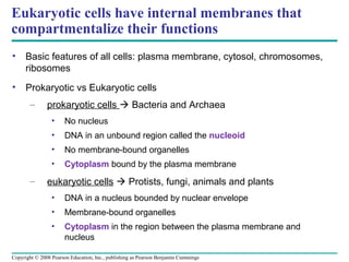
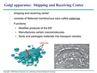
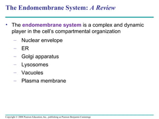
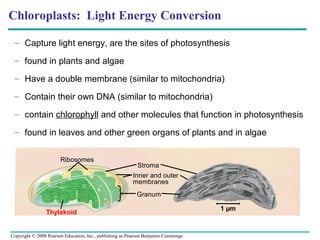
Eukaryotic cells have internal membranes that
compartmentalize their functions
•    Basic features of all cells: plasma membrane, cytosol, chromosomes,
     ribosomes
•    Prokaryotic vs Eukaryotic cells
       –       prokaryotic cells  Bacteria and Archaea
                 •    No nucleus
                 •    DNA in an unbound region called the nucleoid
                 •    No membrane-bound organelles
                 •    Cytoplasm bound by the plasma membrane
       –       eukaryotic cells  Protists, fungi, animals and plants
                 •    DNA in a nucleus bounded by nuclear envelope
                 •    Membrane-bound organelles
                 •    Cytoplasm in the region between the plasma membrane and
                      nucleus

Copyright © 2008 Pearson Education, Inc., publishing as Pearson Benjamin Cummings
Fig. 6-6


                 Prokaryotic cell

                   Fimbriae

                      Nucleoid

                         Ribosomes

                       Plasma membrane

     Bacterial                Cell wall
 chromosome
                                  Capsule
                                            0.5 µm
                                 Flagella
Fig. 6-9a
                      Eukaryotic Animal Cell
                                                                  Nuclear
                                                                  envelope
                          ENDOPLASMIC RETICULUM (ER)
                                                                  Nucleolus      NUCLEUS
                               Rough ER    Smooth ER
             Flagellum***                                         Chromatin
             (not in plants)

               Centrosome
                                                                               Plasma
                                                                               membrane

       CYTOSKELETON:
        Microfilaments
             Intermediate
                 filaments
            Microtubules                                                  Ribosomes

             Microvilli


                                                                Golgi
        Peroxisome                                              apparatus
                               Mitochondrion
                                                 Lysosome***
                                                 (not in plants/prokaryotes)
Fig. 6-9b

                             Eukaryotic Plant Cell
                       Nuclear envelope    Rough endoplasmic reticulum
            NUCLEUS    Nucleolus
                       Chromatin
                                                     Smooth endoplasmic reticulum
                                                                Ribosomes


                                                                Central
                    Golgi                                       vacuole***
                apparatus
                                                                  Microfilaments
                                                                  Intermediate      CYTO-SKELETON
                                                                  filaments
                                                                  Microtubules


             Mitochondrion
               Peroxisome
                                                                         Chloroplast***
                     Plasma
                 membrane
                                 Cell
                                 wall***
                                                     Plasmodesmata***
                   Wall of adjacent cell

                                                                           ***specific to plants
Organelles to know
•   Nucleus – Genetic information
•   Ribosomes – protein factories
•   Endoplasmic Reticulum – protein trafficking and metabolic functions
•   Golgi Apparatus – shipping and receiving center
•   Lysosomes – digestive compartments
•   Vacuoles – maintenance compatments
•   Mitochondria – chemical energy conversion (site of cellular respiration)
•   Chloroplasts – light energy conversion (site of photosynthesis)
•   Peroxisomes - oxidation
•   Cytoskeleton – support, motility and regulation
•   Extracellular Matrix – support, adhesion, movement, regulation
•   Intercellular Junctions – facilitate contact between cells
Nucleus: Information Central

       –       nuclear envelope encloses the nucleus, separating it from the
               cytoplasm
       –       nuclear membrane is a double membrane; each membrane
               consists of a lipid bilayer
       –       Pores regulate the entry and exit of molecules from the nucleus
       –       The shape of the nucleus is maintained by the nuclear lamina,
               which is composed of protein
       –       In the nucleus, DNA and proteins form genetic material called
               chromatin
       –       Chromatin condenses to form discrete chromosomes
       –       The nucleolus is located within the nucleus and is the site of
               ribosomal RNA (rRNA) synthesis


Copyright © 2008 Pearson Education, Inc., publishing as Pearson Benjamin Cummings
Fig. 6-10

                                                                      Nucleus
  1 µm                              Nucleolus
                               Chromatin

                     Nuclear envelope


                     Nuclear pore


                               Pore
                               complex


 Surface of
 nuclear envelope
                    Ribosome                                            1 µm

  0.25 µm

                               Close-up of nuclear
                               envelope


 Pore complexes                                      Nuclear lamina
Ribosomes: Protein factories

   –   particles made of ribosomal RNA and protein
   –   Protein synthesis occurs here
       • Free ribosomes are localized to the cytosol

       • Bound ribosomes are on the ER or the nuclear envelope

                                       Cytosol
                                       Endoplasmic reticulum (ER)

                                       Free ribosomes

                                       Bound ribosomes


                                                         Large subunit


         Fig. 6-11                                       Small subunit

                                        Diagram of a ribosome
Endoplasmic Reticulum (ER): Protein Trafficking

       –       The ER membrane is attached to the nuclear envelope
       –       There are two distinct regions of ER:
                 • Smooth ER  lacks ribosomes
                        –     Synthesizes lipids
                        –     Metabolizes carbohydrates
                        –     Detoxifies poison
                        –     Stores calcium
                 • Rough ER with ribosomes
                        –     Secretes glycoproteins (proteins covalently bonded to
                              carbohydrates)
                        –     Distributes transport vesicles, proteins surrounded by
                              membranes
                        –     membrane factory for the cell

Copyright © 2008 Pearson Education, Inc., publishing as Pearson Benjamin Cummings
Fig. 6-12
                      Smooth ER

                   Rough ER           Nuclear
                                      envelope


                        Ribosomes
                                    Transport vesicle


                                                        Rough ER
       Smooth ER
Golgi apparatus: Shipping and Receiving Center

         –      shipping and receiving center
         –      consists of flattened membranous sacs called cisternae
         –      Functions:
                  • Modifies products of the ER
                  • Manufactures certain macromolecules
                  • Sorts and packages materials into transport vesicles

                                                        Cisternae


Copyright © 2008 Pearson Education, Inc., publishing as Pearson Benjamin Cummings
Lysosome: Digestive Compartment
      –       a membranous sac of hydrolytic enzymes that can digest macromolecules
      –       Lysosomal enzymes can hydrolyze proteins, fats, polysaccharides, and
              nucleic acids
               •       After phagocytosis (engulfing of another cell) lysosomes fuse with the
                       food vacuole and digests the molecules
               •     Autophagy uses enzymes to recycle the cell’s own organelles and
                     macromolecules


 (a) Phagocytosis                                                       (b) Autophagy
                        Digestive
                        enzymes
Lysosome                                                                            Lysosome

    Plasma                                                             Peroxisome
    membrane
                                                        Digestion

                     Food vacuole                                                                             Digestion
                                                                                              Mitochondrion
                                                                                    Vesicle


Copyright © 2008 Pearson Education, Inc., publishing as Pearson Benjamin Cummings
Fig. 6-15
Vacuoles: Maintenance Compartment

      –       Diverse
              Maintenance
              Compartments
                                                                                    Central
      –       Food                                                                  vacuole
              vacuoles are                                                                    Cytosol
              formed by
              phagocytosis
      –       Contractile
              vacuoles                                               Nucleus                   Central
                                                                                               vacuole
              pump excess
              water out of                                           Cell wall
              cells                                                  Chloroplast

      –       Central
              vacuoles (in
              many mature
              plant cells)
              hold organic
Copyright © 2008 Pearson Education, Inc., publishing as Pearson Benjamin Cummings
The Endomembrane System: A Review

• The endomembrane system is a complex and dynamic
  player in the cell’s compartmental organization
       – Nuclear envelope
       – ER
       – Golgi apparatus
       – Lysosomes
       – Vacuoles
       – Plasma membrane


Copyright © 2008 Pearson Education, Inc., publishing as Pearson Benjamin Cummings
Fig. 6-16-3


                                                               Nucleus


                                                               Rough ER


         Smooth                                                   Golgi
         ER     Transport vessicle


                                                                      Transport vessicle


              Lysosome                                                   Plasma
                                                                         membrane
    •Nuclear envelope is connected to rough ER
    •Proteins produced by the ER flow in transport vessicles to the Golgi
    •Golgi pinches off vessicles that give rise to lysosomes, vessicles and vacuoles
    •Lysosomes can fuse with another vessicle for digestion
    •Transport vessicle carries proteins to plasma membrane for secretion
    •Plasma membrane expands by fusion of vessicles; proteins are secreted from the cell
Mitochondria: Chemical Energy Conversion

 –      sites of cellular respiration, a metabolic process that generates ATP
 –      Have a double membrane
 –      Contain their own DNA
 –      Mitochondria are in nearly all eukaryotic cells
 –      They have a smooth outer membrane and an inner membrane folded
        into cristae
               •     Cristae present a large surface area for enzymes that
                     synthesize ATP
 –      The inner membrane creates two compartments: intermembrane
        space and mitochondrial matrix
               •     Some metabolic steps of cellular respiration are catalyzed in the
                     mitochondrial matrix


Copyright © 2008 Pearson Education, Inc., publishing as Pearson Benjamin Cummings
Fig. 6-17


                  Intermembrane space
                                        Outer
                                        membrane


 Free ribosomes
 in the
 mitochondrial
 matrix
                                   Inner
                                   membrane
                                    Cristae
                                        Matrix


                                                   0.1 µm
Chloroplasts: Light Energy Conversion

 – Capture light energy, are the sites of photosynthesis

 – found in plants and algae

 – Have a double membrane (similar to mitochondria)

 – Contain their own DNA (similar to mitochondria)

 – contain chlorophyll and other molecules that function in photosynthesis

 – found in leaves and other green organs of plants and in algae


                       Ribosomes
                                                           Stroma
                                                         Inner and outer
                                                         membranes
                                                           Granum

                                                                                    1 µm
                Thylakoid


Copyright © 2008 Pearson Education, Inc., publishing as Pearson Benjamin Cummings
Peroxisome: Oxidation

    – oxidative organelles
                                                                                    Chloroplast
                                                                                      Peroxisome
    – specialized
                                                                                              Mitochondrion
      metabolic
      compartments
      bounded by a single
      membrane
    – produce hydrogen
      peroxide and convert
      it to water
                                                                                                 1 µm
    – Oxygen is used to
      break down different
      types of molecules

Copyright © 2008 Pearson Education, Inc., publishing as Pearson Benjamin Cummings
Cytoskeleton: Support, Motility and Regulation

      –       a network of fibers extending throughout the cytoplasm
      –       organizes the cell’s structures and activities, anchoring many
              organelles
      –       composed of three types of molecular structures:
               •     Microtubules are the thickest of the three components
               •     Microfilaments, also called actin filaments, are the thinnest
                     components
               •     Intermediate filaments are fibers with diameters in a middle
                     range
      –       helps to support the cell and maintain its shape
      –       interacts with motor proteins to produce motility


Copyright © 2008 Pearson Education, Inc., publishing as Pearson Benjamin Cummings
Microtubules
•      Functions
         –      Shaping the cell
         –      Guiding movement of organelles
         –      Separating chromosomes during cell division

•      Centrosomes
         –      The centrosome is a “microtubule-organizing center”
         –      microtubules grow out from a centrosome near the nucleus and
                attach to chromosomes during mitosis

•      Cilia and Flagella
         –      Microtubules control the beating of cilia and flagella
         –      A core of microtubules sheathed by the plasma membrane


    Copyright © 2008 Pearson Education, Inc., publishing as Pearson Benjamin Cummings
Microtubules separate chromosomes during mitosis

     Centrosome

                                        microtubules
                      Microtubule


                                    centrosome


                                                 Fig. 6-22
Microtubules assist in motility


            (a) EX: Motion of flagella in sperm   (b) EX: Motion of cilia in aquatic life


Fig. 6-23
Fig. 6-24


                     Microtubule structure in cilia
                                                                  Plasma
                                                                  membrane


              Microtubules


                             (b) Cross section of
              Plasma             cilium
              membrane
              Basal body


 (a) Longitudinal
     section of cilium


                                (c) Cross section of basal body
Microfilaments (Actin Filaments)
•      Microfilaments are solid rods                                                Microvillus
       built as a twisted double chain of
       actin subunits
•      The structural role: bear tension
       and resist pulling forces within the
       cell                                                                 Microfilaments
•                                                                           (actin filaments)
       Cellular function: cellular motility
         –      Myosin and actin contribute to
                this

•      Examples
         –      Muscle contraction                                            Intermediate
                                                                              filaments
         –      Ameoboid movement occurs
                through Pseudopodia
         –      Cytoplasmic streaming
    Copyright © 2008 Pearson Education, Inc., publishing as Pearson Benjamin Cummings
Intermediate Filaments
•   They support cell shape and fix organelles in place
•   more permanent cytoskeleton fixtures than the other two classes


Copyright © 2008 Pearson Education, Inc., publishing as Pearson Benjamin Cummings
Cell Walls of Plants

• Prokaryotes, fungi, and some protists also have cell walls
• Functions: protects the plant cell, maintains its shape, prevents
     excessive uptake of water

• made of cellulose fibers

• Plant cell walls may have multiple layers:
       – Primary cell wall: relatively thin and flexible
       – Middle lamella: thin layer between primary walls of
         adjacent cells
       – Secondary cell wall (in some cells): added between the
         plasma membrane and the primary cell wall


Copyright © 2008 Pearson Education, Inc., publishing as Pearson Benjamin Cummings
Fig. 6-28


                                       Secondary
                                       cell wall

                                       Primary
                                       cell wall

                                       Middle
                                       lamella


                                1 µm
                Central vacuole
                            Cytosol
                            Plasma membrane

                            Plant cell walls


            Plasmodesmata
The Extracellular Matrix (ECM) of Animal Cells
•    Functions
      – Support
       –       Adhesion                                                                                     Proteoglycan
                                                  Collagen
                                                                       EXTRACELLULAR MATRIX                 complex
       –       Movement
       –       Regulation
•    made up of                                Fibronectin

     glycoproteins
     (collagen,
     proteoglycans, and
     fibronectin)                                                                               Integrins


•    ECM proteins bind
     to receptor proteins
     in the plasma
                                                                                    Micro-       CYTOPLASM
     membrane called                                                                filaments

     integrins
Copyright © 2008 Pearson Education, Inc., publishing as Pearson Benjamin Cummings
Intercellular Junctions
•    Neighboring cells in tissues, organs, or organ systems often adhere,
     interact, and communicate through direct physical contact
•    Intercellular junctions facilitate this contact
•    There are several types of intercellular junctions
       –       Plasmodesmata – channels that perforate cell walls
       –       Tight junctions - membranes of neighboring cells are pressed
               together, preventing leakage of extracellular fluid
       –       Desmosomes (anchoring junctions) fasten cells together into
               strong sheets
       –       Gap junctions (communicating junctions) provide cytoplasmic
               channels between adjacent cells


Copyright © 2008 Pearson Education, Inc., publishing as Pearson Benjamin Cummings
Fig. 6-32


                                                         Tight                        Tight junctions prevent
                                                         junction                     fluid from moving
                                                                                      across a layer of cells


                                                                             0.5 µm


                               Tight junction                                         Desmosomes fasten
                                                                                      cells together in sheets
                      Intermediate
                      filaments

                               Desmosome


                                     Gap                         Desmosome
                                     junctions


                                                                                      Gap junctions all cells to
                                                                                      communicate with one
            Space
                                                 Extracellular                        another via cytoplasmic
                                                 matrix
            between                                              Gap                  channels
            cells                                                junction
                  Plasma membranes
                  of adjacent cells
                                                                            0.1 µm
Fig. 6-UN1
                                                   Cell Component                       Structure                         Function

             Concept 6.3                     Nucleus                          Surrounded by nuclear          Houses chromosomes, made of
             The eukaryotic cell’s genetic                                    envelope (double membrane)     chromatin (DNA, the genetic
             instructions are housed in                                       perforated by nuclear pores.   material, and proteins); contains
             the nucleus and carried out                                      The nuclear envelope is        nucleoli, where ribosomal
             by the ribosomes                                                 continuous with the            subunits are made. Pores
                                                                              endoplasmic reticulum (ER).    regulate entry and exit of
                                                                                                             materials.


                                                                     (ER)


                                             Ribosome                        Two subunits made of ribo-     Protein synthesis
                                                                             somal RNA and proteins; can be
                                                                             free in cytosol or bound to ER

             Concept 6.4                     Endoplasmic reticulum           Extensive network of             Smooth ER: synthesis of
             The endomembrane system                                         membrane-bound tubules and       lipids, metabolism of carbohy-
             regulates protein traffic and                       (Nuclear    sacs; membrane separates         drates, Ca2+ storage, detoxifica-
             performs metabolic functions                        envelope)   lumen from cytosol;              tion of drugs and poisons
             in the cell                                                     continuous with
                                                                             the nuclear envelope.            Rough ER: Aids in synthesis of
                                                                                                              secretory and other proteins from
                                                                                                              bound ribosomes; adds
                                                                                                              carbohydrates to glycoproteins;
                                                                                                              produces new membrane

                                             Golgi apparatus                 Stacks of flattened              Modification of proteins, carbo-
                                                                             membranous                       hydrates on proteins, and phos-
                                                                             sacs; has polarity               pholipids; synthesis of many
                                                                             (cis and trans                   polysaccharides; sorting of Golgi
                                                                             faces)                           products, which are then
                                                                                                              released in vesicles.


                                             Lysosome                         Membranous sac of hydrolytic   Breakdown of ingested substances,
                                                                              enzymes (in animal cells)      cell macromolecules, and damaged
                                                                                                             organelles for recycling

                                             Vacuole                          Large membrane-bounded          Digestion, storage, waste
                                                                              vesicle in plants               disposal, water balance, cell
                                                                                                              growth, and protection


             Concept 6.5                     Mitochondrion                   Bounded by double                Cellular respiration
             Mitochondria and chloro-                                        membrane;
             plasts change energy from                                       inner membrane has
             one form to another                                             infoldings (cristae)


                                             Chloroplast                     Typically two membranes        Photosynthesis
                                                                             around fluid stroma, which
                                                                             contains membranous thylakoids
                                                                             stacked into grana (in plants)

                                             Peroxisome                       Specialized metabolic           Contains enzymes that transfer
                                                                              compartment bounded by a        hydrogen to water, producing
                                                                              single membrane                 hydrogen peroxide (H2O2) as a
                                                                                                              by-product, which is converted
                                                                                                              to water by other enzymes
                                                                                                              in the peroxisome

Chapter 6

  • 1.
    Chapter 6 ATour of the Cell PowerPoint® Lecture Presentations for Biology Eighth Edition Neil Campbell and Jane Reece Lectures by Chris Romero, updated by Erin Barley with contributions from Joan Sharp Copyright © 2008 Pearson Education, Inc., publishing as Pearson Benjamin Cummings
  • 2.
    Eukaryotic cells haveinternal membranes that compartmentalize their functions • Basic features of all cells: plasma membrane, cytosol, chromosomes, ribosomes • Prokaryotic vs Eukaryotic cells – prokaryotic cells  Bacteria and Archaea • No nucleus • DNA in an unbound region called the nucleoid • No membrane-bound organelles • Cytoplasm bound by the plasma membrane – eukaryotic cells  Protists, fungi, animals and plants • DNA in a nucleus bounded by nuclear envelope • Membrane-bound organelles • Cytoplasm in the region between the plasma membrane and nucleus Copyright © 2008 Pearson Education, Inc., publishing as Pearson Benjamin Cummings
  • 3.
    Fig. 6-6 Prokaryotic cell Fimbriae Nucleoid Ribosomes Plasma membrane Bacterial Cell wall chromosome Capsule 0.5 µm Flagella
  • 4.
    Fig. 6-9a Eukaryotic Animal Cell Nuclear envelope ENDOPLASMIC RETICULUM (ER) Nucleolus NUCLEUS Rough ER Smooth ER Flagellum*** Chromatin (not in plants) Centrosome Plasma membrane CYTOSKELETON: Microfilaments Intermediate filaments Microtubules Ribosomes Microvilli Golgi Peroxisome apparatus Mitochondrion Lysosome*** (not in plants/prokaryotes)
  • 5.
    Fig. 6-9b Eukaryotic Plant Cell Nuclear envelope Rough endoplasmic reticulum NUCLEUS Nucleolus Chromatin Smooth endoplasmic reticulum Ribosomes Central Golgi vacuole*** apparatus Microfilaments Intermediate CYTO-SKELETON filaments Microtubules Mitochondrion Peroxisome Chloroplast*** Plasma membrane Cell wall*** Plasmodesmata*** Wall of adjacent cell ***specific to plants
  • 6.
    Organelles to know • Nucleus – Genetic information • Ribosomes – protein factories • Endoplasmic Reticulum – protein trafficking and metabolic functions • Golgi Apparatus – shipping and receiving center • Lysosomes – digestive compartments • Vacuoles – maintenance compatments • Mitochondria – chemical energy conversion (site of cellular respiration) • Chloroplasts – light energy conversion (site of photosynthesis) • Peroxisomes - oxidation • Cytoskeleton – support, motility and regulation • Extracellular Matrix – support, adhesion, movement, regulation • Intercellular Junctions – facilitate contact between cells
  • 7.
    Nucleus: Information Central – nuclear envelope encloses the nucleus, separating it from the cytoplasm – nuclear membrane is a double membrane; each membrane consists of a lipid bilayer – Pores regulate the entry and exit of molecules from the nucleus – The shape of the nucleus is maintained by the nuclear lamina, which is composed of protein – In the nucleus, DNA and proteins form genetic material called chromatin – Chromatin condenses to form discrete chromosomes – The nucleolus is located within the nucleus and is the site of ribosomal RNA (rRNA) synthesis Copyright © 2008 Pearson Education, Inc., publishing as Pearson Benjamin Cummings
  • 8.
    Fig. 6-10 Nucleus 1 µm Nucleolus Chromatin Nuclear envelope Nuclear pore Pore complex Surface of nuclear envelope Ribosome 1 µm 0.25 µm Close-up of nuclear envelope Pore complexes Nuclear lamina
  • 9.
    Ribosomes: Protein factories – particles made of ribosomal RNA and protein – Protein synthesis occurs here • Free ribosomes are localized to the cytosol • Bound ribosomes are on the ER or the nuclear envelope Cytosol Endoplasmic reticulum (ER) Free ribosomes Bound ribosomes Large subunit Fig. 6-11 Small subunit Diagram of a ribosome
  • 10.
    Endoplasmic Reticulum (ER):Protein Trafficking – The ER membrane is attached to the nuclear envelope – There are two distinct regions of ER: • Smooth ER  lacks ribosomes – Synthesizes lipids – Metabolizes carbohydrates – Detoxifies poison – Stores calcium • Rough ER with ribosomes – Secretes glycoproteins (proteins covalently bonded to carbohydrates) – Distributes transport vesicles, proteins surrounded by membranes – membrane factory for the cell Copyright © 2008 Pearson Education, Inc., publishing as Pearson Benjamin Cummings
  • 11.
    Fig. 6-12 Smooth ER Rough ER Nuclear envelope Ribosomes Transport vesicle Rough ER Smooth ER
  • 12.
    Golgi apparatus: Shippingand Receiving Center – shipping and receiving center – consists of flattened membranous sacs called cisternae – Functions: • Modifies products of the ER • Manufactures certain macromolecules • Sorts and packages materials into transport vesicles Cisternae Copyright © 2008 Pearson Education, Inc., publishing as Pearson Benjamin Cummings
  • 13.
    Lysosome: Digestive Compartment – a membranous sac of hydrolytic enzymes that can digest macromolecules – Lysosomal enzymes can hydrolyze proteins, fats, polysaccharides, and nucleic acids • After phagocytosis (engulfing of another cell) lysosomes fuse with the food vacuole and digests the molecules • Autophagy uses enzymes to recycle the cell’s own organelles and macromolecules (a) Phagocytosis (b) Autophagy Digestive enzymes Lysosome Lysosome Plasma Peroxisome membrane Digestion Food vacuole Digestion Mitochondrion Vesicle Copyright © 2008 Pearson Education, Inc., publishing as Pearson Benjamin Cummings
  • 14.
    Fig. 6-15 Vacuoles: MaintenanceCompartment – Diverse Maintenance Compartments Central – Food vacuole vacuoles are Cytosol formed by phagocytosis – Contractile vacuoles Nucleus Central vacuole pump excess water out of Cell wall cells Chloroplast – Central vacuoles (in many mature plant cells) hold organic Copyright © 2008 Pearson Education, Inc., publishing as Pearson Benjamin Cummings
  • 15.
    The Endomembrane System:A Review • The endomembrane system is a complex and dynamic player in the cell’s compartmental organization – Nuclear envelope – ER – Golgi apparatus – Lysosomes – Vacuoles – Plasma membrane Copyright © 2008 Pearson Education, Inc., publishing as Pearson Benjamin Cummings
  • 16.
    Fig. 6-16-3 Nucleus Rough ER Smooth Golgi ER Transport vessicle Transport vessicle Lysosome Plasma membrane •Nuclear envelope is connected to rough ER •Proteins produced by the ER flow in transport vessicles to the Golgi •Golgi pinches off vessicles that give rise to lysosomes, vessicles and vacuoles •Lysosomes can fuse with another vessicle for digestion •Transport vessicle carries proteins to plasma membrane for secretion •Plasma membrane expands by fusion of vessicles; proteins are secreted from the cell
  • 17.
    Mitochondria: Chemical EnergyConversion – sites of cellular respiration, a metabolic process that generates ATP – Have a double membrane – Contain their own DNA – Mitochondria are in nearly all eukaryotic cells – They have a smooth outer membrane and an inner membrane folded into cristae • Cristae present a large surface area for enzymes that synthesize ATP – The inner membrane creates two compartments: intermembrane space and mitochondrial matrix • Some metabolic steps of cellular respiration are catalyzed in the mitochondrial matrix Copyright © 2008 Pearson Education, Inc., publishing as Pearson Benjamin Cummings
  • 18.
    Fig. 6-17 Intermembrane space Outer membrane Free ribosomes in the mitochondrial matrix Inner membrane Cristae Matrix 0.1 µm
  • 19.
    Chloroplasts: Light EnergyConversion – Capture light energy, are the sites of photosynthesis – found in plants and algae – Have a double membrane (similar to mitochondria) – Contain their own DNA (similar to mitochondria) – contain chlorophyll and other molecules that function in photosynthesis – found in leaves and other green organs of plants and in algae Ribosomes Stroma Inner and outer membranes Granum 1 µm Thylakoid Copyright © 2008 Pearson Education, Inc., publishing as Pearson Benjamin Cummings
  • 20.
    Peroxisome: Oxidation – oxidative organelles Chloroplast Peroxisome – specialized Mitochondrion metabolic compartments bounded by a single membrane – produce hydrogen peroxide and convert it to water 1 µm – Oxygen is used to break down different types of molecules Copyright © 2008 Pearson Education, Inc., publishing as Pearson Benjamin Cummings
  • 21.
    Cytoskeleton: Support, Motilityand Regulation – a network of fibers extending throughout the cytoplasm – organizes the cell’s structures and activities, anchoring many organelles – composed of three types of molecular structures: • Microtubules are the thickest of the three components • Microfilaments, also called actin filaments, are the thinnest components • Intermediate filaments are fibers with diameters in a middle range – helps to support the cell and maintain its shape – interacts with motor proteins to produce motility Copyright © 2008 Pearson Education, Inc., publishing as Pearson Benjamin Cummings
  • 22.
    Microtubules • Functions – Shaping the cell – Guiding movement of organelles – Separating chromosomes during cell division • Centrosomes – The centrosome is a “microtubule-organizing center” – microtubules grow out from a centrosome near the nucleus and attach to chromosomes during mitosis • Cilia and Flagella – Microtubules control the beating of cilia and flagella – A core of microtubules sheathed by the plasma membrane Copyright © 2008 Pearson Education, Inc., publishing as Pearson Benjamin Cummings
  • 23.
    Microtubules separate chromosomesduring mitosis Centrosome microtubules Microtubule centrosome Fig. 6-22
  • 24.
    Microtubules assist inmotility (a) EX: Motion of flagella in sperm (b) EX: Motion of cilia in aquatic life Fig. 6-23
  • 25.
    Fig. 6-24 Microtubule structure in cilia Plasma membrane Microtubules (b) Cross section of Plasma cilium membrane Basal body (a) Longitudinal section of cilium (c) Cross section of basal body
  • 26.
    Microfilaments (Actin Filaments) • Microfilaments are solid rods Microvillus built as a twisted double chain of actin subunits • The structural role: bear tension and resist pulling forces within the cell Microfilaments • (actin filaments) Cellular function: cellular motility – Myosin and actin contribute to this • Examples – Muscle contraction Intermediate filaments – Ameoboid movement occurs through Pseudopodia – Cytoplasmic streaming Copyright © 2008 Pearson Education, Inc., publishing as Pearson Benjamin Cummings
  • 27.
    Intermediate Filaments • They support cell shape and fix organelles in place • more permanent cytoskeleton fixtures than the other two classes Copyright © 2008 Pearson Education, Inc., publishing as Pearson Benjamin Cummings
  • 28.
    Cell Walls ofPlants • Prokaryotes, fungi, and some protists also have cell walls • Functions: protects the plant cell, maintains its shape, prevents excessive uptake of water • made of cellulose fibers • Plant cell walls may have multiple layers: – Primary cell wall: relatively thin and flexible – Middle lamella: thin layer between primary walls of adjacent cells – Secondary cell wall (in some cells): added between the plasma membrane and the primary cell wall Copyright © 2008 Pearson Education, Inc., publishing as Pearson Benjamin Cummings
  • 29.
    Fig. 6-28 Secondary cell wall Primary cell wall Middle lamella 1 µm Central vacuole Cytosol Plasma membrane Plant cell walls Plasmodesmata
  • 30.
    The Extracellular Matrix(ECM) of Animal Cells • Functions – Support – Adhesion Proteoglycan Collagen EXTRACELLULAR MATRIX complex – Movement – Regulation • made up of Fibronectin glycoproteins (collagen, proteoglycans, and fibronectin) Integrins • ECM proteins bind to receptor proteins in the plasma Micro- CYTOPLASM membrane called filaments integrins Copyright © 2008 Pearson Education, Inc., publishing as Pearson Benjamin Cummings
  • 31.
    Intercellular Junctions • Neighboring cells in tissues, organs, or organ systems often adhere, interact, and communicate through direct physical contact • Intercellular junctions facilitate this contact • There are several types of intercellular junctions – Plasmodesmata – channels that perforate cell walls – Tight junctions - membranes of neighboring cells are pressed together, preventing leakage of extracellular fluid – Desmosomes (anchoring junctions) fasten cells together into strong sheets – Gap junctions (communicating junctions) provide cytoplasmic channels between adjacent cells Copyright © 2008 Pearson Education, Inc., publishing as Pearson Benjamin Cummings
  • 32.
    Fig. 6-32 Tight Tight junctions prevent junction fluid from moving across a layer of cells 0.5 µm Tight junction Desmosomes fasten cells together in sheets Intermediate filaments Desmosome Gap Desmosome junctions Gap junctions all cells to communicate with one Space Extracellular another via cytoplasmic matrix between Gap channels cells junction Plasma membranes of adjacent cells 0.1 µm
  • 33.
    Fig. 6-UN1 Cell Component Structure Function Concept 6.3 Nucleus Surrounded by nuclear Houses chromosomes, made of The eukaryotic cell’s genetic envelope (double membrane) chromatin (DNA, the genetic instructions are housed in perforated by nuclear pores. material, and proteins); contains the nucleus and carried out The nuclear envelope is nucleoli, where ribosomal by the ribosomes continuous with the subunits are made. Pores endoplasmic reticulum (ER). regulate entry and exit of materials. (ER) Ribosome Two subunits made of ribo- Protein synthesis somal RNA and proteins; can be free in cytosol or bound to ER Concept 6.4 Endoplasmic reticulum Extensive network of Smooth ER: synthesis of The endomembrane system membrane-bound tubules and lipids, metabolism of carbohy- regulates protein traffic and (Nuclear sacs; membrane separates drates, Ca2+ storage, detoxifica- performs metabolic functions envelope) lumen from cytosol; tion of drugs and poisons in the cell continuous with the nuclear envelope. Rough ER: Aids in synthesis of secretory and other proteins from bound ribosomes; adds carbohydrates to glycoproteins; produces new membrane Golgi apparatus Stacks of flattened Modification of proteins, carbo- membranous hydrates on proteins, and phos- sacs; has polarity pholipids; synthesis of many (cis and trans polysaccharides; sorting of Golgi faces) products, which are then released in vesicles. Lysosome Membranous sac of hydrolytic Breakdown of ingested substances, enzymes (in animal cells) cell macromolecules, and damaged organelles for recycling Vacuole Large membrane-bounded Digestion, storage, waste vesicle in plants disposal, water balance, cell growth, and protection Concept 6.5 Mitochondrion Bounded by double Cellular respiration Mitochondria and chloro- membrane; plasts change energy from inner membrane has one form to another infoldings (cristae) Chloroplast Typically two membranes Photosynthesis around fluid stroma, which contains membranous thylakoids stacked into grana (in plants) Peroxisome Specialized metabolic Contains enzymes that transfer compartment bounded by a hydrogen to water, producing single membrane hydrogen peroxide (H2O2) as a by-product, which is converted to water by other enzymes in the peroxisome

Editor's Notes

  • #4 Figure 6.6 A prokaryotic cell
  • #5 Figure 6.9 Animal and plant cells—animal cell
  • #6 Figure 6.9 Animal and plant cells—plant cell
  • #9 Figure 6.10 The nucleus and its envelope
  • #10 For the Cell Biology Video Staining of Endoplasmic Reticulum, go to Animation and Video Files.
  • #12 Figure 6.12 Endoplasmic reticulum (ER)
  • #13 For the Cell Biology Video ER to Golgi Traffic, go to Animation and Video Files. For the Cell Biology Video Golgi Complex in 3D, go to Animation and Video Files. For the Cell Biology Video Secretion From the Golgi, go to Animation and Video Files.
  • #17 Figure 6.16 Review: relationships among organelles of the endomembrane system
  • #18 For the Cell Biology Video ER and Mitochondria in Leaf Cells, go to Animation and Video Files. For the Cell Biology Video Mitochondria in 3D, go to Animation and Video Files. For the Cell Biology Video Chloroplast Movement, go to Animation and Video Files.
  • #19 Figure 6.17 The mitochondrion, site of cellular respiration
  • #22 For the Cell Biology Video The Cytoskeleton in a Neuron Growth Cone, go to Animation and Video Files For the Cell Biology Video Cytoskeletal Protein Dynamics, go to Animation and Video Files.
  • #24 Figure 6.22 Centrosome containing a pair of centrioles
  • #25 Figure 6.23a A comparison of the beating of flagella and cilia — motion of flagella
  • #26 Figure 6.24 Ultrastructure of a eukaryotic flagellum or motile cilium
  • #28 For the Cell Biology Video Interphase Microtubule Dynamics, go to Animation and Video Files. For the Cell Biology Video Microtubule Sliding in Flagellum Movement, go to Animation and Video Files. For the Cell Biology Video Microtubule Dynamics, go to Animation and Video Files.
  • #30 Figure 6.28 Plant cell walls
  • #31 For the Cell Biology Video Cartoon Model of a Collagen Triple Helix, go to Animation and Video Files. For the Cell Biology Video Staining of the Extracellular Matrix, go to Animation and Video Files. For the Cell Biology Video Fibronectin Fibrils, go to Animation and Video Files.
  • #33 Figure 6.32 Intercellular junctions in animal tissues