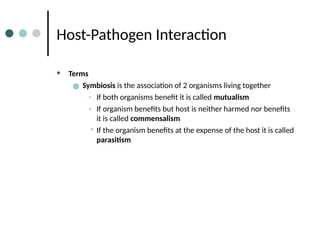
CLINICAL MICROBIOLOGY
Brophy-Martinez
Host-Pathogen Interaction
Origin of Microbial Flora
ā— Fetus in sterile environment until birth
ā— During 1st few days of life newborn introduced to
microorganisms
ā— Some colonize various sites and become the predominant
organism
• Colonization- growth of microbiota in or on a body
site without damage/symptoms
ā— Others are transient or fail to establish themselves
ā— Once established, the microorganisms develop a
relationship with the host
Host-Pathogen Interaction
• Terms
ā— Symbiosis is the association of 2 organisms living together
• If both organisms benefit it is called mutualism
• If organism benefits but host is neither harmed nor benefits
it is called commensalism
• If the organism benefits at the expense of the host it is called
parasitism
Host-Pathogen Interaction
Terms (con’t)
ā— Pathogens are microorganisms that can cause disease
ā— Opportunistic Pathogens are often members of the normal flora
that cause disease when their habitat is damaged, disturbed or
changed OR when the host’s immune system is weakened or
compromised
ā— Opportunistic infections are those infections caused by
opportunistic pathogens
ā— Iatrogenic Infections are infections that result from medical
treatment or procedures
ā— Hospital-acquired Infections are acquired in the hospital or
another health care setting
Host Pathogen Interaction
Terms (con’t)
• Normal, Usual or Indigenous Flora
ā— Resident flora
ā— Transient flora
• ā€œvisitorsā€
ā— Carriers/Carrier State
• Transmits agent
• Lack symptoms
Factors Determining Normal Flora
• Availability of nutrients
• Moisture of anatomical site
• Presence of bile, lysozyme,
fatty acids
• pH
Host Pathogen Interaction
• Colonization
ā— Persistent survival of a microbe on a surface of the human body.
ā— Microbe does not cause infection
ā— Determined by the defenses of the body
ā— Dictated by the microbes ability to survive
Microorganisms Found on the Skin
Common Residents
• Candida spp.
• Cutibacterium acnes
• Micrococcus spp.
• Staphylococcus spp.
• Propionibacterium spp.
• Diphtheroids
(Corynebacterium spp.)
Less Common or Transients
• Streptococcus spp.
• Acinetobacter spp.
• Gram-negative bacilli
• Moraxella spp.
Microorganisms Found in the Mouth
Common Residents
• Staphylococcus epidermidis
• Streptococcus anginosus
group
• Streptococcus mitis group
• Streptococcus salivarius
group
• Streptococcus mutans group
• Peptostreptococcus spp.
• Actinomyces israelii
• Bacteroides spp.
• Prevotella/Porphyromonas
spp.
• Veillonella spp.
Less Common or Transients
• Candida albicans
• Enterococcus spp.
• Eikenella corrodens
• Fusobacterium nucleatum
• Staphylococcus aureus
Microorganisms Found in the Nose &
Nasopharynx
Common Residents
• Diphtheroids
(Corynebacterium spp.)
• Haemophilus parainfluenzae
• Staphylococcus aureus
• Staphylococcus epidermidis
• Streptococcus spp.
Less Common or Transients
• Haemophilus influenzae
• Moraxella spp.
• Neisseria meningitidis
• Streptococcus pneumoniae
Microorganisms Found in the
Oropharynx
Common Residents
• α-Hemolytic and
nonhemolytic streptococci
• Diphtheroids
(Corynebacterium spp.)
• Staphylococcus aureus
• Staphylococcus epidermidis
• Streptococcus pneumoniae
• Streptococcus mutans group
• Streptococcus mitis group
• Streptococcus anginosus
group
• Streptococcus salivarius
group
• Haemophilus parainfluenzae
• Moraxella catarrhalis
• Bacteroides spp.
• Prevotella/Porphyromonas
spp.
• Fusobacterium necrophorum
Microorganisms Found in the
Oropharynx (con’t)
• Haemophilus influenzae
• Neisseria meningitidis
• Gram-negative rod
Less Common or Transients
Microorganisms Found in the GI
Tract
Microorganisms Found in the
Genitourinary Tract
Routes of Transmission
• Airborne
ā— Coughing, sneezing, talking
ā— Droplet nuclei
ā— Airborne pathogens must be resistant to drying and inactivation by ultraviolet
light
ā— Examples: Strep throat, otitis media, diphtheria, rhinoviruses (colds)
Routes of Transmission (cont’d)
• Transmission by Food and Water
• Ingestion of contaminated food or water
• Sometimes oral-fecal route
• Pathogens must be able to survive stomach conditions and
compete with normal flora of the gut
• Preformed toxins (Clostridium botulinum, S. aureus) vs.
toxins produced after infection (C. difficile, V. cholerae)
Routes of Transmission (con’t)
• Close Contact
• Passage of organisms by salivary, skin, and genital contact
• Examples: Infectious mononucleosis caused by EBV; HSV
• Cuts and Bites
• Concerned with infection with mouth flora
• Arthropods ( ticks, fleas)
• Zoonoses are diseases of animals transmitted to humans;
examples: plague, rabies, tularemia
Virulence
• Virulence describes an organism’s relative ability to cause disease, or
the degree of pathogenicity
• Virulence factors refer to a wide variety of mechanisms that allow
them to persist in a host and cause disease. Virulence factors include
functions such as:
• inhibiting phagocytosis
• facilitating adhesion to host cells/tissue
• enhancing intracellular survival AFTER phagocytosis
• damaging tissue via toxins/extracellular enzymes
Virulence Factors
• Resisting Phagocytosis
ā— Macrophages and PMNs, phagocytic cells, defend host
ā— Most common method: Capsule
• Capsule is made of polysaccharides or protein or a combo
• Prevents phagocytosis and activation of complement
• EX. S.pneumoniae, H.influenzae
ā— Another protective bacterial structure: Protein A
• Interferes with host antibodies binding to bacterial cell
surface
• EX. S.aureus
ā— Leukocidins, are potent materials that kill phagocytes
• Lysosomal discharge into cell cytoplasm
• EX. Staph leukocidin or PVL
Virulence Factors (cont’d)
ā—Structures to Promote Adhesion
• Most organisms must attach before establishing infection
• Adhesins mediate attachment
• Host cell receptor necessary for the adhesin
• Main bacterial adhesins are fimbriae (pili)
• Once attached, phagocytosis is less likely
ā—Survive Intracellularly and Proliferate
• If engulfed must be able to avoid being killed
• If survive must be able to replicate
• IgA protease
• Prevent exposure to intracellular digestive processes
• Invasion: Ability to penetrate and grow in tissues
• Dissemination: Disease or organism spreads to distant sites
Virulence Factors (cont’d)
• Produce Extracellular Enzymes or Toxins
ā— Noticeable disease symptoms often caused by toxins, etc.
• Toxins are poisonous substances produced by organism
• Exotoxins
• Both gram pos and gram neg
• have enzymatic activity
• mediate spread through tissue
• preformed toxins - toxins formed before the ingestion (for
example) of the bacteria
• Hyaluronidases/proteases
• Destroy connective tissue matrix to promote
dissemination
• Endotoxins
• LPS of gram neg bacteria exclusively
• no enzyme activity (vs. exotoxins)
• no specificity
• low potency
• dramatic changes to BP, body temp, clotting, immunity
Host Resistance Factors
• First Line of Defense
ā— Physical barriers: Intact skin
ā— Cleansing mechanisms:
• Desquamation (shedding) of skin
• Fluids of the eye (IgA and lysozyme)
• IgA- Serve as opsonins, fix complement
• Lysozyme- breakdowns bacteria cell wall
• Respiratory, digestive, urinary, and genital tracts have
fluids(mucous) and movements (cilia/ peristalsis) to cleanse
the surfaces
Host Resistance Factors (cont’d)
• Second Line of Defense
ā— Inflammatory response
• Vasodilation
• Increased permeability of capillaries
• Arrival of leukocytes
• Chemotaxis
• Phagocytosis
ā— Immune Responses: innate
• Third Line of Defense
• Adaptive/specific immunity
Host Resistance Factors (cont’d)
References
• Engelkirk, P., & Duben-Engelkirk, J. (2008). Laboratory
Diagnosis of Infectious Diseases: Essentials of Diagnostic
Microbiology . Baltimore, MD: Lippincott Williams and Wilkins.
• http://garrisonfive.blogspot.com/2009/05/home-sweet-
home.html
• http://www.5thguy.com/billboards.htm
• Mahon, C. R., Lehman, D. C., & Manuselis, G. (2015). Textbook
of Diagnostic Microbiology (5th ed.). Maryland Heights, MO:
Saunders.

Chapter 02 - Host-Pathogen Interaction.pptx

  • 1.
  • 2.
    Origin of MicrobialFlora ā— Fetus in sterile environment until birth ā— During 1st few days of life newborn introduced to microorganisms ā— Some colonize various sites and become the predominant organism • Colonization- growth of microbiota in or on a body site without damage/symptoms ā— Others are transient or fail to establish themselves ā— Once established, the microorganisms develop a relationship with the host
  • 3.
    Host-Pathogen Interaction • Terms ā—Symbiosis is the association of 2 organisms living together • If both organisms benefit it is called mutualism • If organism benefits but host is neither harmed nor benefits it is called commensalism • If the organism benefits at the expense of the host it is called parasitism
  • 4.
    Host-Pathogen Interaction Terms (con’t) ā—Pathogens are microorganisms that can cause disease ā— Opportunistic Pathogens are often members of the normal flora that cause disease when their habitat is damaged, disturbed or changed OR when the host’s immune system is weakened or compromised ā— Opportunistic infections are those infections caused by opportunistic pathogens ā— Iatrogenic Infections are infections that result from medical treatment or procedures ā— Hospital-acquired Infections are acquired in the hospital or another health care setting
  • 5.
    Host Pathogen Interaction Terms(con’t) • Normal, Usual or Indigenous Flora ā— Resident flora ā— Transient flora • ā€œvisitorsā€ ā— Carriers/Carrier State • Transmits agent • Lack symptoms
  • 6.
    Factors Determining NormalFlora • Availability of nutrients • Moisture of anatomical site • Presence of bile, lysozyme, fatty acids • pH
  • 7.
    Host Pathogen Interaction •Colonization ā— Persistent survival of a microbe on a surface of the human body. ā— Microbe does not cause infection ā— Determined by the defenses of the body ā— Dictated by the microbes ability to survive
  • 8.
    Microorganisms Found onthe Skin Common Residents • Candida spp. • Cutibacterium acnes • Micrococcus spp. • Staphylococcus spp. • Propionibacterium spp. • Diphtheroids (Corynebacterium spp.) Less Common or Transients • Streptococcus spp. • Acinetobacter spp. • Gram-negative bacilli • Moraxella spp.
  • 9.
    Microorganisms Found inthe Mouth Common Residents • Staphylococcus epidermidis • Streptococcus anginosus group • Streptococcus mitis group • Streptococcus salivarius group • Streptococcus mutans group • Peptostreptococcus spp. • Actinomyces israelii • Bacteroides spp. • Prevotella/Porphyromonas spp. • Veillonella spp. Less Common or Transients • Candida albicans • Enterococcus spp. • Eikenella corrodens • Fusobacterium nucleatum • Staphylococcus aureus
  • 10.
    Microorganisms Found inthe Nose & Nasopharynx Common Residents • Diphtheroids (Corynebacterium spp.) • Haemophilus parainfluenzae • Staphylococcus aureus • Staphylococcus epidermidis • Streptococcus spp. Less Common or Transients • Haemophilus influenzae • Moraxella spp. • Neisseria meningitidis • Streptococcus pneumoniae
  • 11.
    Microorganisms Found inthe Oropharynx Common Residents • α-Hemolytic and nonhemolytic streptococci • Diphtheroids (Corynebacterium spp.) • Staphylococcus aureus • Staphylococcus epidermidis • Streptococcus pneumoniae • Streptococcus mutans group • Streptococcus mitis group • Streptococcus anginosus group • Streptococcus salivarius group • Haemophilus parainfluenzae • Moraxella catarrhalis • Bacteroides spp. • Prevotella/Porphyromonas spp. • Fusobacterium necrophorum
  • 12.
    Microorganisms Found inthe Oropharynx (con’t) • Haemophilus influenzae • Neisseria meningitidis • Gram-negative rod Less Common or Transients
  • 13.
  • 14.
    Microorganisms Found inthe Genitourinary Tract
  • 15.
    Routes of Transmission •Airborne ā— Coughing, sneezing, talking ā— Droplet nuclei ā— Airborne pathogens must be resistant to drying and inactivation by ultraviolet light ā— Examples: Strep throat, otitis media, diphtheria, rhinoviruses (colds)
  • 16.
    Routes of Transmission(cont’d) • Transmission by Food and Water • Ingestion of contaminated food or water • Sometimes oral-fecal route • Pathogens must be able to survive stomach conditions and compete with normal flora of the gut • Preformed toxins (Clostridium botulinum, S. aureus) vs. toxins produced after infection (C. difficile, V. cholerae)
  • 17.
    Routes of Transmission(con’t) • Close Contact • Passage of organisms by salivary, skin, and genital contact • Examples: Infectious mononucleosis caused by EBV; HSV • Cuts and Bites • Concerned with infection with mouth flora • Arthropods ( ticks, fleas) • Zoonoses are diseases of animals transmitted to humans; examples: plague, rabies, tularemia
  • 18.
    Virulence • Virulence describesan organism’s relative ability to cause disease, or the degree of pathogenicity • Virulence factors refer to a wide variety of mechanisms that allow them to persist in a host and cause disease. Virulence factors include functions such as: • inhibiting phagocytosis • facilitating adhesion to host cells/tissue • enhancing intracellular survival AFTER phagocytosis • damaging tissue via toxins/extracellular enzymes
  • 19.
    Virulence Factors • ResistingPhagocytosis ā— Macrophages and PMNs, phagocytic cells, defend host ā— Most common method: Capsule • Capsule is made of polysaccharides or protein or a combo • Prevents phagocytosis and activation of complement • EX. S.pneumoniae, H.influenzae ā— Another protective bacterial structure: Protein A • Interferes with host antibodies binding to bacterial cell surface • EX. S.aureus ā— Leukocidins, are potent materials that kill phagocytes • Lysosomal discharge into cell cytoplasm • EX. Staph leukocidin or PVL
  • 20.
    Virulence Factors (cont’d) ā—Structuresto Promote Adhesion • Most organisms must attach before establishing infection • Adhesins mediate attachment • Host cell receptor necessary for the adhesin • Main bacterial adhesins are fimbriae (pili) • Once attached, phagocytosis is less likely ā—Survive Intracellularly and Proliferate • If engulfed must be able to avoid being killed • If survive must be able to replicate • IgA protease • Prevent exposure to intracellular digestive processes • Invasion: Ability to penetrate and grow in tissues • Dissemination: Disease or organism spreads to distant sites
  • 21.
    Virulence Factors (cont’d) •Produce Extracellular Enzymes or Toxins ā— Noticeable disease symptoms often caused by toxins, etc. • Toxins are poisonous substances produced by organism • Exotoxins • Both gram pos and gram neg • have enzymatic activity • mediate spread through tissue • preformed toxins - toxins formed before the ingestion (for example) of the bacteria • Hyaluronidases/proteases • Destroy connective tissue matrix to promote dissemination • Endotoxins • LPS of gram neg bacteria exclusively • no enzyme activity (vs. exotoxins) • no specificity • low potency • dramatic changes to BP, body temp, clotting, immunity
  • 22.
    Host Resistance Factors •First Line of Defense ā— Physical barriers: Intact skin ā— Cleansing mechanisms: • Desquamation (shedding) of skin • Fluids of the eye (IgA and lysozyme) • IgA- Serve as opsonins, fix complement • Lysozyme- breakdowns bacteria cell wall • Respiratory, digestive, urinary, and genital tracts have fluids(mucous) and movements (cilia/ peristalsis) to cleanse the surfaces
  • 23.
    Host Resistance Factors(cont’d) • Second Line of Defense ā— Inflammatory response • Vasodilation • Increased permeability of capillaries • Arrival of leukocytes • Chemotaxis • Phagocytosis ā— Immune Responses: innate • Third Line of Defense • Adaptive/specific immunity
  • 24.
  • 25.
    References • Engelkirk, P.,& Duben-Engelkirk, J. (2008). Laboratory Diagnosis of Infectious Diseases: Essentials of Diagnostic Microbiology . Baltimore, MD: Lippincott Williams and Wilkins. • http://garrisonfive.blogspot.com/2009/05/home-sweet- home.html • http://www.5thguy.com/billboards.htm • Mahon, C. R., Lehman, D. C., & Manuselis, G. (2015). Textbook of Diagnostic Microbiology (5th ed.). Maryland Heights, MO: Saunders.

Editor's Notes

  • #1Ā In this unit we will describe the interactions between the host and infectious agent or pathogen in the pathogenesis of disease. First, we will look at the origin of normal flora and the composition of different body sites. We will also discuss virulence factors that contribute to the invasiveness of organisms, protective defenses the host possesses, and how microbes are able to evade these host defenses.
  • #2Ā During fetal development, the amniotic sac is sterile. After birth, the first few days of life is when the newborn is introduced to microorganisms. Some of these organisms colonize in certain places in the newborn’s body and become the predominant organism. Colonization is the growth of microbiota in or on a body site without the production of damage or notable symptoms. Some other organisms fail to establish themselves, but once they are established, they have a relationship with the host.
  • #3Ā Once established onto or into a particular body site of the host, microorganisms develop a relationship or symbiosis with the host. Symbiosis is the association of two organisms of different species living together. This host-microbe relationship can be one of mutualism, commensalism or parasitism. In mutualism, we see both the host and organism benefiting from one another. For example, Lactobacilli in the female urogenital tract prevents colonization with pathogens while deriving nutrients from host. Commensalism occurs when the organisms benefits but there is no harmful or beneficial effect on the host. For example, Proteus mirabilis is a commensal species in the GI tract and inflicts no harm. Last, if the organism benefits at the expense of the host, that is parasitism.An example of parasitism is Entamoeba histolytica. Entamoeba histolytica is a pathogenic intestinal amoeba that derives nutrients from its host at the expense of the host, causing intestinal ulcers and amoebic dysentery.
  • #4Ā Due to the various relationships of host and organism, defining of terms is important for better understanding of these relationships. Pathogens are microorganisms that cause disease. Staphylococcus aureus in a wound is an example of a pathogen. Opportunistic pathogens are those organisms that cause disease when their habitat is damaged or changed or when the host’s immune system is compromised. Opportunistic infections can happen when trauma, either accidental or surgical, introduces organisms that are part of the normal flora into the traumatized areas of the body where those flora don’t normally live. An example of an opportunity for a pathogen would be a perforation of the colon. Once perforated, say as in an auto accident, the colon contents are emptied into the peritoneal cavity and an overwhelming infection with organisms in the normally sterile peritoneal cavity occurs. Iatrogenic procedures such as indwelling urinary catheters may be necessary for someone who is incapacitated. Many patients with indwelling catheters develop UTIs. The catheter was a necessary medical procedure but ultimately resulted in an infection. Hospital or healthcare acquired infections (HAIs) occur after the patient arrives at the facility, generally NOT within the first 48 hours, and were not incubating in the community/patient before the patient arrived at the facility.
  • #5Ā Microorganisms that are found on or in body sites of healthy persons are called normal, usual or indigenous flora. Microorganisms that colonize an area for months or years represent resident microbial flora. Microorganisms that are present temporarily are transient flora. Transient flora may ā€œvisitā€ but don’t stay. They are either eliminated by the host’s immune defenses or due to competition with the resident flora. Some pathogenic organisms may establish themselves in a host WITHOUT manifesting symptoms. However, these hosts, called carriers, can transmit the infection. This condition is called the carrier state. The carrier state can be acute (short-lived or transient) or chronic (lasting for months, years, or permanently).
  • #6Ā Nutritional and environmental factors influence which microorganisms are present in particular body sites. For example, most organisms inhabit moist areas of the body versus dry areas. Lipids and fatty acids are usually bactericidal but Propionibacterium spp colonize hair follicles because they can breakdown these lipids. These organisms are not easily removed by washing or scrubbing. Which organisms end up in a particular site depends on how resistant they are to the antibacterial effects of bile, lysozyme and fatty acids. The pH of vagina of women of childbearing age is approximately 4-5. Most bacteria cannot withstand this acidic extreme. Human breast milk is at a pH of ~ 5-5.5, while cows are less acidic as that leads to different fecal flora in infants. All of these environmental conditions may change with age, nutritional status, disease states, and drug/antibiotic effect. Changes in these environmental conditions can lead to opportunistic infections.
  • #7Ā Earlier in the presentation we mentioned the term colonization. Colonization is defined as the formation of a population of microorganisms in the host that does not cause disease. The human body is colonized by approximately 100 different species of microorganisms. Host defenses keep normal flora in check which reduces infections with normal flora in immunocompetent people. Immunocompromised patients can become easily infected with normal flora. A microbiologist must be able to determine when an organism is a pathogen versus when it is a colonizer/normal flora. The next few slides include common flora for different anatomical sites. I recommend you bookmark this information to help with your lab unknowns.
  • #8Ā The next series of slides include common flora for different anatomical sites. Bookmark this information to help with your lab unknowns.
  • #9Ā The next series of slides include common flora for different anatomical sites. Bookmark this information to help with your lab unknowns.
  • #10Ā The next series of slides include common flora for different anatomical sites. Bookmark this information to help with your lab unknowns.
  • #11Ā The next series of slides include common flora for different anatomical sites. Bookmark this information to help with your lab unknowns.
  • #12Ā The next series of slides include common flora for different anatomical sites. Bookmark this information to help with your lab unknowns.
  • #13Ā The next series of slides include common flora for different anatomical sites. Bookmark this information to help with your lab unknowns.
  • #15Ā The first step in starting an infection is for the infectious agent to gain access to the host. The route by which a pathogen can be transmitted to a host is an important part in the establishment of the infection. Although some organisms may be naturally transmitted by more than one route, most have a preferred portal of entry. These portals include airborne, via food and water, close contact, cuts and bites, arthropods, and zoonoses. AIRBORNE, where respiratory spread is common. Secretions are aerosolized by coughing, sneezing, and talking. Droplet nuclei (very small particles) are the residue from the evaporation of fluid from larger droplets and are light enough to remain airborne for long periods. Pathogens that are transmitted this way must be resistant to drying and inactivation by UV.
  • #16Ā GI infections are usually the result of ingestion of contaminated food or water. Sometimes the route is fecal-oral. Gastric juices and enzymes usually prevent survival of organisms but if they are able to survive they compete with the normal flora and produce damage to tissues. The damage cause can be due to a a) pre-formed toxin or b) a toxin produced from within the intestine or c) disruption of the normal functioning of the cells due to invasion by the organism. PREFORMED TOXINS are produced outside the body and include food poisoning by C.botulinum, B.cereus, S.aureus. Other bacteria produce a toxin after infection and include enterotoxigenic E.coli (aka traveler’s diarrhea). Classic intestinal pathogen is V.cholera, produces an enterotoxin that causes the outpouring of fluid from the cells into the lumen of the intestine. Massive amounts (20L/day) of fluid lost. Patients will have rice water stools.
  • #17Ā Typically, the routes of transmission of infectious diseases require close contact. However, for this discussion close contact refers to passage of organisms by salivary, skin, and genital contact. Skin-to-skin transfer of an infectious disease is not as common but diseases such as warts (HPV), syphilis, and impetigo result when material from infectious lesions inoculate the skin of a susceptible host. The most common STDs in North America are gonorrhea, HSV, HepB, HepC, Chlamydia, syphilis, Trichomonas, and HIV. Dog and cat bites often lead to Pasteurella multocida infections. Human bites are dangerous because they are difficult to treat and because human mouth flora is so diverse and numerous. Infection as a result of an arthropod bite (tick, flea or mite bite) is common in many parts of the world. In most cases, the infectious agent multiplies in the arthropod which then feeds on a human host and transmits the microorganism. Zoonosis is the route of transmission dependent on contact with animals or animal products. The infectious organism may be passed by animal bites (rabies), arthropod vectors (plague), and contact with animal carcasses (tularemia). Examples include malaria (infected human-Anopheles mosquito-uninfected human), Lyme disease (deer-tick-human), plague (rodent-flea-dog-human), and hemorrhagic fevers.
  • #18Ā Virulence is an organism’s ability to cause a disease. In some cases, it takes a low infective dose of an organism to cause disease, such as in Shigella. Whereas in Salmonella infections, a large infectious dose is required. There are many virulence factors, which is the mechanism that an organism uses to persist and cause disease. This slide lists a few of those factors.
  • #19Ā Macrophages and PMNs (polymorphonuclear) play a major role in defending the host from microbial invasion. An invading pathogen must evade phagocytosis in order to survive. The most common tool for the organism to evade phagocytosis is a polysaccharide capsule, which makes the organisms highly virulent. However, if the capsule is removed, the organism’s virulence becomes very low. That capsule masks the cell surface structures recognized by phagocytic cell receptors=avoid phagocytosis. The capsule also prevents the activation of complement by masking structures to which the complement proteins bind. For example, encapsulated strains of S. pneumoniae and H. influenzae can cause highly invasive infections more often vs. the non-encapsulated strains. Protein A in the cell wall of S. aureus helps avoid phagocytosis by interfering with the binding of the host’s antibodies to the surface of the organism. Protein A binds to the Fc portion of the antibody, preventing opsonization and phagocytosis by turning the antibody around on the surface. Staph leukocidin, also known as Panton-Valentine leukocidin (PVL), is lethal to WBCs and contributes to the invasiveness of this organism. Other organisms may inhibit chemotaxis so the host is less able to call PMNs and macrophages to the site of infection.
  • #20Ā Most infectious agents must attach to host cells before infection occurs. Without adhesion, the pathogen can be carried away in fluid, such as urine or mucus). Adhesions mediate attachment. Host cells must possess the necessary receptors for the adhesion, usually a carbohydrate. Once the organism is attached, phagocytosis by the host cell is unlikely. Examples of adhesion include: E. coli which causes traveler’s diarrhea and uses fimbriae to attach to small intestine cells, it then secretes toxins that cause the disease symptoms. Another example is N. gonorrhoeae, which uses fimbriae that are essential to attach to the genitourinary epithelial cells. Numerous pathogens produce an IgA protease to degrade the IgA found on host mucosal surfaces. Some may prevent fusion of the phagosome and lysosome to avoid exposure to digestion. Pathogens may exhibit an ability to penetrate and grow in tissues, as in the GC, which can infect fallopian tubes. Some organisms that survive phagocytosis may be disseminated to many body sites, but the organism itself is not invasive. The phagocyte simply carried the organism to the distant site.
  • #21Ā Disease from infection is noticeable if tissue damage occurs either from toxins (endo- or exo-) or inflammatory substances that cause host-driven immunologically mediated damage. Toxins are poisonous substances produced by organisms that interact with host cells, disrupting metabolism and causing harm. Exotoxins are a toxin released by a living bacterial cell into its surroundings. Exotoxins are produced from both gram negative and gram positive bacteria. Examples include Staph and Strep, which produce exfoliatin that causes massive skin peeling/exfoliation. Some produce soluble substances, hyaluronidases and proteases (extracellular enzymes) that liquefy the hyaluronic acid of the connective tissue matrix, helping to spread bacteria in tissues, promoting the dissemination of infection. Endotoxins are toxins that present inside a bacterial cell and are released when the cell disintegrates. Endotoxins are a component of the LPS (lipopolysaccharide) of the outer cell membrane of only gram negative bacteria. They do not have enzyme activity, have low potency, and have no specificity. Endotoxins are sometimes responsible for the characteristic symptoms of the disease, such as body temperature changes, blood pressure, etc.
  • #22Ā Few microorganisms can penetrate unbroken skin. Skin also has high numbers of normal flora, a lower pH, flora that compete for nutrients, and produce bactericidal substances, preventing colonization by transient possible pathogenic organisms. Squamous epithelial cells, the outer layer of skin, is being shed continuously. Many microorganisms shed along with the skin. The eyes have tears that lubricate the eye, wash foreign matter/pathogens out of the eye, and contain IgA and lysozyme. IgA is found in saliva, and mucous membranes of the respiratory, genital, and digestive tracts. IgA can serve as opsonins, fix complement and can neutralize infecting organisms. Lysozyme is found in serum, mucus, tissue fluids, tears, breast milk, saliva, and sweat. Lysozyme is an enzyme that breaks down the bacteria cell wall, resulting in cell death. The respiratory tract is protected by nasal hairs, ciliary epithelium, and mucous membranes. The GOAL is to prevent infectious agents and other particles from reaching the bronchioles and lungs. Smokers have reduced ciliated epithelial cells and therefore, are more susceptible to resp infections. The gastrointestinal tract has a low pH which allows for easy destruction of bacteria. Mucus in GI tract and peristalsis also prevent attaching. Secretory antibody (IgA) and phagocytic cells lining the mucosa are also defense mechanisms. The genitourinary tract is cleansed easily by voiding urine. The vagina contains a large population of indigenous flora and acidity of the vagina inhibits colonizing.
  • #23Ā In the second line of defense we see the inflammatory response and innate immunity being involved. Inflammation is the body’s response to injury or a foreign body. The inflammatory process includes: INJURY: Inflammatory response can be triggered by physical, chemical or biological agents. VASODILATION: increase capillary diameter to increase blood flow to provide plasma proteins, neutrophils, and phagocytes to the affected area. INCREASED PERMEABILITY: allows the escape of plasma and plasma proteins such as antibodies and complement. MIGRATION OF WBCs: Neutrophils and macrophages squeeze through gaps in endothelial cells (living blood vessels) and arrive at the site of injury. (called diapedesis) CHEMOTAXIS: certain mediators are released, a chemical ā€œtaxiā€ occurs, directing more cells to the site of injury OPSONIZATION/PHAGOCYTOSIS: bacteria coated with antibodies or complement, flagging the bacterium for destruction Innate immunity is also part of the second line of defense. Innate or nonspecific immunity consists of: #1 physical and chemical barriers, such as the skin, #2 blood proteins that act as mediators of infections, and #3 a mechanism capable of phagocytosis. The third line of defense is adaptive or specific immunity. Adaptive immunity enhances the capabilities of innate immunity. Adaptive immunity is responsible for responding in particular ways to different foreign substances and developing a memory, so that future encounters are remembered and responded to with a quicker response time.
  • #24Ā Phagocytosis is an essential component in the resistance of the host to infectious agents. It is the primary mechanisms in the host defense against extracellular bacteria, fungi , and some viruses. Once the bacterium is opsonized, it is rapidly ingested by the phagocyte. Opsonization is the process where the bacteria is coated with antibody or complement to enhance phagocytosis. Once the bacterium has been ingested, the WBC cell membrane invaginates (folds back on itself to form a cavity or pouch) and surrounds the bacterium. The particle is taken into the cytoplasm and enclosed within a vacuole called a phagosome. The phagosome fuses with a lysosome (vacuole containing hydrolytic enzymes). The lysosome releases its contents into the phagosome (degranulation), killing/digesting the bacteria.