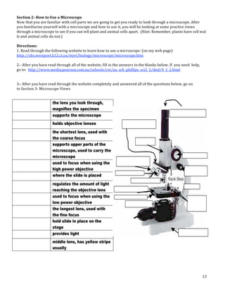
1
Name: ____________________________________________________ Section: _____________
CELL EXPLORATION ACTIVITIES
FIRST THIS IMPORTANT MESSAGE FROM YOUR TEACHER:
This packet contains different activities that are all about cells. The activities may be done in any order unless
I say otherwise, EXCEPT FOR # 12—DO THAT ONE LAST! They will pretty much follow what we do as a
class so you might want to do them in order. These activities will be done in class but you can also do them at
home. They are written so that you can do them INDEPENDENTLY and WITHOUT me. If you do get stuck, go
on to another activity or ask a classmate for help until you can meet with me. Make sure you do your best
since these will count as a TEST GRADE. Some of these activities you will do by yourself or with others (see
key below). You must have a NEW PARTNER each time and BEWARE, if I don’t get a sense that you are doing
the work (not watching others do it), talking about the work (not your social life) or making progress on the
work, (not just havin’ a good time) you’ll be asked to do it by yourself or after school under my supervision.
Remember, choices come with consequences.
Use the checklist below to make sure you keep track of completed activities.
DONE ACTIVITY DONE ACIVITY
#1. HOW BIG IS IT? #7. THE MIXED UP CELL
#2. SEEING CELLS FIRST #8. CELL FOLDABLE
#3. CELLS ORGANELLES CHECK #9. THE CELL AS A SCHOOL
#4. CELL QUIZOLAS #10. CELL INSPECTOR
#5. A TYPICAL ANIMAL CELL #11. CELL STRUCTURE
#6. PLANT & ANIMAL CELLS QUIZ #12. WHAT I’VE LEARNED
Activity Key:
Packet #2
Activity and Study Guide Name ________________________________________
September 15!19
Cells
This packet is due by the end of the class period Friday, September 19, 2003. You may complete the work in
this packet in any order. Be sure to ask for help from your partner or from Mrs. Rowan if you run into problems.
This packet is also online at:
http://www.teachersgarden.com/sgs/
Use the checklist below to make sure you have finished all parts of this packet.
Title of Activity Type
Check
if Done
Journal Observation #2 Performance
Organization of Life Concept Cards Practice
Cell Scientist Cartoon or Mini!Poster Performance
"Elephant!Sized Amoebas?" Activity Practice
Journal Reflection #3 Performance
Journal Observation #3 Performance
"Not All Cells are Cubes" Models Practice
Completed Cell Model, Phase I Practice
Cell Plan Sheet Practice
Journal Reflection #4 Performance
100% on the online cell quiz Practice
Scientist of the Week ! Partner Quiz Performance
Journal Reflection #5 Performance
The whole packet is done (YAY!) Performance
Explore the Cell Movies Clip Gallery at : http://www.cytographics.com/gallery/clips/cg.html
• Reproduction in Volvox
• The rotifer Collotheca feeding on Chlamydomonas
• This basic little amoeba is a predator!
• Three eggs of the African toad Xenopus
• Morphogenesis in the desmid Micrasterias sp.
• Euglena writhing
Pick one of the clips and describe what you see for
Journal Observation #2
ellular
cube
Surface area divided
by Volume
ovide oxygen to a volume
ed on
work on
w the
!!!!!!!!!!!!!!!!!!
ARGER, and there can be a variety
ular,
cellsalive.com/howbig.htm
will let you see that object or
. A magnification of "1" means life
hing looks ten times bigger than it
ed by "1,000" to be seen is one
must be magnified by "10" to be
ale that appear at some of the
gs at the tops of the columns:
The Text: Organization of Life (pages 4!8) of Cells, Heredity, a
by Holt.
Materials Needed: 8 pieces of paper cut to 3 inches by 5 inche
The Task: Create concept cards for each of the following
cells, tissues, organs, organ systems, organism
populations, communities, and ecosystems
Concepts cards take the following form:
The definition of the term
A drawing
Term Illustrate the
A sentence from the text that uses the term A sentence in your own word
Front Back
When completed, the concept cards should be stapled to the back of this p
!!> You may use the concept cards on the exam for this chapter.
!!!!!!!!!!!!!!!!!!!!!!!!!!!!!!!!!!!!!!!!!!!!!!!!!!!!!!!!!!!!!
Journal Reflection #3
First, imagine you have invented the microscope and
are the first person to see a plant cell. Assume your
microscope is so good that you can see cell walls, the
nucleus, vacuoles, and even chloroplasts in the cells.
Describe how you feel as you see parts of living
things never before seen by another human.
Now, read "Seeing the First Cells" on page 9 of your
textbook and examine the photograph of Robert
Hooke’s microscope and his drawing of cells
!!!!!!!!!!!!!!!!!!!!!!!!!!!!!!!!!!!!!!!!!!!!!!!!!!!!!!!!!!!!!
In the space below, copy the three parts of the Cell Theory from page 10
textbook.
1. _____________________________________________
2. _____________________________________________
3. _____________________________________________
!!!!!!!!!!!!!!!!!!!!!!!!!!!!!!!!!!!!!!!!!!!!!!!!!!!!!!!!!!!!!
Cell Scientist Cartoon or Mini!Poste
Leeuwenhoek, Schleiden, Schwann, and Virchow are describe
your textbook.
Pick one of these scientists and make a cartoon or min
their discoveries. Turn in the cartoons and mini!posters
to be graded and posted on the wall.
In your journal, complete R
describing which objects
were most often studi
microscopists, an
The Text: Organization of Life (pages 4!8) of Cells, Heredity, and Classification
by Holt.
Materials Needed: 8 pieces of paper cut to 3 inches by 5 inches.
The Task: Create concept cards for each of the following terms
cells, tissues, organs, organ systems, organisms.
populations, communities, and ecosystems
Concepts cards take the following form:
The definition of the term
A drawing to
Term Illustrate the term
A sentence from the text that uses the term A sentence in your own words using the term
Front Back
When completed, the concept cards should be stapled to the back of this packet.
!!> You may use the concept cards on the exam for this chapter.
!!!!!!!!!!!!!!!!!!!!!!!!!!!!!!!!!!!!!!!!!!!!!!!!!!!!!!!!!!!!!!!!!!
Journal Reflection #3
First, imagine you have invented the microscope and
are the first person to see a plant cell. Assume your
microscope is so good that you can see cell walls, the
nucleus, vacuoles, and even chloroplasts in the cells.
Describe how you feel as you see parts of living
things never before seen by another human.
Now, read "Seeing the First Cells" on page 9 of your
textbook and examine the photograph of Robert
Hooke’s microscope and his drawing of cells
!!!!!!!!!!!!!!!!!!!!!!!!!!!!!!!!!!!!!!!!!!!!!!!!!!!!!!!!!!!!!!!!!!
In the space below, copy the three parts of the Cell Theory from page 10 of your
textbook.
1. _____________________________________________________
2. _____________________________________________________
3. _____________________________________________________
!!!!!!!!!!!!!!!!!!!!!!!!!!!!!!!!!!!!!!!!!!!!!!!!!!!!!!!!!!!!!!!!!!
Cell Scientist Cartoon or Mini!Poster
Leeuwenhoek, Schleiden, Schwann, and Virchow are described on page 10 of
your textbook.
Pick one of these scientists and make a cartoon or mini!poster about
In your journal, complete Reflection #3 by
describing which objects and materials
were most often studied by early
microscopists, and WHY.
These activities require a
computer and possibly my
web page
These activities require some
writing and reflection.
These activities require you to
make something to
demonstrate your knowledge.
These activities require you to
follow procedures and use
science materials.
e REVIEW?
ching Game" at:
/mc/65947.html
and Function Quiz on:
ence/sbi3a1/Cells/cellquiz.htm
e trouble recognizing the diagrams,
ells/cells.htm
!!!!!!!!!!!!!!!!!!!
of the Week
e Week
of the
ect your
what he
the back of this packet.
!!!!!!!!!!!!!!!!!!!
om smallest to largest:
ssue community organ system
Requires a partner to
work with.
Ahhh! Alien Invasion!!!!!
View the movie of Bacteria Invading an Animal Cell at:
http://cmgm.stanford.edu/theriot/seq1LmPtK2.mov
Now read "Where Did They Come from?" on page 20.
Journal Reflection #4
In your own words, describe the process that is dia
described in the text on page 20 of your textbook.
Suppose the scientific idea about the origins of mitochon
Describe in your journal how this mitochondrion origin idea ma
!!!!!!!!!!!!!!!!!!!!!!!!!!!!!!!!!!!!!!!!!!!!!!!!!!!!!
Follow the Protein
Proteins are extremely important to living things!
structures has something to do with proteins. First,
its protein function. You can check 16!22 of your tex
Nucleus Some proteins needs to be alter
cell. This organelle handles alter
Ribosome Tiny organelles that make protei
as their instructions.
Requires a textbook or print
materials.
2
ACTIVITY 1: HOW BIG IS IT? My Partner(s): _______________________________________________________
1. Explore the "How Big is a...?" web page at http://www.cellsalive.com/howbig.htm (or click the link on my
web page). Here you will look at objects found on the head of a pin. Your job is to rank them in order of size
on the chart below and estimate the length of each (in nanometers, micrometers, or millimeters). The line in
the bottom right corner of the screen is used to help you estimate. Sketch each of the objects.
Use COMPLETE SENTENCES to write a brief summary of this activity, what you did and what you learned.
______________________________________________________________________________________________________________________________
______________________________________________________________________________________________________________________________
______________________________________________________________________________________________________________________________
______________________________________________________________________________________________________________________________
______________________________________________________________________________________________________________________________
Scientist of the Week ! Partner Quiz
Journal Reflection #5 Performance
The whole packet is done (YAY!) Performance
Explore the Cell Movies Clip Gallery at : http://www.cytographics.com/gallery/clips/cg.html
• Reproduction in Volvox
• The rotifer Collotheca feeding on Chlamydomonas
• This basic little amoeba is a predator!
• Three eggs of the African toad Xenopus
• Morphogenesis in the desmid Micrasterias sp.
• Euglena writhing
Pick one of the clips and describe what you see for
Journal Observation #2
Back
he concept cards should be stapled to the back of this packet.
may use the concept cards on the exam for this chapter.
!!!!!!!!!!!!!!!!!!!!!!!!!!!!!!!!!!!!!!!!!!!!!!!!!!!
ed the microscope and
lant cell. Assume your
u can see cell walls, the
oroplasts in the cells.
ou see parts of living
nother human.
ells" on page 9 of your
photograph of Robert
wing of cells
!!!!!!!!!!!!!!!!!!!!!!!!!!!!!!!!!!!!!!!!!!!!!!!!!!!
the three parts of the Cell Theory from page 10 of your
_______________________________________
_______________________________________
_______________________________________
!!!!!!!!!!!!!!!!!!!!!!!!!!!!!!!!!!!!!!!!!!!!!!!!!!!
Cell Scientist Cartoon or Mini!Poster
Leeuwenhoek, Schleiden, Schwann, and Virchow are described on page 10 of
your textbook.
Pick one of these scientists and make a cartoon or mini!poster about
their discoveries. Turn in the cartoons and mini!posters to the "In!Box"
to be graded and posted on the wall.
In your journal, complete Reflection #3 by
describing which objects and materials
were most often studied by early
microscopists, and WHY.
all of the questions on the quiz correct. If you have trouble recognizing the diagrams,
Structure and Processes Overview at:
http://www.tvdsb.on.ca/westmin/science/sbi3a1/Cells/cells.htm
!!!!!!!!!!!!!!!!!!!!!!!!!!!!!!!!!!!!!!!!!!!!!!!!!!!!!!!!
each your partner about the Scientist of the Week
(van Helmont)
divide up the information on the Scientist of the Week
partner should become an expert on their part of the
uiz for your partner and let them take it. Correct your
udents learn more about who van Helmont is and what he
s.
leted, the quiz you wrote should be stapled to the back of this packet.
!!!!!!!!!!!!!!!!!!!!!!!!!!!!!!!!!!!!!!!!!!!!!!!!!!!!!!!!
he following list so they are in order, from smallest to largest:
ecosystem cell atom organ molecule tissue community organ system
Structure
...
ection #5, explain why plant and animal cells have so many
n even though plants and animals seem so different.
URL: www.cellsalive.com
Objective: You will look at computer models of cells, learn the functions and the descriptions of the cells and their
components.
Navigating the site: Cellsalive.com has a navigation bar at the left. After accessing the page, click on CELL BIOLOGY
on the left side navigation bar. From here, you will access the links: "How Big is a..", the animal cell model, the plant
cell model, and the bacterial cell model.
Part A. "HOW BIG IS A...."
Here you will look at objects found on the head of a pin. Your job is to rank them in order of size on the chart below
and estimate the length of each (in nanometers, micrometers, or millimeters). The line in the bottom right cornerof
the screen is used to help you estimate. Sketch each of the objects.
Object Sketch
Size in nanometers, micrometers or
millimeters
Human hair
Dust Mite
Red Blood Cells
E. coli
Staphylococcus
Ebola virus
Rhinovirus
3
Part B: Bacterial Cell Model ‐ (you will need to return to the "Cell Biology" link to access this page, or hit
your back button). Label the numbered parts on each line.
Part C; Animal Cell Model ‐ (you will need to return to the "Cell Biology" link to access this page, or hit your
back button) ‐ For this model, you will need to click on the various parts of the cell to go to a screen that tells
you about the parts. Answers to the following questions are found there.
Part B: Bacterial Cell Model - (you will need to return to the "Cell Biology" link to access this page, or hit your
back button)
Part C; Animal Cell Model - (you will need to return to the "Cell Biology" link to access this page, or hit your back
button) - For this model, you will need to click on the various parts of the cell to go to a screen that tells you about the
parts. Answers to the following questions are found there.
1. What do mitochondria do?
2. How big are mitochondria?
3. What does the Golgi Apparatus do?
4. What is the difference between smooth and rough ER?
5. Where is the nucleolus found?
6. What does the nucleolus do?
Sketch each of the following.
Mitochondria
Ribosome
4
7. What does the cytoskeleton do?
8. Cytosol goes by what other name?
9. What is the function of the cytosol?
10. What is the function of the lysosome?
Nucleus
Cell Membrane
Part D: Plant Cell Model - (you will need to return to the "Cell Biology" link to access this page, or hit your back
button)
1. What other type of cell has a cell wall?
2. What makes the plant cells green?
3. In plant cells, what does the vacuole do?
Sketch the following
Chloroplast
Vacuole
Part E: Overview
For the chart below, place a check in the box if the cell has that component.
Organelle Plant Animal Bacteria
Vacuole
Chloroplast
Ribosome
Mitochondria
DNA
Endoplasmic Reticulum
Cell Wall
Golgi Apparatus
5
ACTIVITY 2: SEEING CELLS FIRST
Directions:
1. Go to http://www.kbears.com/sciences/microscope.html(or my web page)
2. Click on any five organisms and use the information to complete the chart below.
Organism Sketch Facts/Information
3. Click on “What is a Microscope” and use the info to label the diagram below by drawing arrows from the
terms to their location on the microscope (USE A RULER). Next, write a fact or some info about each one.
Base­
Eyepiece ­
Coarse focus knob
Arm­
Fine focus knob­
Aperture­
Light­
Stage­
Diaphragm­
Objective lens­
Back
d, the concept cards should be stapled to the back of this packet.
ou may use the concept cards on the exam for this chapter.
!!!!!!!!!!!!!!!!!!!!!!!!!!!!!!!!!!!!!!!!!!!!!!!!!!!!!
ented the microscope and
a plant cell. Assume your
you can see cell walls, the
chloroplasts in the cells.
you see parts of living
y another human.
Cells" on page 9 of your
e photograph of Robert
drawing of cells
!!!!!!!!!!!!!!!!!!!!!!!!!!!!!!!!!!!!!!!!!!!!!!!!!!!!!
py the three parts of the Cell Theory from page 10 of your
_________________________________________
_________________________________________
_________________________________________
!!!!!!!!!!!!!!!!!!!!!!!!!!!!!!!!!!!!!!!!!!!!!!!!!!!!!
Cell Scientist Cartoon or Mini!Poster
Leeuwenhoek, Schleiden, Schwann, and Virchow are described on page 10 of
your textbook.
Pick one of these scientists and make a cartoon or mini!poster about
their discoveries. Turn in the cartoons and mini!posters to the "In!Box"
to be graded and posted on the wall.
In your journal, complete Reflection #3 by
describing which objects and materials
were most often studied by early
microscopists, and WHY.
Scientist of the Week ! Partner Quiz Performance
Journal Reflection #5 Performance
The whole packet is done (YAY!) Performance
Explore the Cell Movies Clip Gallery at : http://www.cytographics.com/gallery/clips/cg.html
• Reproduction in Volvox
• The rotifer Collotheca feeding on Chlamydomonas
• This basic little amoeba is a predator!
• Three eggs of the African toad Xenopus
• Morphogenesis in the desmid Micrasterias sp.
• Euglena writhing
Pick one of the clips and describe what you see for
Journal Observation #2
Life Science NAME_______________________________
Chapter 02 Cell Structure S/G DATE _______________ PERIOD _________
V 1.0
Instructions Identify each of the cell and microscope structures by drawing arrows to them and
labeling them. In the space provided give the function of each structure. Many of these questions
will appear on you next quiz!! (You will find the answer to these questions in the text book or your
notes)
golgi bodies, mitochondria, lisosome, cell membrane,
nucleus, vacuole, chromosome, endoplasmic reticulum,
ribosome, cytoplasm, chloroplasts
fine adjustment, ocular lens, coarse adjustment, stage clips,
diaphragm, stage, objective lens, base
6
ACTIVITY 3: CELL ORGANELLES CHECK My Partner(s): _______________________________________________________
1. Go to http://www.quia.com/jg/65947.html (or my web page)
2. Play the Flashcard and Matching games Time how long it takes you (you can do it more than once) .
3. Enter you scores below.
My Best Matching Time: ______________
My Best Flashcard Time: ______________
4. Next, play the Concentration game and put your best score here: ___________________
ACTIVITY 4: CELL QUIZOLAS
1. Go to http://www.biology4kids.com/extras/quiz_cellorgan/index.html (or my web page)
Take the quiz and put your score here: ______________
2. Go to http://www.zerobio.com/target_practice_quiz/target_practice_quiz_cells.htm (or my web page)
Take the quiz and put your score here: ______________
3. Go to http://www.proprofs.com/quiz‐school/quizshow.php?title=3rd‐block‐group‐2_1&quesnum=1 (or
my web page)
Take the quiz and put your score here: ______________
4. Go to http://www.schools.utah.gov/curr/science/sciber00/7th/cells/Quiz/index.htm (or my web page)
Take the quiz and put your score here: ______________
ACTIVITY 5: A TYPICAL ANIMAL CELL My Partner(s): _______________________________________________________
1. Go to http://www.wisc‐online.com/Objects/ViewObject.aspx?ID=AP11403 (or my web page)
2. Follow the directions and see how many organelles you can find.
3. Go to http://www.funtrivia.com/playquiz/quiz14606510ba900.html (or my web page)
4. Take the quiz and put your score here: ___________________ (each one is worth 10 points)
ence/sbi3a1/Cells/cellquiz.htm
e trouble recognizing the diagrams,
ells/cells.htm
!!!!!!!!!!!!!!!!!!!
of the Week
e Week
of the
ect your
what he
the back of this packet.
!!!!!!!!!!!!!!!!!!!
om smallest to largest:
ssue community organ system
ve so many
nt.
Cell Plan Sheet Practice
Journal Reflection #4 Performance
100% on the online cell quiz Practice
Scientist of the Week ! Partner Quiz Performance
Journal Reflection #5 Performance
The whole packet is done (YAY!) Performance
Explore the Cell Movies Clip Gallery at : http://www.cytographics.com/gallery/clips/cg.html
• Reproduction in Volvox
• The rotifer Collotheca feeding on Chlamydomonas
• This basic little amoeba is a predator!
• Three eggs of the African toad Xenopus
• Morphogenesis in the desmid Micrasterias sp.
• Euglena writhing
Pick one of the clips and describe what you see for
Journal Observation #2
Packet #2
Activity and Study Guide Name ________________________________________
September 15!19
Cells
This packet is due by the end of the class period Friday, September 19, 2003. You may complete the work in
this packet in any order. Be sure to ask for help from your partner or from Mrs. Rowan if you run into problems.
This packet is also online at:
http://www.teachersgarden.com/sgs/
Use the checklist below to make sure you have finished all parts of this packet.
Title of Activity Type
Check
if Done
Journal Observation #2 Performance
Organization of Life Concept Cards Practice
Cell Scientist Cartoon or Mini!Poster Performance
"Elephant!Sized Amoebas?" Activity Practice
Journal Reflection #3 Performance
Journal Observation #3 Performance
"Not All Cells are Cubes" Models Practice
Completed Cell Model, Phase I Practice
Cell Plan Sheet Practice
Journal Reflection #4 Performance
100% on the online cell quiz Practice
Scientist of the Week ! Partner Quiz Performance
Journal Reflection #5 Performance
The whole packet is done (YAY!) Performance
Explore the Cell Movies Clip Gallery at : http://www.cytographics.com/gallery/clips/cg.html
• Reproduction in Volvox
• The rotifer Collotheca feeding on Chlamydomonas
• This basic little amoeba is a predator!
• Three eggs of the African toad Xenopus
• Morphogenesis in the desmid Micrasterias sp.
• Euglena writhing
Pick one of the clips and describe what you see for
Journal Observation #2
ady for Some REVIEW?
Organelles Matching Game" at:
://www.quia.com/mc/65947.html
e Cell Structure and Function Quiz on:
n.ca/westmin/science/sbi3a1/Cells/cellquiz.htm
z correct. If you have trouble recognizing the diagrams,
iew at:
min/science/sbi3a1/Cells/cells.htm
!!!!!!!!!!!!!!!!!!!!!!!!!!!!!!!!!!
t the Scientist of the Week
Helmont)
the Scientist of the Week
expert on their part of the
hem take it. Correct your
van Helmont is and what he
ould be stapled to the back of this packet.
!!!!!!!!!!!!!!!!!!!!!!!!!!!!!!!!!!
are in order, from smallest to largest:
gan molecule tissue community organ system
Structure
100% on the online cell quiz Practice
Scientist of the Week ! Partner Quiz Performance
Journal Reflection #5 Performance
The whole packet is done (YAY!) Performance
Explore the Cell Movies Clip Gallery at : http://www.cytographics.com/gallery/clips/cg.html
• Reproduction in Volvox
• The rotifer Collotheca feeding on Chlamydomonas
• This basic little amoeba is a predator!
• Three eggs of the African toad Xenopus
• Morphogenesis in the desmid Micrasterias sp.
• Euglena writhing
Pick one of the clips and describe what you see for
Journal Observation #2
z correct. If you have trouble recognizing the diagrams,
iew at:
min/science/sbi3a1/Cells/cells.htm
!!!!!!!!!!!!!!!!!!!!!!!!!!!!!!!!!!
t the Scientist of the Week
Helmont)
the Scientist of the Week
expert on their part of the
hem take it. Correct your
van Helmont is and what he
ould be stapled to the back of this packet.
!!!!!!!!!!!!!!!!!!!!!!!!!!!!!!!!!!
are in order, from smallest to largest:
gan molecule tissue community organ system
Structure
and animal cells have so many
mals seem so different.
7
ACTIVITY 6: PLANT AND ANIMAL CELL ORGANELLES QUIZ
My Partner(s): __________________________________________________________
1. Go to http://www.lahc.cc.ca.us/biology/bio3/mchernoff/quiz.html (or my web page)
2. Fill in your answers on the diagrams below and then compare them to the correct answer page.
Check the questions you got correct: 3 4 5 6 7 8 9 10 11 12
Scientist of the Week ! Partner Quiz Performance
Journal Reflection #5 Performance
The whole packet is done (YAY!) Performance
Explore the Cell Movies Clip Gallery at : http://www.cytographics.com/gallery/clips/cg.html
• Reproduction in Volvox
• The rotifer Collotheca feeding on Chlamydomonas
• This basic little amoeba is a predator!
• Three eggs of the African toad Xenopus
• Morphogenesis in the desmid Micrasterias sp.
• Euglena writhing
Pick one of the clips and describe what you see for
Journal Observation #2
z correct. If you have trouble recognizing the diagrams,
iew at:
min/science/sbi3a1/Cells/cells.htm
!!!!!!!!!!!!!!!!!!!!!!!!!!!!!!!!!!
t the Scientist of the Week
Helmont)
the Scientist of the Week
expert on their part of the
hem take it. Correct your
van Helmont is and what he
ould be stapled to the back of this packet.
!!!!!!!!!!!!!!!!!!!!!!!!!!!!!!!!!!
are in order, from smallest to largest:
gan molecule tissue community organ system
Structure
and animal cells have so many
mals seem so different.
8
ACTIVITY 7: THE MIXED UP CELLS My Partner(s): __________________________________________________________
1. Go to http://www.beaconlearningcenter.com/WebLessons/MixedUpCells/default.htm (or my web page)
2. Follow the directions and record the data in the space below.
100% on the online cell quiz Practice
Scientist of the Week ! Partner Quiz Performance
Journal Reflection #5 Performance
The whole packet is done (YAY!) Performance
Explore the Cell Movies Clip Gallery at : http://www.cytographics.com/gallery/clips/cg.html
• Reproduction in Volvox
• The rotifer Collotheca feeding on Chlamydomonas
• This basic little amoeba is a predator!
• Three eggs of the African toad Xenopus
• Morphogenesis in the desmid Micrasterias sp.
• Euglena writhing
Pick one of the clips and describe what you see for
Journal Observation #2
z correct. If you have trouble recognizing the diagrams,
iew at:
min/science/sbi3a1/Cells/cells.htm
!!!!!!!!!!!!!!!!!!!!!!!!!!!!!!!!!!
t the Scientist of the Week
Helmont)
the Scientist of the Week
expert on their part of the
hem take it. Correct your
van Helmont is and what he
ould be stapled to the back of this packet.
!!!!!!!!!!!!!!!!!!!!!!!!!!!!!!!!!!
are in order, from smallest to largest:
gan molecule tissue community organ system
Structure
and animal cells have so many
mals seem so different.
A drawing to
Term Illustrate the term
A sentence from the text that uses the term A sentence in your own words using the term
Front Back
When completed, the concept cards should be stapled to the back of this packet.
!!> You may use the concept cards on the exam for this chapter.
!!!!!!!!!!!!!!!!!!!!!!!!!!!!!!!!!!!!!!!!!!!!!!!!!!!!!!!!!!!!!!!!!!
Journal Reflection #3
First, imagine you have invented the microscope and
are the first person to see a plant cell. Assume your
microscope is so good that you can see cell walls, the
nucleus, vacuoles, and even chloroplasts in the cells.
Describe how you feel as you see parts of living
things never before seen by another human.
Now, read "Seeing the First Cells" on page 9 of your
textbook and examine the photograph of Robert
Hooke’s microscope and his drawing of cells
!!!!!!!!!!!!!!!!!!!!!!!!!!!!!!!!!!!!!!!!!!!!!!!!!!!!!!!!!!!!!!!!!!
In the space below, copy the three parts of the Cell Theory from page 10 of your
textbook.
1. _____________________________________________________
2. _____________________________________________________
3. _____________________________________________________
!!!!!!!!!!!!!!!!!!!!!!!!!!!!!!!!!!!!!!!!!!!!!!!!!!!!!!!!!!!!!!!!!!
Cell Scientist Cartoon or Mini!Poster
Leeuwenhoek, Schleiden, Schwann, and Virchow are described on page 10 of
your textbook.
Pick one of these scientists and make a cartoon or mini!poster about
their discoveries. Turn in the cartoons and mini!posters to the "In!Box"
to be graded and posted on the wall.
In your journal, complete Reflection #3 by
describing which objects and materials
were most often studied by early
microscopists, and WHY.
9
ACTIVITY 8: CELL FOLDABLE
Goal: To create a pictorial representation of the parts of cells, with the name of the major organelles, and a description of
their functions.
Materials:
• 12 X 18 White “poster” paper
• Markers or colored pencils
• Scissors
• Textbook
Procedures:
STEP 1: Fold poster paper in a way so that it is divided into 16 sections
STEP 2: Cut the folds on one side of the paper only
STEP 3: Draw each of the cell parts on the front of the flap as it is closed down.
STEP 4: On the back of the flap write the function of the organelle.
Example: Nucleus – This is the part of the cell that is the control
center and directs all activity.
STEP 5: Put your name on the last flap on the front.
8 organelles (1 per flap)
1. _____________________________________________________
2. _____________________________________________________
3. _____________________________________________________
!!!!!!!!!!!!!!!!!!!!!!!!!!!!!!!!!!!!!!!!!!!!!!!!!!!!!!!!!!!!!!!!!!
Cell Scientist Cartoon or Mini!Poster
Leeuwenhoek, Schleiden, Schwann, and Virchow are described on page 10 of
your textbook.
Pick one of these scientists and make a cartoon or mini!poster about
their discoveries. Turn in the cartoons and mini!posters to the "In!Box"
to be graded and posted on the wall.
10
ACTIVITY 9: THE CELL AS A SCHOOL
A cell is like our school. Each part of the cell (and school) has responsibilities that must be done and certain organelles
(people or places) to do them. Identify the function of the following parts of the cell. Then, identify which person does the
same job (or a place like it) in the school. The first one is done for you as an example to follow.
Organelle Function Part of our school that has
a similar function
Cell Membrane Controls what goes in and out of the cell Front Office
Cytoplasm
Golgi Body
Lysosome
Mitochondria
Nucleus
Nucleolus
Ribosome
Rough ER
Smooth ER
Vacuole
Cell Wall
(Plant only)
Chloroplast
(Plant only)
Central Vacuole
(Plant only)
A sentence in your own words using the term
Back
should be stapled to the back of this packet.
pt cards on the exam for this chapter.
!!!!!!!!!!!!!!!!!!!!!!!!!!!!!!!!!!!!
and
your
the
s.
ving
your
bert
!!!!!!!!!!!!!!!!!!!!!!!!!!!!!!!!!!!!
of the Cell Theory from page 10 of your
_____________________________
_____________________________
_____________________________
!!!!!!!!!!!!!!!!!!!!!!!!!!!!!!!!!!!!
st Cartoon or Mini!Poster
iden, Schwann, and Virchow are described on page 10 of
e scientists and make a cartoon or mini!poster about
Turn in the cartoons and mini!posters to the "In!Box"
to be graded and posted on the wall.
In your journal, complete Reflection #3 by
describing which objects and materials
were most often studied by early
microscopists, and WHY.
In your own words, describe the process that is diagrammed in Figure 24 and
described in the text on page 20 of your textbook.
Suppose the scientific idea about the origins of mitochondria is true.
Describe in your journal how this mitochondrion origin idea makes you feel.
!!!!!!!!!!!!!!!!!!!!!!!!!!!!!!!!!!!!!!!!!!!!!!!!!!!!!!!!!!!!!!!!!!
Follow the Protein
Proteins are extremely important to living things! Each of the following cell
structures has something to do with proteins. First, match the cell structure with
its protein function. You can check 16!22 of your textbook for more information.
Nucleus Some proteins needs to be altered and shipped out of the
cell. This organelle handles alterations and shipping.
Ribosome Tiny organelles that make proteins using coded messages
as their instructions.
Endoplasmic reticulum This organelle carries coded copies of the information to
make proteins.
Golgi complex When proteins are first made, many pass into this structure
for transport to other areas.
After connecting the organelles with their functions above, draw a cell below and
show the path of protein from beginning to end.
Be sure to label the cell parts that are involved!
11
ACTIVITY 10: CELL INSPECTOR My Partner(s): _______________________________________________________
In this activity, you will identify cell parts and microscope. Follow the directions below to receive credit for
this activity. Make sure you answer all of the questions and fill in all blanks to receive credit. Partial credit
will not be given.
Directions: Section 1
1. Go to the Cell Inspector website: http://www.harcourtschool.com/activity/cell/cell.html (or my web page).
2. Use the arrow keys provided to read the instructions in the box on the upper right. Click on the help key to
get further instructions. Follow the instructions. Once you get 100 percent on all four cells, go on to step 3.
3. Record your correct answers for each cell in the space provided on this worksheet. (Make sure you have
the right answers for the right cell. Be sure to answer the check questions. Now go on to Section 2.
!"##$%&'(")*+,$%&*",&"*$-)*./.*0$
123"45555555555555$
6",.+74555555$
!"#$%&'#()$&*&$+,#+-.#/&00#&12"$&3+#)200#4(5$'#("1#6&)5-')-42#*&2/'#&"#4524(5($&-"#3-5#$%2#6&)5-')-42#
()$&*&$+#$%($#/2#/&00#72#1-&"8#&"#(#32/#1(+'9#:-00-/#$%2#1&52)$&-"'#720-/#$-#52)2&*2#)521&$#3-5#$%&'#()$&*&$+9#
;(<2#'.52#+-.#("'/25#(00#-3#$%2#=.2'$&-"'#("1#3&00#&"#(00#70("<'#$-#52)2&*2#)521&$9##>(5$&(0#)521&$#/&00#"-$#72#
8&*2"?$
8.,")*.+&'4$$9")*.+&$:$
@9A#B-#$-#$%2#C200#!"'42)$-5#/27'&$2D#
%$$4DEE///9%(5)-.5$')%--09)-6E()$&*&$+E)200E)2009%$60#
F9A#G'2#$%2#(55-/#<2+'#45-*&121#$-#52(1#$%2#&"'$5.)$&-"'#&"#$%2#7-H#-"#$%2#.4425#5&8%$9#C0&)<#-"#$%2#%204#<2+#$-#
82$#3.5$%25#&"'$5.)$&-"'9#:-00-/#$%2#&"'$5.)$&-"'9#I")2#+-.#82$#@JJ#425)2"$#-"#(00#3-.5#)200',#8-#-"#$-#'$24#K9#
K9A#L2)-51#+-.5#)-552)$#("'/25'#3-5#2()%#)200#&"#$%2#'4()2#45-*&121#-"#$%&'#/-5<'%22$9#M;(<2#'.52#+-.#%(*2#$%2#
5&8%$#("'/25'#3-5#$%2#5&8%$#)2009N##O2#'.52#$-#("'/25#$%2#)%2)<#=.2'$&-"'9##P-/#8-#-"#$-#Q2)$&-"#F9!
"#$%&'($)************************!
*********************************!
*********************************!
"#$%&'($)************************!
*********************************!
*********************************!
"#$%&'($)************************!
*********************************!
*********************************!
"#!$%&#!'!()'*$!+,))!-.!'*&/')!+,))0!
+,-%.!/#-0&'($)!1%'$!&#!$%,!*'/,!-2!
$%,!+,))!$%'$!'((,'.,3!4%,*!5-6!7-$!'))!-2!
$%,!86,#$&-*#!.&7%$0!
"#$%&'($)************************!
*********************************!
*********************************!
"#$%&'($)************************!
*********************************!
*********************************!
"#$%&'($)************************!
*********************************!
*********************************!
"#!$%&#!'!()'*$!+,))!-.!'*&/')!+,))0!
+,-%.!/#-0&'($)!1%'$!&#!$%,!*'/,!-2!
$%,!+,))!$%'$!'((,'.,3!4%,*!5-6!7-$!'))!-2!
$%,!86,#$&-*#!.&7%$0
Scientist of the Week ! Partner Quiz Performance
Journal Reflection #5 Performance
The whole packet is done (YAY!) Performance
Explore the Cell Movies Clip Gallery at : http://www.cytographics.com/gallery/clips/cg.html
• Reproduction in Volvox
• The rotifer Collotheca feeding on Chlamydomonas
• This basic little amoeba is a predator!
• Three eggs of the African toad Xenopus
• Morphogenesis in the desmid Micrasterias sp.
• Euglena writhing
Pick one of the clips and describe what you see for
Journal Observation #2
e quiz correct. If you have trouble recognizing the diagrams,
Overview at:
westmin/science/sbi3a1/Cells/cells.htm
!!!!!!!!!!!!!!!!!!!!!!!!!!!!!!!!!!!!!!
about the Scientist of the Week
an Helmont)
on on the Scientist of the Week
an expert on their part of the
let them take it. Correct your
who van Helmont is and what he
te should be stapled to the back of this packet.
!!!!!!!!!!!!!!!!!!!!!!!!!!!!!!!!!!!!!!
they are in order, from smallest to largest:
m organ molecule tissue community organ system
Structure
plant and animal cells have so many
d animals seem so different.
12
!"#$%&'#())))))))))))))))))))))))*
)))))))))))))))))))))))))))))))))*
)))))))))))))))))))))))))))))))))*
!"#$%&'#())))))))))))))))))))))))*
)))))))))))))))))))))))))))))))))*
)))))))))))))))))))))))))))))))))*
!"#$%&'#())))))))))))))))))))))))*
)))))))))))))))))))))))))))))))))*
)))))))))))))))))))))))))))))))))!
"#!$%&#!'!()'*$!+,))!-.!'*&/')!+,))0*
+,-$.*/"-0%&'#(!1%'$!&#!$%,!*'/,!-2!
$%,!+,))!$%'$!'((,'.,3!4%,*!5-6!7-$!'))!-2!
$%,!86,#$&-*#!.&7%$0*
!"#$%&'#())))))))))))))))))))))))*
)))))))))))))))))))))))))))))))))*
)))))))))))))))))))))))))))))))))*
!"#$%&'#())))))))))))))))))))))))*
)))))))))))))))))))))))))))))))))*
)))))))))))))))))))))))))))))))))*
!"#$%&'#())))))))))))))))))))))))*
)))))))))))))))))))))))))))))))))*
)))))))))))))))))))))))))))))))))!
"#!$%&#!'!()'*$!+,))!-.!'*&/')!+,))0*
+,-$.*/"-0%&'#(!1%'$!&#!$%,!*'/,!-2!
$%,!+,))!$%'$!'((,'.,3!4%,*!5-6!7-$!'))!
-2!$%,!86,#$&-*#!.&7%$0
13
Section 2­ How to Use a Microscope
Now that you are familiar with cell parts we are going to get you ready to look through a microscope. After
you familiarize yourself with a microscope and how to use it, you will be looking at some practice views
through a microscope to see if you can tell plant and animal cells apart. (Hint: Remember, plants have cell wal
ls and animal cells do not.)
Directions:
1. Read through the following website to learn how to use a microscope: (on my web page)
http://shs.westport.k12.ct.us/mjvl/biology/microscope/microscope.htm
2.‐ After you have read through all of the website, fill in the answers to the blanks below. If you need help,
go to: http://www.media.pearson.com.au/schools/cw/au_sch_phillips_sci2_1/dnd/4_1_2.html
3.‐ After you have read through the website completely and answered all of the questions below, go on
to Section 3‐ Microscope Views
<.+#2$%&*")3&$'$-.0*"20"1+$%"$2++$.,$(")$0'4$%+//$1/'4%$'45$'4.-'/$0+//2$'1'*%7$$=>.4%?$@+-+-;+*:$1/'4%2$
&'<+$0+//$#'//2$'45$'4.-'/$0+//2$5"$4"%7$$A&.46$;'06$%"$#&'%$@";+*%$>""6+$2'#BC(
3%1"#$%&'.4(
D7E$@+'5$%&*")3&$%&+$,"//"#.43$#+;2.%+2$%"$/+'*4$&"#$%"$)2+$'$-.0*"20"1+?$
&%%1?FF2&27#+2%1"*%76DG70%7)2F-H</F;."/"3(F-.0*"20"1+F-.0*"20"1+7&%-$
&%%1?FF###7-.0*"20"1+E-.0*"20"1+7"*3F;'2.0F-.0*"20"1+E1'*%27&%-$
&%%1?FF###7-.0*"20"1+E-.0*"20"1+7"*3F'0%.<.%.+2F20&""/F-.0*"20"1+E)2+7&%-$
&%%1?FF###7-.0*"20"1+E-.0*"20"1+7"*3F'0%.<.%.+2F20&""/F1*+1'*.43E2/.5+27&%-$
&%%1?FF###7;"*370"-FI/);+&'#6F-20"1+7&%-J,.+/5KGLM)+2KGLGKGL'42$
G7E$8,%+*$(")$&'<+$*+'5$%&*")3&$'//$",$%&+$#+;2.%+2:$,.//$.4$%&+$'42#+*2$%"$%&+$;/'462$;+/"#7$$=N,$(")$4++5$
&+/1:$%)*4$%"$1'3+2$OOL$'45$OOD$.4$(")*$;""6$"*$3"$%"?$
&%%1?FF###7-+5.'71+'*2"470"-7')F20&""/2F0#F')P20&P1&.//.12P20.GPDF545FQPDPG7&%-/$C$
R7E$8,%+*$(")$&'<+$*+'5$%&*")3&$%&+$#+;2.%+$0"-1/+%+/($'45$'42#+*+5$'//$",$%&+$M)+2%."42$;+/"#:$3"$"4$
%"$S+0%."4$RE$T.0*"20"1+$U.+#2$
Q7E$VW1/'.4$.4$(")*$"#4$#"*52$&"#$%"$-'6+$'$XY+%$T")4%Z$
!"#$%#&'$()*$%))+$!",)*-".$
/0-&121#'$!"#$'3#41/#&$
'*33),!'$!"#$/14,)'4)3#$
")%5'$)67#4!18#$%#&'#'$
!"#$'"),!#'!$%#&'.$*'#5$91!"$
!"#$4)0,'#$2)4*'$
'*33),!'$*33#,$30,!'$)2$!"#$
/14,)'4)3#.$*'#5$!)$40,,($!"#$
/14,)'4)3#$
*'#5$!)$2)4*'$9"#&$*'1&-$!"#$
"1-"$3)9#,$)67#4!18#$
9"#,#$!"#$'%15#$1'$3%04#5$
,#-*%0!#'$!"#$0/)*&!$)2$%1-"!$
,#04"1&-$!"#$)67#4!18#$%#&'$
*'#5$!)$2)4*'$9"#&$*'1&-$!"#$
%)9$3)9#,$)67#4!18#$
!"#$%)&-#'!$%#&'.$*'#5$91!"$
!"#$21&#$2)4*'$
")%5$'%15#$1&$3%04#$)&$!"#$
'!0-#$
3,)815#'$%1-"!$
/155%#$%#&'.$"0'$(#%%)9$'!,13#$
*'*0%%(
14
Section 3­ Microscope Views
To prepare you for the next lab, in this section you will be looking at slides of plant and animal cells. Follow
the directions below to complete this internet activity. (Make sure you have filled in all of the
blanks in the previous sections before you start this section.)
Directions:
1.Go to: http://www.usoe.k12.ut.us/curr/science/sciber00/7th/cells/sciber/cellphot.htm
2.‐ Draw the cells you see the microscope field in the circles below. The numbers on this worksheet match the
pictures.
3. Label on the first line whether it is a Plant or an Animal cells.
(Hint: Remember that plant cells have some things that animal cells don’t.
4.‐ Explain what major characteristic(s) causes you think that they are a plant or an animal cell.
!"#$%&'()*(+%#,&-#&."(/%"0-(
!"#$%&$'%&#(")#*"%#+,&#-&.+#/'01#2-#+,23#3&4+2"-#(")#52//#0&#/""62-7#'+#3/28&3#"*#$/'-+#'-8#'-29'/#4&//3:
;"//"5#+,&#82%&4+2"-3#0&/"5#+"#4"9$/&+&#+,23#2-+&%-&+#'4+2<2+(:##=>'6&#3)%&#(")#,'<&#*2//&8#2-#'//#"*#+,&
0/'-63#2-#+,&#$%&<2")3#3&4+2"-3#0&*"%&#(")#3+'%+#+,23#3&4+2"-:?!
"#$%&'(#&)!
*+,!-$!.$)!
/..0)11222+(3$&+4*5+(.+(31%(##13%6&7%&13%68&#991:./1%&;;313%68&#1%&;;0/$.+/.<!
5+,!=#>2!./&!%&;;3!?$(!3&&!./&!<6%#$3%$0&!@6&;'!67!./&!%6#%;&3!8&;$2+!!A/&!7(<8&#3!$7!./63!2$#43/&&.!<>.%
./&!06%.(#&3+!
B+,!C>8&;!$7!./&!@6#3.!;67&!2/&./&# 6.!63!>!";>7.!$#!>7!D76<>; %&;;3+!EF67.)!G&<&<8&#!./>.!0;>7.!%&;;3!/>H&!
3$<&!./67I3!./>.!>76<>;!%&;;3!'$7J.+!!A/674!>8$(.!2/>.!G$8&#.!F$$4&!3>2!2/&7!/&!;$$4&'!>.!>76<>;!%&;;3
L+,!MN0;>67!2/>.!<>O$#!%/>#>%.&#63.6%E3K!%>(3&3!?$(!./674!./>.!./&?!>#&!>!0;>7.!$#!>7!>76<>;!%&;;+!
*K! 5K!
"#$%&!'(!)%*+$#,------------------! "#$%&!'(!)%*+$#,------------------!
./0#$*%1!------------------------! ./0#$*%1!------------------------!
BK! LK!
"#$%&!'(!)%*+$#,------------------! "#$%&!'(!)%*+$#,------------------!
./0#$*%1!------------------------! ./0#$*%1!------------------------!
PK! QK!
"#$%&!'(!)%*+$#,------------------! "#$%&!'(!)%*+$#,------------------!
./0#$*%1!------------------------! ./0#$*%1!------------------------
15
ACTIVITY 11: CELL STRUCTURE
In this activity you will learn about the different types of cells and get to create your very own.
Directions:
1. Go to http://www.wiley.com/legacy/college/boyer/0470003790/animations/cell_structure/cell_structure.htm (or
my web page).
2. Click on INTRO and then continue on by clicking the arrows.
3. Fill in the information in the organizer below.
Scientist of the Week ! Partner Quiz Performance
Journal Reflection #5 Performance
The whole packet is done (YAY!) Performance
Explore the Cell Movies Clip Gallery at : http://www.cytographics.com/gallery/clips/cg.html
• Reproduction in Volvox
• The rotifer Collotheca feeding on Chlamydomonas
• This basic little amoeba is a predator!
• Three eggs of the African toad Xenopus
• Morphogenesis in the desmid Micrasterias sp.
• Euglena writhing
Pick one of the clips and describe what you see for
Journal Observation #2
rom the text that uses the term A sentence in your own words using the term
Front Back
en completed, the concept cards should be stapled to the back of this packet.
!!> You may use the concept cards on the exam for this chapter.
!!!!!!!!!!!!!!!!!!!!!!!!!!!!!!!!!!!!!!!!!!!!!!!!!!!!!!!!!!!!!!!
lection #3
you have invented the microscope and
rson to see a plant cell. Assume your
so good that you can see cell walls, the
es, and even chloroplasts in the cells.
you feel as you see parts of living
efore seen by another human.
eing the First Cells" on page 9 of your
examine the photograph of Robert
cope and his drawing of cells
!!!!!!!!!!!!!!!!!!!!!!!!!!!!!!!!!!!!!!!!!!!!!!!!!!!!!!!!!!!!!!!
below, copy the three parts of the Cell Theory from page 10 of your
________________________________________________
________________________________________________
________________________________________________
!!!!!!!!!!!!!!!!!!!!!!!!!!!!!!!!!!!!!!!!!!!!!!!!!!!!!!!!!!!!!!!
Cell Scientist Cartoon or Mini!Poster
Leeuwenhoek, Schleiden, Schwann, and Virchow are described on page 10 of
your textbook.
Pick one of these scientists and make a cartoon or mini!poster about
their discoveries. Turn in the cartoons and mini!posters to the "In!Box"
to be graded and posted on the wall.
In your journal, complete Reflection #3 by
describing which objects and materials
were most often studied by early
microscopists, and WHY.
!"#$#%&'()*+',-."-/'
01'23"4%'56-'7-44'8"9%:9;<'=#;>9:-?=#45:935'9'>:#@9:&#5"='94A'9'-B@9:&#5"='=-$$1'
'
'
'
C1'D4'56-'E#$$#/"4%';95:"F<'>B5'9'=6-=@'"4'56-'G#F'5#'36#/'56-'#:%94-$$-'"3'>:-3-45'"4'
>:#@9:&#5"='#:'-B@9:&#5"='=-$$3<'#:'G#56<'94A'3595-'"4'0'3-45-4=-'/695'56-'EB4=5"#4'#E'56-'
#:%94-$$-'"31'
'
):%94-$$-' H:#@9:&#5-' (B@9:&#5-' IB4=5"#4'
*-$$'
;-;G:94-'
' ' '
*-$$'/9$$' ' ' '
JB=$-B3' ' ' '
K"5#=6#4A:"9' ' ' '
*6$#:#>$935' ' ' '
(4A#>$93;"='
:-5"=B$B;'
L3;##56M'
' ' '
N#$%"'
9>>9:95B3'
' ' '
O&3#3#;-3' ' ' '
,"G#3#;-3' ' ' '
(4A#>$93;"='
:-5"=B$B;'
L:#B%6M'
' ' '
79=B#$-' ' ' '
'
16
ACTIVITY 12: WHAT I’VE LEARNED (DO THIS LAST!)
Directions: On the lines below, write about what you’ve learned by doing the activities in this packet. Be as
specific as possible and use COMPLETE SENTENCES.
Let me tell you about some of the important things I’ve learned about cells. First, I’ll start with…
______________________________________________________________________________________________________________________________
______________________________________________________________________________________________________________________________
______________________________________________________________________________________________________________________________
______________________________________________________________________________________________________________________________
______________________________________________________________________________________________________________________________
Next, I’ll tell you about…
______________________________________________________________________________________________________________________________
______________________________________________________________________________________________________________________________
______________________________________________________________________________________________________________________________
______________________________________________________________________________________________________________________________
______________________________________________________________________________________________________________________________
______________________________________________________________________________________________________________________________
______________________________________________________________________________________________________________________________
Something else I learned was…
______________________________________________________________________________________________________________________________
______________________________________________________________________________________________________________________________
______________________________________________________________________________________________________________________________
______________________________________________________________________________________________________________________________
______________________________________________________________________________________________________________________________
Lastly, I now really know about and understand…
______________________________________________________________________________________________________________________________
______________________________________________________________________________________________________________________________
______________________________________________________________________________________________________________________________
______________________________________________________________________________________________________________________________
______________________________________________________________________________________________________________________________
A sentence in your own words using the term
Back
should be stapled to the back of this packet.
pt cards on the exam for this chapter.
!!!!!!!!!!!!!!!!!!!!!!!!!!!!!!!!!!!!
and
your
the
s.
ving
your
bert
!!!!!!!!!!!!!!!!!!!!!!!!!!!!!!!!!!!!
of the Cell Theory from page 10 of your
_____________________________
_____________________________
_____________________________
!!!!!!!!!!!!!!!!!!!!!!!!!!!!!!!!!!!!
st Cartoon or Mini!Poster
iden, Schwann, and Virchow are described on page 10 of
e scientists and make a cartoon or mini!poster about
Turn in the cartoons and mini!posters to the "In!Box"
to be graded and posted on the wall.
In your journal, complete Reflection #3 by
describing which objects and materials
were most often studied by early
microscopists, and WHY.

cells_exploration_activities.pdf

  • 1.
    1 Name: ____________________________________________________ Section:_____________ CELL EXPLORATION ACTIVITIES FIRST THIS IMPORTANT MESSAGE FROM YOUR TEACHER: This packet contains different activities that are all about cells. The activities may be done in any order unless I say otherwise, EXCEPT FOR # 12—DO THAT ONE LAST! They will pretty much follow what we do as a class so you might want to do them in order. These activities will be done in class but you can also do them at home. They are written so that you can do them INDEPENDENTLY and WITHOUT me. If you do get stuck, go on to another activity or ask a classmate for help until you can meet with me. Make sure you do your best since these will count as a TEST GRADE. Some of these activities you will do by yourself or with others (see key below). You must have a NEW PARTNER each time and BEWARE, if I don’t get a sense that you are doing the work (not watching others do it), talking about the work (not your social life) or making progress on the work, (not just havin’ a good time) you’ll be asked to do it by yourself or after school under my supervision. Remember, choices come with consequences. Use the checklist below to make sure you keep track of completed activities. DONE ACTIVITY DONE ACIVITY #1. HOW BIG IS IT? #7. THE MIXED UP CELL #2. SEEING CELLS FIRST #8. CELL FOLDABLE #3. CELLS ORGANELLES CHECK #9. THE CELL AS A SCHOOL #4. CELL QUIZOLAS #10. CELL INSPECTOR #5. A TYPICAL ANIMAL CELL #11. CELL STRUCTURE #6. PLANT & ANIMAL CELLS QUIZ #12. WHAT I’VE LEARNED Activity Key: Packet #2 Activity and Study Guide Name ________________________________________ September 15!19 Cells This packet is due by the end of the class period Friday, September 19, 2003. You may complete the work in this packet in any order. Be sure to ask for help from your partner or from Mrs. Rowan if you run into problems. This packet is also online at: http://www.teachersgarden.com/sgs/ Use the checklist below to make sure you have finished all parts of this packet. Title of Activity Type Check if Done Journal Observation #2 Performance Organization of Life Concept Cards Practice Cell Scientist Cartoon or Mini!Poster Performance "Elephant!Sized Amoebas?" Activity Practice Journal Reflection #3 Performance Journal Observation #3 Performance "Not All Cells are Cubes" Models Practice Completed Cell Model, Phase I Practice Cell Plan Sheet Practice Journal Reflection #4 Performance 100% on the online cell quiz Practice Scientist of the Week ! Partner Quiz Performance Journal Reflection #5 Performance The whole packet is done (YAY!) Performance Explore the Cell Movies Clip Gallery at : http://www.cytographics.com/gallery/clips/cg.html • Reproduction in Volvox • The rotifer Collotheca feeding on Chlamydomonas • This basic little amoeba is a predator! • Three eggs of the African toad Xenopus • Morphogenesis in the desmid Micrasterias sp. • Euglena writhing Pick one of the clips and describe what you see for Journal Observation #2 ellular cube Surface area divided by Volume ovide oxygen to a volume ed on work on w the !!!!!!!!!!!!!!!!!! ARGER, and there can be a variety ular, cellsalive.com/howbig.htm will let you see that object or . A magnification of "1" means life hing looks ten times bigger than it ed by "1,000" to be seen is one must be magnified by "10" to be ale that appear at some of the gs at the tops of the columns: The Text: Organization of Life (pages 4!8) of Cells, Heredity, a by Holt. Materials Needed: 8 pieces of paper cut to 3 inches by 5 inche The Task: Create concept cards for each of the following cells, tissues, organs, organ systems, organism populations, communities, and ecosystems Concepts cards take the following form: The definition of the term A drawing Term Illustrate the A sentence from the text that uses the term A sentence in your own word Front Back When completed, the concept cards should be stapled to the back of this p !!> You may use the concept cards on the exam for this chapter. !!!!!!!!!!!!!!!!!!!!!!!!!!!!!!!!!!!!!!!!!!!!!!!!!!!!!!!!!!!!! Journal Reflection #3 First, imagine you have invented the microscope and are the first person to see a plant cell. Assume your microscope is so good that you can see cell walls, the nucleus, vacuoles, and even chloroplasts in the cells. Describe how you feel as you see parts of living things never before seen by another human. Now, read "Seeing the First Cells" on page 9 of your textbook and examine the photograph of Robert Hooke’s microscope and his drawing of cells !!!!!!!!!!!!!!!!!!!!!!!!!!!!!!!!!!!!!!!!!!!!!!!!!!!!!!!!!!!!! In the space below, copy the three parts of the Cell Theory from page 10 textbook. 1. _____________________________________________ 2. _____________________________________________ 3. _____________________________________________ !!!!!!!!!!!!!!!!!!!!!!!!!!!!!!!!!!!!!!!!!!!!!!!!!!!!!!!!!!!!! Cell Scientist Cartoon or Mini!Poste Leeuwenhoek, Schleiden, Schwann, and Virchow are describe your textbook. Pick one of these scientists and make a cartoon or min their discoveries. Turn in the cartoons and mini!posters to be graded and posted on the wall. In your journal, complete R describing which objects were most often studi microscopists, an The Text: Organization of Life (pages 4!8) of Cells, Heredity, and Classification by Holt. Materials Needed: 8 pieces of paper cut to 3 inches by 5 inches. The Task: Create concept cards for each of the following terms cells, tissues, organs, organ systems, organisms. populations, communities, and ecosystems Concepts cards take the following form: The definition of the term A drawing to Term Illustrate the term A sentence from the text that uses the term A sentence in your own words using the term Front Back When completed, the concept cards should be stapled to the back of this packet. !!> You may use the concept cards on the exam for this chapter. !!!!!!!!!!!!!!!!!!!!!!!!!!!!!!!!!!!!!!!!!!!!!!!!!!!!!!!!!!!!!!!!!! Journal Reflection #3 First, imagine you have invented the microscope and are the first person to see a plant cell. Assume your microscope is so good that you can see cell walls, the nucleus, vacuoles, and even chloroplasts in the cells. Describe how you feel as you see parts of living things never before seen by another human. Now, read "Seeing the First Cells" on page 9 of your textbook and examine the photograph of Robert Hooke’s microscope and his drawing of cells !!!!!!!!!!!!!!!!!!!!!!!!!!!!!!!!!!!!!!!!!!!!!!!!!!!!!!!!!!!!!!!!!! In the space below, copy the three parts of the Cell Theory from page 10 of your textbook. 1. _____________________________________________________ 2. _____________________________________________________ 3. _____________________________________________________ !!!!!!!!!!!!!!!!!!!!!!!!!!!!!!!!!!!!!!!!!!!!!!!!!!!!!!!!!!!!!!!!!! Cell Scientist Cartoon or Mini!Poster Leeuwenhoek, Schleiden, Schwann, and Virchow are described on page 10 of your textbook. Pick one of these scientists and make a cartoon or mini!poster about In your journal, complete Reflection #3 by describing which objects and materials were most often studied by early microscopists, and WHY. These activities require a computer and possibly my web page These activities require some writing and reflection. These activities require you to make something to demonstrate your knowledge. These activities require you to follow procedures and use science materials. e REVIEW? ching Game" at: /mc/65947.html and Function Quiz on: ence/sbi3a1/Cells/cellquiz.htm e trouble recognizing the diagrams, ells/cells.htm !!!!!!!!!!!!!!!!!!! of the Week e Week of the ect your what he the back of this packet. !!!!!!!!!!!!!!!!!!! om smallest to largest: ssue community organ system Requires a partner to work with. Ahhh! Alien Invasion!!!!! View the movie of Bacteria Invading an Animal Cell at: http://cmgm.stanford.edu/theriot/seq1LmPtK2.mov Now read "Where Did They Come from?" on page 20. Journal Reflection #4 In your own words, describe the process that is dia described in the text on page 20 of your textbook. Suppose the scientific idea about the origins of mitochon Describe in your journal how this mitochondrion origin idea ma !!!!!!!!!!!!!!!!!!!!!!!!!!!!!!!!!!!!!!!!!!!!!!!!!!!!! Follow the Protein Proteins are extremely important to living things! structures has something to do with proteins. First, its protein function. You can check 16!22 of your tex Nucleus Some proteins needs to be alter cell. This organelle handles alter Ribosome Tiny organelles that make protei as their instructions. Requires a textbook or print materials.
  • 2.
    2 ACTIVITY 1: HOWBIG IS IT? My Partner(s): _______________________________________________________ 1. Explore the "How Big is a...?" web page at http://www.cellsalive.com/howbig.htm (or click the link on my web page). Here you will look at objects found on the head of a pin. Your job is to rank them in order of size on the chart below and estimate the length of each (in nanometers, micrometers, or millimeters). The line in the bottom right corner of the screen is used to help you estimate. Sketch each of the objects. Use COMPLETE SENTENCES to write a brief summary of this activity, what you did and what you learned. ______________________________________________________________________________________________________________________________ ______________________________________________________________________________________________________________________________ ______________________________________________________________________________________________________________________________ ______________________________________________________________________________________________________________________________ ______________________________________________________________________________________________________________________________ Scientist of the Week ! Partner Quiz Journal Reflection #5 Performance The whole packet is done (YAY!) Performance Explore the Cell Movies Clip Gallery at : http://www.cytographics.com/gallery/clips/cg.html • Reproduction in Volvox • The rotifer Collotheca feeding on Chlamydomonas • This basic little amoeba is a predator! • Three eggs of the African toad Xenopus • Morphogenesis in the desmid Micrasterias sp. • Euglena writhing Pick one of the clips and describe what you see for Journal Observation #2 Back he concept cards should be stapled to the back of this packet. may use the concept cards on the exam for this chapter. !!!!!!!!!!!!!!!!!!!!!!!!!!!!!!!!!!!!!!!!!!!!!!!!!!! ed the microscope and lant cell. Assume your u can see cell walls, the oroplasts in the cells. ou see parts of living nother human. ells" on page 9 of your photograph of Robert wing of cells !!!!!!!!!!!!!!!!!!!!!!!!!!!!!!!!!!!!!!!!!!!!!!!!!!! the three parts of the Cell Theory from page 10 of your _______________________________________ _______________________________________ _______________________________________ !!!!!!!!!!!!!!!!!!!!!!!!!!!!!!!!!!!!!!!!!!!!!!!!!!! Cell Scientist Cartoon or Mini!Poster Leeuwenhoek, Schleiden, Schwann, and Virchow are described on page 10 of your textbook. Pick one of these scientists and make a cartoon or mini!poster about their discoveries. Turn in the cartoons and mini!posters to the "In!Box" to be graded and posted on the wall. In your journal, complete Reflection #3 by describing which objects and materials were most often studied by early microscopists, and WHY. all of the questions on the quiz correct. If you have trouble recognizing the diagrams, Structure and Processes Overview at: http://www.tvdsb.on.ca/westmin/science/sbi3a1/Cells/cells.htm !!!!!!!!!!!!!!!!!!!!!!!!!!!!!!!!!!!!!!!!!!!!!!!!!!!!!!!! each your partner about the Scientist of the Week (van Helmont) divide up the information on the Scientist of the Week partner should become an expert on their part of the uiz for your partner and let them take it. Correct your udents learn more about who van Helmont is and what he s. leted, the quiz you wrote should be stapled to the back of this packet. !!!!!!!!!!!!!!!!!!!!!!!!!!!!!!!!!!!!!!!!!!!!!!!!!!!!!!!! he following list so they are in order, from smallest to largest: ecosystem cell atom organ molecule tissue community organ system Structure ... ection #5, explain why plant and animal cells have so many n even though plants and animals seem so different. URL: www.cellsalive.com Objective: You will look at computer models of cells, learn the functions and the descriptions of the cells and their components. Navigating the site: Cellsalive.com has a navigation bar at the left. After accessing the page, click on CELL BIOLOGY on the left side navigation bar. From here, you will access the links: "How Big is a..", the animal cell model, the plant cell model, and the bacterial cell model. Part A. "HOW BIG IS A...." Here you will look at objects found on the head of a pin. Your job is to rank them in order of size on the chart below and estimate the length of each (in nanometers, micrometers, or millimeters). The line in the bottom right cornerof the screen is used to help you estimate. Sketch each of the objects. Object Sketch Size in nanometers, micrometers or millimeters Human hair Dust Mite Red Blood Cells E. coli Staphylococcus Ebola virus Rhinovirus
  • 3.
    3 Part B: BacterialCell Model ‐ (you will need to return to the "Cell Biology" link to access this page, or hit your back button). Label the numbered parts on each line. Part C; Animal Cell Model ‐ (you will need to return to the "Cell Biology" link to access this page, or hit your back button) ‐ For this model, you will need to click on the various parts of the cell to go to a screen that tells you about the parts. Answers to the following questions are found there. Part B: Bacterial Cell Model - (you will need to return to the "Cell Biology" link to access this page, or hit your back button) Part C; Animal Cell Model - (you will need to return to the "Cell Biology" link to access this page, or hit your back button) - For this model, you will need to click on the various parts of the cell to go to a screen that tells you about the parts. Answers to the following questions are found there. 1. What do mitochondria do? 2. How big are mitochondria? 3. What does the Golgi Apparatus do? 4. What is the difference between smooth and rough ER? 5. Where is the nucleolus found? 6. What does the nucleolus do? Sketch each of the following. Mitochondria Ribosome Part B: Bacterial Cell Model - (you will need to return to the "Cell Biology" link to access this page, or hit your back button) Part C; Animal Cell Model - (you will need to return to the "Cell Biology" link to access this page, or hit your back button) - For this model, you will need to click on the various parts of the cell to go to a screen that tells you about the parts. Answers to the following questions are found there. 1. What do mitochondria do? 2. How big are mitochondria? 3. What does the Golgi Apparatus do? 4. What is the difference between smooth and rough ER? 5. Where is the nucleolus found? 6. What does the nucleolus do? Sketch each of the following. Mitochondria Ribosome
  • 4.
    4 7. What doesthe cytoskeleton do? 8. Cytosol goes by what other name? 9. What is the function of the cytosol? 10. What is the function of the lysosome? Nucleus Cell Membrane Part D: Plant Cell Model - (you will need to return to the "Cell Biology" link to access this page, or hit your back button) 1. What other type of cell has a cell wall? 2. What makes the plant cells green? 3. In plant cells, what does the vacuole do? Sketch the following Chloroplast Vacuole 7. What does the cytoskeleton do? 8. Cytosol goes by what other name? 9. What is the function of the cytosol? 10. What is the function of the lysosome? Nucleus Cell Membrane Part D: Plant Cell Model - (you will need to return to the "Cell Biology" link to access this page, or hit your back button) 1. What other type of cell has a cell wall? 2. What makes the plant cells green? 3. In plant cells, what does the vacuole do? Sketch the following Chloroplast Vacuole Part E: Overview For the chart below, place a check in the box if the cell has that component. Organelle Plant Animal Bacteria Vacuole Chloroplast Ribosome Mitochondria DNA Endoplasmic Reticulum Cell Wall Golgi Apparatus
  • 5.
    5 ACTIVITY 2: SEEINGCELLS FIRST Directions: 1. Go to http://www.kbears.com/sciences/microscope.html(or my web page) 2. Click on any five organisms and use the information to complete the chart below. Organism Sketch Facts/Information 3. Click on “What is a Microscope” and use the info to label the diagram below by drawing arrows from the terms to their location on the microscope (USE A RULER). Next, write a fact or some info about each one. Base­ Eyepiece ­ Coarse focus knob Arm­ Fine focus knob­ Aperture­ Light­ Stage­ Diaphragm­ Objective lens­ Back d, the concept cards should be stapled to the back of this packet. ou may use the concept cards on the exam for this chapter. !!!!!!!!!!!!!!!!!!!!!!!!!!!!!!!!!!!!!!!!!!!!!!!!!!!!! ented the microscope and a plant cell. Assume your you can see cell walls, the chloroplasts in the cells. you see parts of living y another human. Cells" on page 9 of your e photograph of Robert drawing of cells !!!!!!!!!!!!!!!!!!!!!!!!!!!!!!!!!!!!!!!!!!!!!!!!!!!!! py the three parts of the Cell Theory from page 10 of your _________________________________________ _________________________________________ _________________________________________ !!!!!!!!!!!!!!!!!!!!!!!!!!!!!!!!!!!!!!!!!!!!!!!!!!!!! Cell Scientist Cartoon or Mini!Poster Leeuwenhoek, Schleiden, Schwann, and Virchow are described on page 10 of your textbook. Pick one of these scientists and make a cartoon or mini!poster about their discoveries. Turn in the cartoons and mini!posters to the "In!Box" to be graded and posted on the wall. In your journal, complete Reflection #3 by describing which objects and materials were most often studied by early microscopists, and WHY. Scientist of the Week ! Partner Quiz Performance Journal Reflection #5 Performance The whole packet is done (YAY!) Performance Explore the Cell Movies Clip Gallery at : http://www.cytographics.com/gallery/clips/cg.html • Reproduction in Volvox • The rotifer Collotheca feeding on Chlamydomonas • This basic little amoeba is a predator! • Three eggs of the African toad Xenopus • Morphogenesis in the desmid Micrasterias sp. • Euglena writhing Pick one of the clips and describe what you see for Journal Observation #2 Life Science NAME_______________________________ Chapter 02 Cell Structure S/G DATE _______________ PERIOD _________ V 1.0 Instructions Identify each of the cell and microscope structures by drawing arrows to them and labeling them. In the space provided give the function of each structure. Many of these questions will appear on you next quiz!! (You will find the answer to these questions in the text book or your notes) golgi bodies, mitochondria, lisosome, cell membrane, nucleus, vacuole, chromosome, endoplasmic reticulum, ribosome, cytoplasm, chloroplasts fine adjustment, ocular lens, coarse adjustment, stage clips, diaphragm, stage, objective lens, base
  • 6.
    6 ACTIVITY 3: CELLORGANELLES CHECK My Partner(s): _______________________________________________________ 1. Go to http://www.quia.com/jg/65947.html (or my web page) 2. Play the Flashcard and Matching games Time how long it takes you (you can do it more than once) . 3. Enter you scores below. My Best Matching Time: ______________ My Best Flashcard Time: ______________ 4. Next, play the Concentration game and put your best score here: ___________________ ACTIVITY 4: CELL QUIZOLAS 1. Go to http://www.biology4kids.com/extras/quiz_cellorgan/index.html (or my web page) Take the quiz and put your score here: ______________ 2. Go to http://www.zerobio.com/target_practice_quiz/target_practice_quiz_cells.htm (or my web page) Take the quiz and put your score here: ______________ 3. Go to http://www.proprofs.com/quiz‐school/quizshow.php?title=3rd‐block‐group‐2_1&quesnum=1 (or my web page) Take the quiz and put your score here: ______________ 4. Go to http://www.schools.utah.gov/curr/science/sciber00/7th/cells/Quiz/index.htm (or my web page) Take the quiz and put your score here: ______________ ACTIVITY 5: A TYPICAL ANIMAL CELL My Partner(s): _______________________________________________________ 1. Go to http://www.wisc‐online.com/Objects/ViewObject.aspx?ID=AP11403 (or my web page) 2. Follow the directions and see how many organelles you can find. 3. Go to http://www.funtrivia.com/playquiz/quiz14606510ba900.html (or my web page) 4. Take the quiz and put your score here: ___________________ (each one is worth 10 points) ence/sbi3a1/Cells/cellquiz.htm e trouble recognizing the diagrams, ells/cells.htm !!!!!!!!!!!!!!!!!!! of the Week e Week of the ect your what he the back of this packet. !!!!!!!!!!!!!!!!!!! om smallest to largest: ssue community organ system ve so many nt. Cell Plan Sheet Practice Journal Reflection #4 Performance 100% on the online cell quiz Practice Scientist of the Week ! Partner Quiz Performance Journal Reflection #5 Performance The whole packet is done (YAY!) Performance Explore the Cell Movies Clip Gallery at : http://www.cytographics.com/gallery/clips/cg.html • Reproduction in Volvox • The rotifer Collotheca feeding on Chlamydomonas • This basic little amoeba is a predator! • Three eggs of the African toad Xenopus • Morphogenesis in the desmid Micrasterias sp. • Euglena writhing Pick one of the clips and describe what you see for Journal Observation #2 Packet #2 Activity and Study Guide Name ________________________________________ September 15!19 Cells This packet is due by the end of the class period Friday, September 19, 2003. You may complete the work in this packet in any order. Be sure to ask for help from your partner or from Mrs. Rowan if you run into problems. This packet is also online at: http://www.teachersgarden.com/sgs/ Use the checklist below to make sure you have finished all parts of this packet. Title of Activity Type Check if Done Journal Observation #2 Performance Organization of Life Concept Cards Practice Cell Scientist Cartoon or Mini!Poster Performance "Elephant!Sized Amoebas?" Activity Practice Journal Reflection #3 Performance Journal Observation #3 Performance "Not All Cells are Cubes" Models Practice Completed Cell Model, Phase I Practice Cell Plan Sheet Practice Journal Reflection #4 Performance 100% on the online cell quiz Practice Scientist of the Week ! Partner Quiz Performance Journal Reflection #5 Performance The whole packet is done (YAY!) Performance Explore the Cell Movies Clip Gallery at : http://www.cytographics.com/gallery/clips/cg.html • Reproduction in Volvox • The rotifer Collotheca feeding on Chlamydomonas • This basic little amoeba is a predator! • Three eggs of the African toad Xenopus • Morphogenesis in the desmid Micrasterias sp. • Euglena writhing Pick one of the clips and describe what you see for Journal Observation #2 Packet #2 Activity and Study Guide Name ________________________________________ September 15!19 Cells This packet is due by the end of the class period Friday, September 19, 2003. You may complete the work in this packet in any order. Be sure to ask for help from your partner or from Mrs. Rowan if you run into problems. This packet is also online at: http://www.teachersgarden.com/sgs/ Use the checklist below to make sure you have finished all parts of this packet. Title of Activity Type Check if Done Journal Observation #2 Performance Organization of Life Concept Cards Practice Cell Scientist Cartoon or Mini!Poster Performance "Elephant!Sized Amoebas?" Activity Practice Journal Reflection #3 Performance Journal Observation #3 Performance "Not All Cells are Cubes" Models Practice Completed Cell Model, Phase I Practice Cell Plan Sheet Practice Journal Reflection #4 Performance 100% on the online cell quiz Practice Scientist of the Week ! Partner Quiz Performance Journal Reflection #5 Performance The whole packet is done (YAY!) Performance Explore the Cell Movies Clip Gallery at : http://www.cytographics.com/gallery/clips/cg.html • Reproduction in Volvox • The rotifer Collotheca feeding on Chlamydomonas • This basic little amoeba is a predator! • Three eggs of the African toad Xenopus • Morphogenesis in the desmid Micrasterias sp. • Euglena writhing Pick one of the clips and describe what you see for Journal Observation #2 ady for Some REVIEW? Organelles Matching Game" at: ://www.quia.com/mc/65947.html e Cell Structure and Function Quiz on: n.ca/westmin/science/sbi3a1/Cells/cellquiz.htm z correct. If you have trouble recognizing the diagrams, iew at: min/science/sbi3a1/Cells/cells.htm !!!!!!!!!!!!!!!!!!!!!!!!!!!!!!!!!! t the Scientist of the Week Helmont) the Scientist of the Week expert on their part of the hem take it. Correct your van Helmont is and what he ould be stapled to the back of this packet. !!!!!!!!!!!!!!!!!!!!!!!!!!!!!!!!!! are in order, from smallest to largest: gan molecule tissue community organ system Structure 100% on the online cell quiz Practice Scientist of the Week ! Partner Quiz Performance Journal Reflection #5 Performance The whole packet is done (YAY!) Performance Explore the Cell Movies Clip Gallery at : http://www.cytographics.com/gallery/clips/cg.html • Reproduction in Volvox • The rotifer Collotheca feeding on Chlamydomonas • This basic little amoeba is a predator! • Three eggs of the African toad Xenopus • Morphogenesis in the desmid Micrasterias sp. • Euglena writhing Pick one of the clips and describe what you see for Journal Observation #2 z correct. If you have trouble recognizing the diagrams, iew at: min/science/sbi3a1/Cells/cells.htm !!!!!!!!!!!!!!!!!!!!!!!!!!!!!!!!!! t the Scientist of the Week Helmont) the Scientist of the Week expert on their part of the hem take it. Correct your van Helmont is and what he ould be stapled to the back of this packet. !!!!!!!!!!!!!!!!!!!!!!!!!!!!!!!!!! are in order, from smallest to largest: gan molecule tissue community organ system Structure and animal cells have so many mals seem so different.
  • 7.
    7 ACTIVITY 6: PLANTAND ANIMAL CELL ORGANELLES QUIZ My Partner(s): __________________________________________________________ 1. Go to http://www.lahc.cc.ca.us/biology/bio3/mchernoff/quiz.html (or my web page) 2. Fill in your answers on the diagrams below and then compare them to the correct answer page. Check the questions you got correct: 3 4 5 6 7 8 9 10 11 12 Scientist of the Week ! Partner Quiz Performance Journal Reflection #5 Performance The whole packet is done (YAY!) Performance Explore the Cell Movies Clip Gallery at : http://www.cytographics.com/gallery/clips/cg.html • Reproduction in Volvox • The rotifer Collotheca feeding on Chlamydomonas • This basic little amoeba is a predator! • Three eggs of the African toad Xenopus • Morphogenesis in the desmid Micrasterias sp. • Euglena writhing Pick one of the clips and describe what you see for Journal Observation #2 z correct. If you have trouble recognizing the diagrams, iew at: min/science/sbi3a1/Cells/cells.htm !!!!!!!!!!!!!!!!!!!!!!!!!!!!!!!!!! t the Scientist of the Week Helmont) the Scientist of the Week expert on their part of the hem take it. Correct your van Helmont is and what he ould be stapled to the back of this packet. !!!!!!!!!!!!!!!!!!!!!!!!!!!!!!!!!! are in order, from smallest to largest: gan molecule tissue community organ system Structure and animal cells have so many mals seem so different.
  • 8.
    8 ACTIVITY 7: THEMIXED UP CELLS My Partner(s): __________________________________________________________ 1. Go to http://www.beaconlearningcenter.com/WebLessons/MixedUpCells/default.htm (or my web page) 2. Follow the directions and record the data in the space below. 100% on the online cell quiz Practice Scientist of the Week ! Partner Quiz Performance Journal Reflection #5 Performance The whole packet is done (YAY!) Performance Explore the Cell Movies Clip Gallery at : http://www.cytographics.com/gallery/clips/cg.html • Reproduction in Volvox • The rotifer Collotheca feeding on Chlamydomonas • This basic little amoeba is a predator! • Three eggs of the African toad Xenopus • Morphogenesis in the desmid Micrasterias sp. • Euglena writhing Pick one of the clips and describe what you see for Journal Observation #2 z correct. If you have trouble recognizing the diagrams, iew at: min/science/sbi3a1/Cells/cells.htm !!!!!!!!!!!!!!!!!!!!!!!!!!!!!!!!!! t the Scientist of the Week Helmont) the Scientist of the Week expert on their part of the hem take it. Correct your van Helmont is and what he ould be stapled to the back of this packet. !!!!!!!!!!!!!!!!!!!!!!!!!!!!!!!!!! are in order, from smallest to largest: gan molecule tissue community organ system Structure and animal cells have so many mals seem so different. A drawing to Term Illustrate the term A sentence from the text that uses the term A sentence in your own words using the term Front Back When completed, the concept cards should be stapled to the back of this packet. !!> You may use the concept cards on the exam for this chapter. !!!!!!!!!!!!!!!!!!!!!!!!!!!!!!!!!!!!!!!!!!!!!!!!!!!!!!!!!!!!!!!!!! Journal Reflection #3 First, imagine you have invented the microscope and are the first person to see a plant cell. Assume your microscope is so good that you can see cell walls, the nucleus, vacuoles, and even chloroplasts in the cells. Describe how you feel as you see parts of living things never before seen by another human. Now, read "Seeing the First Cells" on page 9 of your textbook and examine the photograph of Robert Hooke’s microscope and his drawing of cells !!!!!!!!!!!!!!!!!!!!!!!!!!!!!!!!!!!!!!!!!!!!!!!!!!!!!!!!!!!!!!!!!! In the space below, copy the three parts of the Cell Theory from page 10 of your textbook. 1. _____________________________________________________ 2. _____________________________________________________ 3. _____________________________________________________ !!!!!!!!!!!!!!!!!!!!!!!!!!!!!!!!!!!!!!!!!!!!!!!!!!!!!!!!!!!!!!!!!! Cell Scientist Cartoon or Mini!Poster Leeuwenhoek, Schleiden, Schwann, and Virchow are described on page 10 of your textbook. Pick one of these scientists and make a cartoon or mini!poster about their discoveries. Turn in the cartoons and mini!posters to the "In!Box" to be graded and posted on the wall. In your journal, complete Reflection #3 by describing which objects and materials were most often studied by early microscopists, and WHY.
  • 9.
    9 ACTIVITY 8: CELLFOLDABLE Goal: To create a pictorial representation of the parts of cells, with the name of the major organelles, and a description of their functions. Materials: • 12 X 18 White “poster” paper • Markers or colored pencils • Scissors • Textbook Procedures: STEP 1: Fold poster paper in a way so that it is divided into 16 sections STEP 2: Cut the folds on one side of the paper only STEP 3: Draw each of the cell parts on the front of the flap as it is closed down. STEP 4: On the back of the flap write the function of the organelle. Example: Nucleus – This is the part of the cell that is the control center and directs all activity. STEP 5: Put your name on the last flap on the front. 8 organelles (1 per flap) 1. _____________________________________________________ 2. _____________________________________________________ 3. _____________________________________________________ !!!!!!!!!!!!!!!!!!!!!!!!!!!!!!!!!!!!!!!!!!!!!!!!!!!!!!!!!!!!!!!!!! Cell Scientist Cartoon or Mini!Poster Leeuwenhoek, Schleiden, Schwann, and Virchow are described on page 10 of your textbook. Pick one of these scientists and make a cartoon or mini!poster about their discoveries. Turn in the cartoons and mini!posters to the "In!Box" to be graded and posted on the wall.
  • 10.
    10 ACTIVITY 9: THECELL AS A SCHOOL A cell is like our school. Each part of the cell (and school) has responsibilities that must be done and certain organelles (people or places) to do them. Identify the function of the following parts of the cell. Then, identify which person does the same job (or a place like it) in the school. The first one is done for you as an example to follow. Organelle Function Part of our school that has a similar function Cell Membrane Controls what goes in and out of the cell Front Office Cytoplasm Golgi Body Lysosome Mitochondria Nucleus Nucleolus Ribosome Rough ER Smooth ER Vacuole Cell Wall (Plant only) Chloroplast (Plant only) Central Vacuole (Plant only) A sentence in your own words using the term Back should be stapled to the back of this packet. pt cards on the exam for this chapter. !!!!!!!!!!!!!!!!!!!!!!!!!!!!!!!!!!!! and your the s. ving your bert !!!!!!!!!!!!!!!!!!!!!!!!!!!!!!!!!!!! of the Cell Theory from page 10 of your _____________________________ _____________________________ _____________________________ !!!!!!!!!!!!!!!!!!!!!!!!!!!!!!!!!!!! st Cartoon or Mini!Poster iden, Schwann, and Virchow are described on page 10 of e scientists and make a cartoon or mini!poster about Turn in the cartoons and mini!posters to the "In!Box" to be graded and posted on the wall. In your journal, complete Reflection #3 by describing which objects and materials were most often studied by early microscopists, and WHY. In your own words, describe the process that is diagrammed in Figure 24 and described in the text on page 20 of your textbook. Suppose the scientific idea about the origins of mitochondria is true. Describe in your journal how this mitochondrion origin idea makes you feel. !!!!!!!!!!!!!!!!!!!!!!!!!!!!!!!!!!!!!!!!!!!!!!!!!!!!!!!!!!!!!!!!!! Follow the Protein Proteins are extremely important to living things! Each of the following cell structures has something to do with proteins. First, match the cell structure with its protein function. You can check 16!22 of your textbook for more information. Nucleus Some proteins needs to be altered and shipped out of the cell. This organelle handles alterations and shipping. Ribosome Tiny organelles that make proteins using coded messages as their instructions. Endoplasmic reticulum This organelle carries coded copies of the information to make proteins. Golgi complex When proteins are first made, many pass into this structure for transport to other areas. After connecting the organelles with their functions above, draw a cell below and show the path of protein from beginning to end. Be sure to label the cell parts that are involved!
  • 11.
    11 ACTIVITY 10: CELLINSPECTOR My Partner(s): _______________________________________________________ In this activity, you will identify cell parts and microscope. Follow the directions below to receive credit for this activity. Make sure you answer all of the questions and fill in all blanks to receive credit. Partial credit will not be given. Directions: Section 1 1. Go to the Cell Inspector website: http://www.harcourtschool.com/activity/cell/cell.html (or my web page). 2. Use the arrow keys provided to read the instructions in the box on the upper right. Click on the help key to get further instructions. Follow the instructions. Once you get 100 percent on all four cells, go on to step 3. 3. Record your correct answers for each cell in the space provided on this worksheet. (Make sure you have the right answers for the right cell. Be sure to answer the check questions. Now go on to Section 2. !"##$%&'(")*+,$%&*",&"*$-)*./.*0$ 123"45555555555555$ 6",.+74555555$ !"#$%&'#()$&*&$+,#+-.#/&00#&12"$&3+#)200#4(5$'#("1#6&)5-')-42#*&2/'#&"#4524(5($&-"#3-5#$%2#6&)5-')-42# ()$&*&$+#$%($#/2#/&00#72#1-&"8#&"#(#32/#1(+'9#:-00-/#$%2#1&52)$&-"'#720-/#$-#52)2&*2#)521&$#3-5#$%&'#()$&*&$+9# ;(<2#'.52#+-.#("'/25#(00#-3#$%2#=.2'$&-"'#("1#3&00#&"#(00#70("<'#$-#52)2&*2#)521&$9##>(5$&(0#)521&$#/&00#"-$#72# 8&*2"?$ 8.,")*.+&'4$$9")*.+&$:$ @9A#B-#$-#$%2#C200#!"'42)$-5#/27'&$2D# %$$4DEE///9%(5)-.5$')%--09)-6E()$&*&$+E)200E)2009%$60# F9A#G'2#$%2#(55-/#<2+'#45-*&121#$-#52(1#$%2#&"'$5.)$&-"'#&"#$%2#7-H#-"#$%2#.4425#5&8%$9#C0&)<#-"#$%2#%204#<2+#$-# 82$#3.5$%25#&"'$5.)$&-"'9#:-00-/#$%2#&"'$5.)$&-"'9#I")2#+-.#82$#@JJ#425)2"$#-"#(00#3-.5#)200',#8-#-"#$-#'$24#K9# K9A#L2)-51#+-.5#)-552)$#("'/25'#3-5#2()%#)200#&"#$%2#'4()2#45-*&121#-"#$%&'#/-5<'%22$9#M;(<2#'.52#+-.#%(*2#$%2# 5&8%$#("'/25'#3-5#$%2#5&8%$#)2009N##O2#'.52#$-#("'/25#$%2#)%2)<#=.2'$&-"'9##P-/#8-#-"#$-#Q2)$&-"#F9! "#$%&'($)************************! *********************************! *********************************! "#$%&'($)************************! *********************************! *********************************! "#$%&'($)************************! *********************************! *********************************! "#!$%&#!'!()'*$!+,))!-.!'*&/')!+,))0! +,-%.!/#-0&'($)!1%'$!&#!$%,!*'/,!-2! $%,!+,))!$%'$!'((,'.,3!4%,*!5-6!7-$!'))!-2! $%,!86,#$&-*#!.&7%$0! "#$%&'($)************************! *********************************! *********************************! "#$%&'($)************************! *********************************! *********************************! "#$%&'($)************************! *********************************! *********************************! "#!$%&#!'!()'*$!+,))!-.!'*&/')!+,))0! +,-%.!/#-0&'($)!1%'$!&#!$%,!*'/,!-2! $%,!+,))!$%'$!'((,'.,3!4%,*!5-6!7-$!'))!-2! $%,!86,#$&-*#!.&7%$0 Scientist of the Week ! Partner Quiz Performance Journal Reflection #5 Performance The whole packet is done (YAY!) Performance Explore the Cell Movies Clip Gallery at : http://www.cytographics.com/gallery/clips/cg.html • Reproduction in Volvox • The rotifer Collotheca feeding on Chlamydomonas • This basic little amoeba is a predator! • Three eggs of the African toad Xenopus • Morphogenesis in the desmid Micrasterias sp. • Euglena writhing Pick one of the clips and describe what you see for Journal Observation #2 e quiz correct. If you have trouble recognizing the diagrams, Overview at: westmin/science/sbi3a1/Cells/cells.htm !!!!!!!!!!!!!!!!!!!!!!!!!!!!!!!!!!!!!! about the Scientist of the Week an Helmont) on on the Scientist of the Week an expert on their part of the let them take it. Correct your who van Helmont is and what he te should be stapled to the back of this packet. !!!!!!!!!!!!!!!!!!!!!!!!!!!!!!!!!!!!!! they are in order, from smallest to largest: m organ molecule tissue community organ system Structure plant and animal cells have so many d animals seem so different.
  • 12.
    12 !"#$%&'#())))))))))))))))))))))))* )))))))))))))))))))))))))))))))))* )))))))))))))))))))))))))))))))))* !"#$%&'#())))))))))))))))))))))))* )))))))))))))))))))))))))))))))))* )))))))))))))))))))))))))))))))))* !"#$%&'#())))))))))))))))))))))))* )))))))))))))))))))))))))))))))))* )))))))))))))))))))))))))))))))))! "#!$%&#!'!()'*$!+,))!-.!'*&/')!+,))0* +,-$.*/"-0%&'#(!1%'$!&#!$%,!*'/,!-2! $%,!+,))!$%'$!'((,'.,3!4%,*!5-6!7-$!'))!-2! $%,!86,#$&-*#!.&7%$0* !"#$%&'#())))))))))))))))))))))))* )))))))))))))))))))))))))))))))))* )))))))))))))))))))))))))))))))))* !"#$%&'#())))))))))))))))))))))))* )))))))))))))))))))))))))))))))))* )))))))))))))))))))))))))))))))))* !"#$%&'#())))))))))))))))))))))))* )))))))))))))))))))))))))))))))))* )))))))))))))))))))))))))))))))))! "#!$%&#!'!()'*$!+,))!-.!'*&/')!+,))0* +,-$.*/"-0%&'#(!1%'$!&#!$%,!*'/,!-2! $%,!+,))!$%'$!'((,'.,3!4%,*!5-6!7-$!'))! -2!$%,!86,#$&-*#!.&7%$0
  • 13.
    13 Section 2­ Howto Use a Microscope Now that you are familiar with cell parts we are going to get you ready to look through a microscope. After you familiarize yourself with a microscope and how to use it, you will be looking at some practice views through a microscope to see if you can tell plant and animal cells apart. (Hint: Remember, plants have cell wal ls and animal cells do not.) Directions: 1. Read through the following website to learn how to use a microscope: (on my web page) http://shs.westport.k12.ct.us/mjvl/biology/microscope/microscope.htm 2.‐ After you have read through all of the website, fill in the answers to the blanks below. If you need help, go to: http://www.media.pearson.com.au/schools/cw/au_sch_phillips_sci2_1/dnd/4_1_2.html 3.‐ After you have read through the website completely and answered all of the questions below, go on to Section 3‐ Microscope Views <.+#2$%&*")3&$'$-.0*"20"1+$%"$2++$.,$(")$0'4$%+//$1/'4%$'45$'4.-'/$0+//2$'1'*%7$$=>.4%?$@+-+-;+*:$1/'4%2$ &'<+$0+//$#'//2$'45$'4.-'/$0+//2$5"$4"%7$$A&.46$;'06$%"$#&'%$@";+*%$>""6+$2'#BC( 3%1"#$%&'.4( D7E$@+'5$%&*")3&$%&+$,"//"#.43$#+;2.%+2$%"$/+'*4$&"#$%"$)2+$'$-.0*"20"1+?$ &%%1?FF2&27#+2%1"*%76DG70%7)2F-H</F;."/"3(F-.0*"20"1+F-.0*"20"1+7&%-$ &%%1?FF###7-.0*"20"1+E-.0*"20"1+7"*3F;'2.0F-.0*"20"1+E1'*%27&%-$ &%%1?FF###7-.0*"20"1+E-.0*"20"1+7"*3F'0%.<.%.+2F20&""/F-.0*"20"1+E)2+7&%-$ &%%1?FF###7-.0*"20"1+E-.0*"20"1+7"*3F'0%.<.%.+2F20&""/F1*+1'*.43E2/.5+27&%-$ &%%1?FF###7;"*370"-FI/);+&'#6F-20"1+7&%-J,.+/5KGLM)+2KGLGKGL'42$ G7E$8,%+*$(")$&'<+$*+'5$%&*")3&$'//$",$%&+$#+;2.%+2:$,.//$.4$%&+$'42#+*2$%"$%&+$;/'462$;+/"#7$$=N,$(")$4++5$ &+/1:$%)*4$%"$1'3+2$OOL$'45$OOD$.4$(")*$;""6$"*$3"$%"?$ &%%1?FF###7-+5.'71+'*2"470"-7')F20&""/2F0#F')P20&P1&.//.12P20.GPDF545FQPDPG7&%-/$C$ R7E$8,%+*$(")$&'<+$*+'5$%&*")3&$%&+$#+;2.%+$0"-1/+%+/($'45$'42#+*+5$'//$",$%&+$M)+2%."42$;+/"#:$3"$"4$ %"$S+0%."4$RE$T.0*"20"1+$U.+#2$ Q7E$VW1/'.4$.4$(")*$"#4$#"*52$&"#$%"$-'6+$'$XY+%$T")4%Z$ !"#$%#&'$()*$%))+$!",)*-".$ /0-&121#'$!"#$'3#41/#&$ '*33),!'$!"#$/14,)'4)3#$ ")%5'$)67#4!18#$%#&'#'$ !"#$'"),!#'!$%#&'.$*'#5$91!"$ !"#$4)0,'#$2)4*'$ '*33),!'$*33#,$30,!'$)2$!"#$ /14,)'4)3#.$*'#5$!)$40,,($!"#$ /14,)'4)3#$ *'#5$!)$2)4*'$9"#&$*'1&-$!"#$ "1-"$3)9#,$)67#4!18#$ 9"#,#$!"#$'%15#$1'$3%04#5$ ,#-*%0!#'$!"#$0/)*&!$)2$%1-"!$ ,#04"1&-$!"#$)67#4!18#$%#&'$ *'#5$!)$2)4*'$9"#&$*'1&-$!"#$ %)9$3)9#,$)67#4!18#$ !"#$%)&-#'!$%#&'.$*'#5$91!"$ !"#$21&#$2)4*'$ ")%5$'%15#$1&$3%04#$)&$!"#$ '!0-#$ 3,)815#'$%1-"!$ /155%#$%#&'.$"0'$(#%%)9$'!,13#$ *'*0%%(
  • 14.
    14 Section 3­ MicroscopeViews To prepare you for the next lab, in this section you will be looking at slides of plant and animal cells. Follow the directions below to complete this internet activity. (Make sure you have filled in all of the blanks in the previous sections before you start this section.) Directions: 1.Go to: http://www.usoe.k12.ut.us/curr/science/sciber00/7th/cells/sciber/cellphot.htm 2.‐ Draw the cells you see the microscope field in the circles below. The numbers on this worksheet match the pictures. 3. Label on the first line whether it is a Plant or an Animal cells. (Hint: Remember that plant cells have some things that animal cells don’t. 4.‐ Explain what major characteristic(s) causes you think that they are a plant or an animal cell. !"#$%&'()*(+%#,&-#&."(/%"0-( !"#$%&$'%&#(")#*"%#+,&#-&.+#/'01#2-#+,23#3&4+2"-#(")#52//#0&#/""62-7#'+#3/28&3#"*#$/'-+#'-8#'-29'/#4&//3: ;"//"5#+,&#82%&4+2"-3#0&/"5#+"#4"9$/&+&#+,23#2-+&%-&+#'4+2<2+(:##=>'6&#3)%&#(")#,'<&#*2//&8#2-#'//#"*#+,& 0/'-63#2-#+,&#$%&<2")3#3&4+2"-3#0&*"%&#(")#3+'%+#+,23#3&4+2"-:?! "#$%&'(#&)! *+,!-$!.$)! /..0)11222+(3$&+4*5+(.+(31%(##13%6&7%&13%68&#991:./1%&;;313%68&#1%&;;0/$.+/.<! 5+,!=#>2!./&!%&;;3!?$(!3&&!./&!<6%#$3%$0&!@6&;'!67!./&!%6#%;&3!8&;$2+!!A/&!7(<8&#3!$7!./63!2$#43/&&.!<>.% ./&!06%.(#&3+! B+,!C>8&;!$7!./&!@6#3.!;67&!2/&./&# 6.!63!>!";>7.!$#!>7!D76<>; %&;;3+!EF67.)!G&<&<8&#!./>.!0;>7.!%&;;3!/>H&! 3$<&!./67I3!./>.!>76<>;!%&;;3!'$7J.+!!A/674!>8$(.!2/>.!G$8&#.!F$$4&!3>2!2/&7!/&!;$$4&'!>.!>76<>;!%&;;3 L+,!MN0;>67!2/>.!<>O$#!%/>#>%.&#63.6%E3K!%>(3&3!?$(!./674!./>.!./&?!>#&!>!0;>7.!$#!>7!>76<>;!%&;;+! *K! 5K! "#$%&!'(!)%*+$#,------------------! "#$%&!'(!)%*+$#,------------------! ./0#$*%1!------------------------! ./0#$*%1!------------------------! BK! LK! "#$%&!'(!)%*+$#,------------------! "#$%&!'(!)%*+$#,------------------! ./0#$*%1!------------------------! ./0#$*%1!------------------------! PK! QK! "#$%&!'(!)%*+$#,------------------! "#$%&!'(!)%*+$#,------------------! ./0#$*%1!------------------------! ./0#$*%1!------------------------
  • 15.
    15 ACTIVITY 11: CELLSTRUCTURE In this activity you will learn about the different types of cells and get to create your very own. Directions: 1. Go to http://www.wiley.com/legacy/college/boyer/0470003790/animations/cell_structure/cell_structure.htm (or my web page). 2. Click on INTRO and then continue on by clicking the arrows. 3. Fill in the information in the organizer below. Scientist of the Week ! Partner Quiz Performance Journal Reflection #5 Performance The whole packet is done (YAY!) Performance Explore the Cell Movies Clip Gallery at : http://www.cytographics.com/gallery/clips/cg.html • Reproduction in Volvox • The rotifer Collotheca feeding on Chlamydomonas • This basic little amoeba is a predator! • Three eggs of the African toad Xenopus • Morphogenesis in the desmid Micrasterias sp. • Euglena writhing Pick one of the clips and describe what you see for Journal Observation #2 rom the text that uses the term A sentence in your own words using the term Front Back en completed, the concept cards should be stapled to the back of this packet. !!> You may use the concept cards on the exam for this chapter. !!!!!!!!!!!!!!!!!!!!!!!!!!!!!!!!!!!!!!!!!!!!!!!!!!!!!!!!!!!!!!! lection #3 you have invented the microscope and rson to see a plant cell. Assume your so good that you can see cell walls, the es, and even chloroplasts in the cells. you feel as you see parts of living efore seen by another human. eing the First Cells" on page 9 of your examine the photograph of Robert cope and his drawing of cells !!!!!!!!!!!!!!!!!!!!!!!!!!!!!!!!!!!!!!!!!!!!!!!!!!!!!!!!!!!!!!! below, copy the three parts of the Cell Theory from page 10 of your ________________________________________________ ________________________________________________ ________________________________________________ !!!!!!!!!!!!!!!!!!!!!!!!!!!!!!!!!!!!!!!!!!!!!!!!!!!!!!!!!!!!!!! Cell Scientist Cartoon or Mini!Poster Leeuwenhoek, Schleiden, Schwann, and Virchow are described on page 10 of your textbook. Pick one of these scientists and make a cartoon or mini!poster about their discoveries. Turn in the cartoons and mini!posters to the "In!Box" to be graded and posted on the wall. In your journal, complete Reflection #3 by describing which objects and materials were most often studied by early microscopists, and WHY. !"#$#%&'()*+',-."-/' 01'23"4%'56-'7-44'8"9%:9;<'=#;>9:-?=#45:935'9'>:#@9:&#5"='94A'9'-B@9:&#5"='=-$$1' ' ' ' C1'D4'56-'E#$$#/"4%';95:"F<'>B5'9'=6-=@'"4'56-'G#F'5#'36#/'56-'#:%94-$$-'"3'>:-3-45'"4' >:#@9:&#5"='#:'-B@9:&#5"='=-$$3<'#:'G#56<'94A'3595-'"4'0'3-45-4=-'/695'56-'EB4=5"#4'#E'56-' #:%94-$$-'"31' ' ):%94-$$-' H:#@9:&#5-' (B@9:&#5-' IB4=5"#4' *-$$' ;-;G:94-' ' ' ' *-$$'/9$$' ' ' ' JB=$-B3' ' ' ' K"5#=6#4A:"9' ' ' ' *6$#:#>$935' ' ' ' (4A#>$93;"=' :-5"=B$B;' L3;##56M' ' ' ' N#$%"' 9>>9:95B3' ' ' ' O&3#3#;-3' ' ' ' ,"G#3#;-3' ' ' ' (4A#>$93;"=' :-5"=B$B;' L:#B%6M' ' ' ' 79=B#$-' ' ' ' '
  • 16.
    16 ACTIVITY 12: WHATI’VE LEARNED (DO THIS LAST!) Directions: On the lines below, write about what you’ve learned by doing the activities in this packet. Be as specific as possible and use COMPLETE SENTENCES. Let me tell you about some of the important things I’ve learned about cells. First, I’ll start with… ______________________________________________________________________________________________________________________________ ______________________________________________________________________________________________________________________________ ______________________________________________________________________________________________________________________________ ______________________________________________________________________________________________________________________________ ______________________________________________________________________________________________________________________________ Next, I’ll tell you about… ______________________________________________________________________________________________________________________________ ______________________________________________________________________________________________________________________________ ______________________________________________________________________________________________________________________________ ______________________________________________________________________________________________________________________________ ______________________________________________________________________________________________________________________________ ______________________________________________________________________________________________________________________________ ______________________________________________________________________________________________________________________________ Something else I learned was… ______________________________________________________________________________________________________________________________ ______________________________________________________________________________________________________________________________ ______________________________________________________________________________________________________________________________ ______________________________________________________________________________________________________________________________ ______________________________________________________________________________________________________________________________ Lastly, I now really know about and understand… ______________________________________________________________________________________________________________________________ ______________________________________________________________________________________________________________________________ ______________________________________________________________________________________________________________________________ ______________________________________________________________________________________________________________________________ ______________________________________________________________________________________________________________________________ A sentence in your own words using the term Back should be stapled to the back of this packet. pt cards on the exam for this chapter. !!!!!!!!!!!!!!!!!!!!!!!!!!!!!!!!!!!! and your the s. ving your bert !!!!!!!!!!!!!!!!!!!!!!!!!!!!!!!!!!!! of the Cell Theory from page 10 of your _____________________________ _____________________________ _____________________________ !!!!!!!!!!!!!!!!!!!!!!!!!!!!!!!!!!!! st Cartoon or Mini!Poster iden, Schwann, and Virchow are described on page 10 of e scientists and make a cartoon or mini!poster about Turn in the cartoons and mini!posters to the "In!Box" to be graded and posted on the wall. In your journal, complete Reflection #3 by describing which objects and materials were most often studied by early microscopists, and WHY.