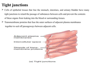
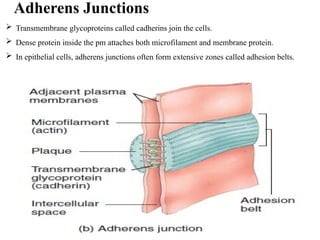
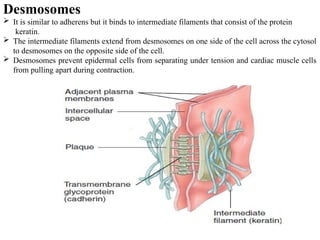
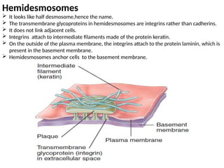
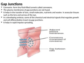
Tight junctions
 Cells of epithelial tissues that line the stomach, intestines, and urinary bladder have many
tight junctions to retard the passage of substances between cells and prevent the contents
of these organs from leaking into the blood or surrounding tissues.
 Transmembrane proteins that fuse the outer surfaces of adjacent plasma membranes
together to seal off passageways between adjacent cells.
Adherens Junctions
 Transmembrane glycoproteins called cadherins join the cells.
 Dense protein inside the pm attaches both microfilament and membrane protein.
 In epithelial cells, adherens junctions often form extensive zones called adhesion belts.
Desmosomes
 It is similar to adherens but it binds to intermediate filaments that consist of the protein
keratin.
 The intermediate filaments extend from desmosomes on one side of the cell across the cytosol
to desmosomes on the opposite side of the cell.
 Desmosomes prevent epidermal cells from separating under tension and cardiac muscle cells
from pulling apart during contraction.
Hemidesmosomes
 It looks like half desmosome,hence the name.
 The transmembrane glycoproteins in hemidesmosomes are integrins rather than cadherins.
 It does not link adjacent cells.
 Integrins attach to intermediate filaments made of the protein keratin.
 On the outside of the plasma membrane, the integrins attach to the protein laminin, which is
present in the basement membrane.
 Hemidesmosomes anchor cells to the basement membrane.
Gap Junctions
 Connexins form tiny fluid-filled tunnels called connexons.
 The plasma membranes of gap junctions are not fused.
 It helps in the transfer of ions, small molecules, nutrients and wastes in avascular tissues
such as the lens and cornea of the eye.
 In a developing embryo, some of the chemical and electrical signals that regulate growth
and cell differentiation travel via gap junctions.
 It helps in rapid impulse spreading.

Gap junctions - its location and its functions.pptx

  • 1.
    Tight junctions  Cellsof epithelial tissues that line the stomach, intestines, and urinary bladder have many tight junctions to retard the passage of substances between cells and prevent the contents of these organs from leaking into the blood or surrounding tissues.  Transmembrane proteins that fuse the outer surfaces of adjacent plasma membranes together to seal off passageways between adjacent cells.
  • 2.
    Adherens Junctions  Transmembraneglycoproteins called cadherins join the cells.  Dense protein inside the pm attaches both microfilament and membrane protein.  In epithelial cells, adherens junctions often form extensive zones called adhesion belts.
  • 3.
    Desmosomes  It issimilar to adherens but it binds to intermediate filaments that consist of the protein keratin.  The intermediate filaments extend from desmosomes on one side of the cell across the cytosol to desmosomes on the opposite side of the cell.  Desmosomes prevent epidermal cells from separating under tension and cardiac muscle cells from pulling apart during contraction.
  • 4.
    Hemidesmosomes  It lookslike half desmosome,hence the name.  The transmembrane glycoproteins in hemidesmosomes are integrins rather than cadherins.  It does not link adjacent cells.  Integrins attach to intermediate filaments made of the protein keratin.  On the outside of the plasma membrane, the integrins attach to the protein laminin, which is present in the basement membrane.  Hemidesmosomes anchor cells to the basement membrane.
  • 5.
    Gap Junctions  Connexinsform tiny fluid-filled tunnels called connexons.  The plasma membranes of gap junctions are not fused.  It helps in the transfer of ions, small molecules, nutrients and wastes in avascular tissues such as the lens and cornea of the eye.  In a developing embryo, some of the chemical and electrical signals that regulate growth and cell differentiation travel via gap junctions.  It helps in rapid impulse spreading.