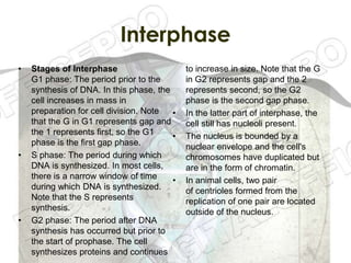
Cell Division
MITOSIS & MEIOSIS
Interphase
•   Stages of Interphase                     to increase in size. Note that the G
    G1 phase: The period prior to the        in G2 represents gap and the 2
    synthesis of DNA. In this phase, the     represents second, so the G2
    cell increases in mass in                phase is the second gap phase.
    preparation for cell division. Note  •   In the latter part of interphase, the
    that the G in G1 represents gap and      cell still has nucleoli present.
    the 1 represents first, so the G1    •   The nucleus is bounded by a
    phase is the first gap phase.            nuclear envelope and the cell's
•   S phase: The period during which         chromosomes have duplicated but
    DNA is synthesized. In most cells,       are in the form of chromatin.
    there is a narrow window of time     •   In animal cells, two pair
    during which DNA is synthesized.         of centrioles formed from the
    Note that the S represents               replication of one pair are located
    synthesis.                               outside of the nucleus.
•   G2 phase: The period after DNA
    synthesis has occurred but prior to
    the start of prophase. The cell
    synthesizes proteins and continues
Prophase
Changes that occur in a cell during prophase:


• Chromatin fibers become           • In animal cells, the mitotic
  coiled into chromosomes with        spindle initially appears as
  each chromosome having two          structures called asters which
  chromatids joined at                surround each centriole pair.
  a centromere.

• The mitotic spindle, composed     • The two pair
  of microtubules and proteins, f     of centrioles (formed from the
  orms in the cytoplasm.              replication of one pair in
                                      Interphase) move away from
                                      one another toward opposite
                                      ends of the cell due to the
                                      lengthening of the
                                      microtubules that form
                                      between them.
In late prophase:


• The nuclear envelope breaks       • The kinetochore fibers "interact"
  up.                                 with the spindle polar fibers
                                      connecting the kinetochores to
• Polar fibers, which are             the polar fibers.
  microtubules that make up the
  spindle fibers, reach from each • The chromosomes begin to
  cell pole to the cell's equator.  migrate toward the cell center.

• Kinetochores, which are
  specialized regions in the
  centromeres of chromosomes,
  attach to a type of microtubule
  called kinetochore fibers.
Metaphase
•The nuclear membrane
disappears completely.
In animal cells, the two
pair of centrioles align at
opposite poles of the
cell.
                              •Chromosomes move randomly until
•Polar fibers                 they attach (at their kinetochores) to
(microtubules that make       polar fibers from both sides of
up the spindle fibers)        their centromeres.
                              Chromosomes align at the
continue to extend from
                              metaphase plate at right angles to the
the poles to the              spindle poles.

                              •Chromosomes are held at the
                              metaphase plate by the equal forces
                              of the polar fibers pushing on the
                              centromeres of the chromosomes.
Anaphase
•The paired centromeres in
each distinct chromosome
begin to move apart.

•Once the paired sister
chromatids separate from
one another, each is
considered a "full"
chromosome. They are         •The daughter chromosomes migrate
referred to as daughter      centromere first and
chromosomes.                 the kinetochore fibers become shorter
                             as the chromosomes near a pole.
•Through the spindle
apparatus, the daughter      •In preparation for telophase, the two
chromosomes move to the      cell poles also move further apart during
poles at opposite ends of    the course of anaphase. At the end of
the cell.                    anaphase, each pole contains a
                             complete compilation of chromosomes.
Telophase
• In telophase, the chromosomes
  are cordoned off in distinct new
  nuclei in the emerging daughter
  cells.
Interphase
•   G1 phase: The period prior to the         to increase in size. Note that the G
    synthesis of DNA. In this phase, the      in G2 represents gap and the 2
    cell increases in mass in                 represents second, so the G2
    preparation for cell division. Note       phase is the second gap phase.
    that the G in G1 represents gap and
    the 1 represents first, so the G1    •    In the latter part of interphase, the
    phase is the first gap phase.             cell still has nucleoli present.

•   S phase: The period during which      •   The nucleus is bounded by a
    DNA is synthesized. In most cells,        nuclear envelope and the cell's
    there is a narrow window of time          chromosomes have duplicated but
    during which DNA is synthesized.          are in the form of chromatin.
    Note that the S represents
    synthesis.                            •   In animal cells, two pair
                                              of centrioles formed from the
•   G2 phase: The period after DNA            replication of one pair are located
    synthesis has occurred but prior to       outside of the nucleus.
    the start of prophase. The cell
    synthesizes proteins and continues
Interphase
Prophase I:
                              •Chromosomes thicken and
                              detach from the nuclear
                              envelope.

                              •Similar to mitosis,
•Chromosomes condense         the centrioles migrate away
and attach to the nuclear     from one another and both
envelope.                     the nuclear envelope and
                              nucleoli break down.
•Synapsis occurs (a pair of
                              •Likewise, the
homologous chromosomes
                              chromosomes begin their
lines up closely together)
                              migration to the metaphase
and a tetrad is formed.
                              plate.
Each tetrad is composed of
four chromatids.
Crossing over may occur.
Metaphase I:
•Tetrads align at the
metaphase plate.

•Note that
the centromeres of
homologous
chromosomes are
oriented toward the
opposite cell poles.
Anaphase I:
              •Chromosomes move to
              the opposite cell poles.
              Similar to mitosis, the
              microtubules and the
              kinetochore fibers
              interact to cause the
              movement.

              •Unlike in mitosis, the
              homologous
              chromosomes move to
              opposite poles yet
              the sister
              chromatids remain
              together.
Telophase I:
•The spindles continue to
move the homologous
chromosomes to the poles.
Once movement is
complete, each pole has a
haploid number of
chromosomes.                  •At the end of telophase I and
                              cytokinesis, two daughter cells are
•In most cases, cytokinesis   produced, each with one half the
occurs at the same time as    number of chromosomes of the original
telophase I.                  parent cell.

                              •Depending on the kind of cell, various
                              processes occur in preparation for
                              meiosis II. There is however a
                              constant: The genetic material does not
                              replicate again.
Prophase II:


•The nuclear membrane and nuclei break up while the spindle
network appears.

•Chromosomes do not replicate any further in this phase of
meiosis.

•The chromosomes begin migrating to the metaphase II plate
(at the cell's equator).
Metaphase II:

•The chromosomes line up at
the metaphase II plate at the
cell's center.

•The kinetochores of the
sister chromatids point
toward opposite poles.
Anaphase II:


The sister chromatids separate and move toward the
                opposite cell poles.
Telophase II:
  •Distinct nuclei form at the
  opposite poles
  and cytokinesis occurs.

  •At the end of meiosis II, there
  are four daughter cells each
  with one half the number of
  chromosomes of the original
  parent cell.
Barro, Kevin Winge B.
Casas, Gregorio Jr. A.

CELL DIVISION:
MITOSIS & MEIOSIS

Cell division

  • 1.
  • 3.
    Interphase • Stages of Interphase to increase in size. Note that the G G1 phase: The period prior to the in G2 represents gap and the 2 synthesis of DNA. In this phase, the represents second, so the G2 cell increases in mass in phase is the second gap phase. preparation for cell division. Note • In the latter part of interphase, the that the G in G1 represents gap and cell still has nucleoli present. the 1 represents first, so the G1 • The nucleus is bounded by a phase is the first gap phase. nuclear envelope and the cell's • S phase: The period during which chromosomes have duplicated but DNA is synthesized. In most cells, are in the form of chromatin. there is a narrow window of time • In animal cells, two pair during which DNA is synthesized. of centrioles formed from the Note that the S represents replication of one pair are located synthesis. outside of the nucleus. • G2 phase: The period after DNA synthesis has occurred but prior to the start of prophase. The cell synthesizes proteins and continues
  • 4.
  • 5.
    Changes that occurin a cell during prophase: • Chromatin fibers become • In animal cells, the mitotic coiled into chromosomes with spindle initially appears as each chromosome having two structures called asters which chromatids joined at surround each centriole pair. a centromere. • The mitotic spindle, composed • The two pair of microtubules and proteins, f of centrioles (formed from the orms in the cytoplasm. replication of one pair in Interphase) move away from one another toward opposite ends of the cell due to the lengthening of the microtubules that form between them.
  • 6.
    In late prophase: •The nuclear envelope breaks • The kinetochore fibers "interact" up. with the spindle polar fibers connecting the kinetochores to • Polar fibers, which are the polar fibers. microtubules that make up the spindle fibers, reach from each • The chromosomes begin to cell pole to the cell's equator. migrate toward the cell center. • Kinetochores, which are specialized regions in the centromeres of chromosomes, attach to a type of microtubule called kinetochore fibers.
  • 7.
    Metaphase •The nuclear membrane disappearscompletely. In animal cells, the two pair of centrioles align at opposite poles of the cell. •Chromosomes move randomly until •Polar fibers they attach (at their kinetochores) to (microtubules that make polar fibers from both sides of up the spindle fibers) their centromeres. Chromosomes align at the continue to extend from metaphase plate at right angles to the the poles to the spindle poles. •Chromosomes are held at the metaphase plate by the equal forces of the polar fibers pushing on the centromeres of the chromosomes.
  • 8.
    Anaphase •The paired centromeresin each distinct chromosome begin to move apart. •Once the paired sister chromatids separate from one another, each is considered a "full" chromosome. They are •The daughter chromosomes migrate referred to as daughter centromere first and chromosomes. the kinetochore fibers become shorter as the chromosomes near a pole. •Through the spindle apparatus, the daughter •In preparation for telophase, the two chromosomes move to the cell poles also move further apart during poles at opposite ends of the course of anaphase. At the end of the cell. anaphase, each pole contains a complete compilation of chromosomes.
  • 9.
    Telophase • In telophase,the chromosomes are cordoned off in distinct new nuclei in the emerging daughter cells.
  • 11.
    Interphase • G1 phase: The period prior to the to increase in size. Note that the G synthesis of DNA. In this phase, the in G2 represents gap and the 2 cell increases in mass in represents second, so the G2 preparation for cell division. Note phase is the second gap phase. that the G in G1 represents gap and the 1 represents first, so the G1 • In the latter part of interphase, the phase is the first gap phase. cell still has nucleoli present. • S phase: The period during which • The nucleus is bounded by a DNA is synthesized. In most cells, nuclear envelope and the cell's there is a narrow window of time chromosomes have duplicated but during which DNA is synthesized. are in the form of chromatin. Note that the S represents synthesis. • In animal cells, two pair of centrioles formed from the • G2 phase: The period after DNA replication of one pair are located synthesis has occurred but prior to outside of the nucleus. the start of prophase. The cell synthesizes proteins and continues
  • 12.
  • 13.
    Prophase I: •Chromosomes thicken and detach from the nuclear envelope. •Similar to mitosis, •Chromosomes condense the centrioles migrate away and attach to the nuclear from one another and both envelope. the nuclear envelope and nucleoli break down. •Synapsis occurs (a pair of •Likewise, the homologous chromosomes chromosomes begin their lines up closely together) migration to the metaphase and a tetrad is formed. plate. Each tetrad is composed of four chromatids. Crossing over may occur.
  • 14.
    Metaphase I: •Tetrads alignat the metaphase plate. •Note that the centromeres of homologous chromosomes are oriented toward the opposite cell poles.
  • 15.
    Anaphase I: •Chromosomes move to the opposite cell poles. Similar to mitosis, the microtubules and the kinetochore fibers interact to cause the movement. •Unlike in mitosis, the homologous chromosomes move to opposite poles yet the sister chromatids remain together.
  • 16.
    Telophase I: •The spindlescontinue to move the homologous chromosomes to the poles. Once movement is complete, each pole has a haploid number of chromosomes. •At the end of telophase I and cytokinesis, two daughter cells are •In most cases, cytokinesis produced, each with one half the occurs at the same time as number of chromosomes of the original telophase I. parent cell. •Depending on the kind of cell, various processes occur in preparation for meiosis II. There is however a constant: The genetic material does not replicate again.
  • 17.
    Prophase II: •The nuclearmembrane and nuclei break up while the spindle network appears. •Chromosomes do not replicate any further in this phase of meiosis. •The chromosomes begin migrating to the metaphase II plate (at the cell's equator).
  • 18.
    Metaphase II: •The chromosomesline up at the metaphase II plate at the cell's center. •The kinetochores of the sister chromatids point toward opposite poles.
  • 19.
    Anaphase II: The sisterchromatids separate and move toward the opposite cell poles.
  • 20.
    Telophase II: •Distinct nuclei form at the opposite poles and cytokinesis occurs. •At the end of meiosis II, there are four daughter cells each with one half the number of chromosomes of the original parent cell.
  • 21.
    Barro, Kevin WingeB. Casas, Gregorio Jr. A. CELL DIVISION: MITOSIS & MEIOSIS