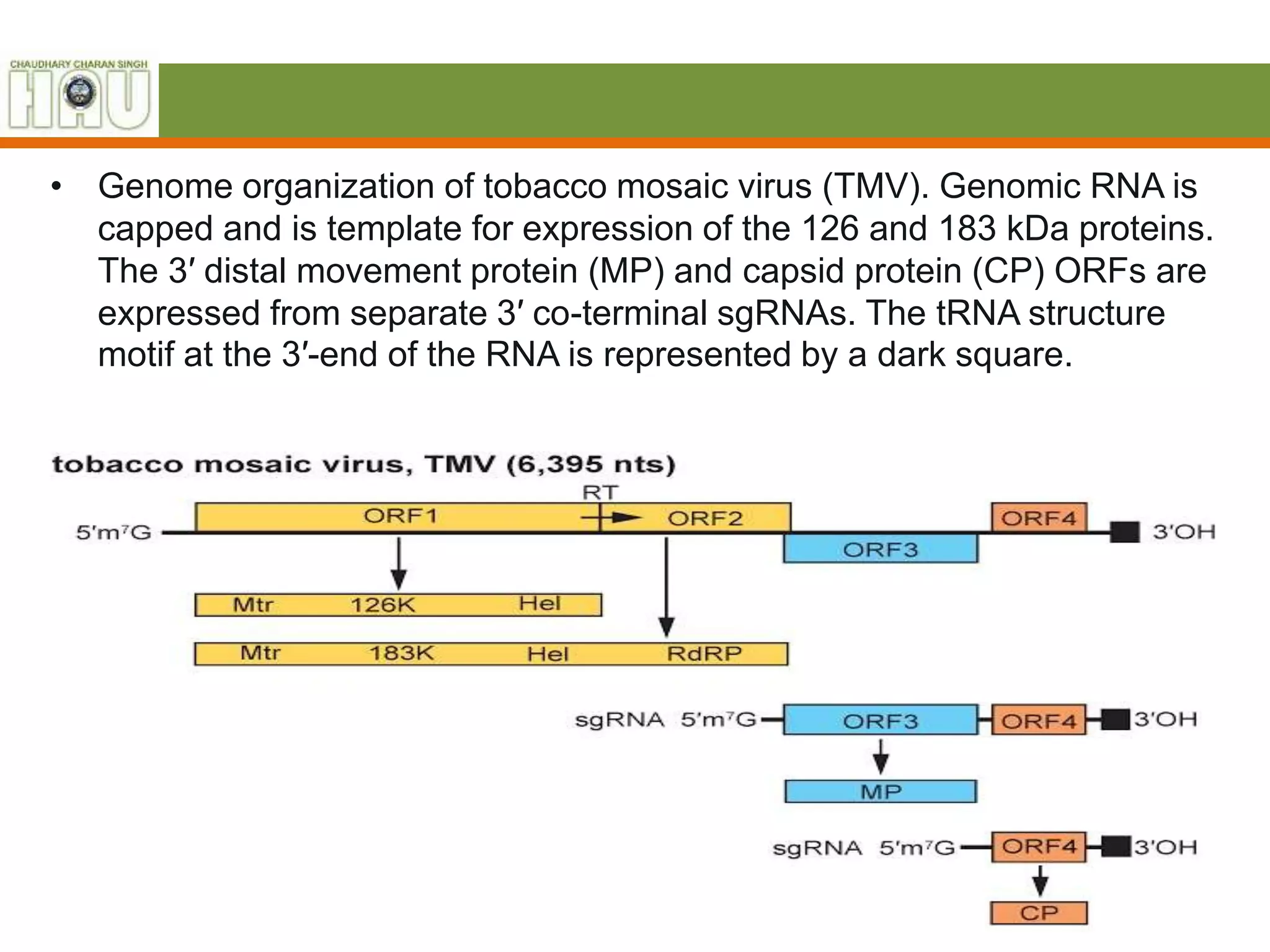
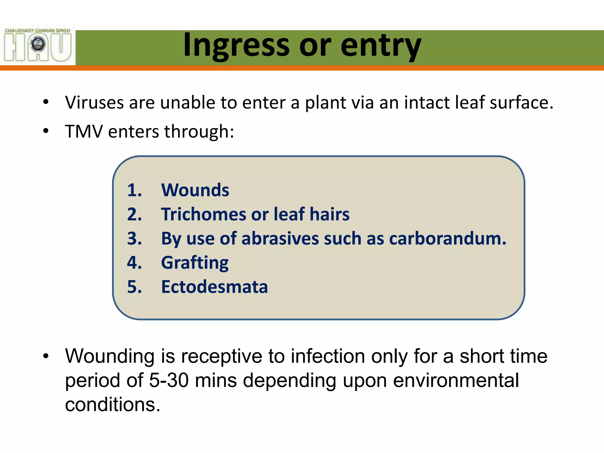
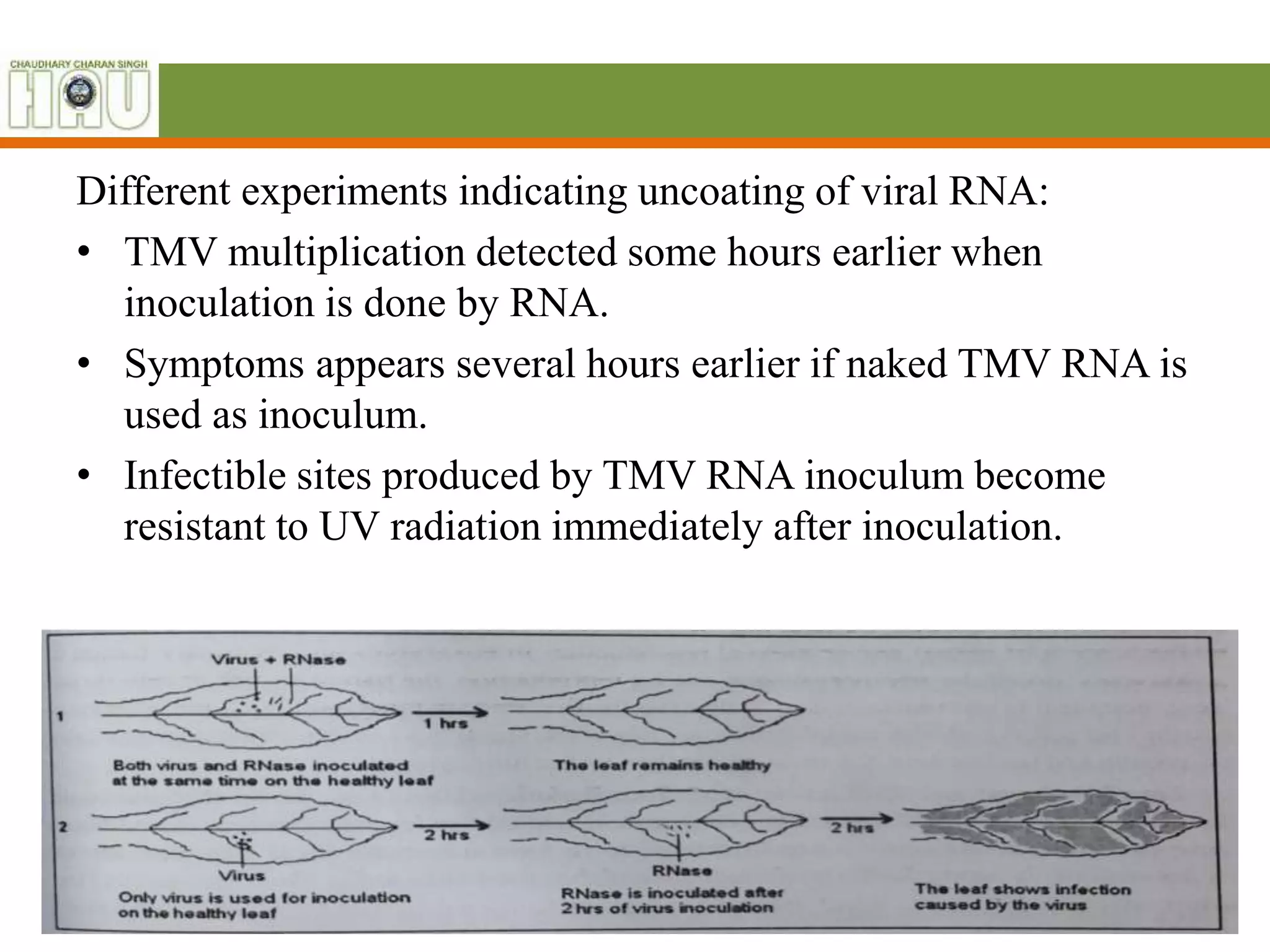
2. Tobamovirus (TMV)
• Tobamoviruses are the only members of the family to
have a non-segmented genome.
• They have a “30K”-like cell-to-cell movement protein, are
not vector-transmissible and when seed transmitted, the
embryo is not affected.
• It is the largest genus in the family and the literature is
extensive.
3. Morphology
• Virions are 18 nm in diameter and have a predominant
length of 300–310 nm
• Shorter virions produced by the encapsidation of
subgenome-sized RNA are usually a minor component of
the virion population, although virions of two species
produce an abundant short virion of 32–34 nm.
• Virions often form large crystalline arrays visible by light
microscopy.
4. Genome organization of TMV
• The single genomic RNA encodes at least four proteins.
• The 124–132 kDa and 181–189 kDa replication proteins
are translated directly from the 5' proximal ORF of the
genomic RNA.
• The 124–132 kDa replication protein contains the Mtr and
Hel domains.
• The 181–189 kDa replication protein additionally contains
the polymerase domain, synthesized by occasional
readthrough of the leaky termination codon of the 124–
132 kDa protein encoding ORF.
• The 181–189 kDa replication protein is the only protein
required for replication in single cells, although the 124–
132 kDa replication protein is also required for efficient
replication.
5. • The downstream ORFs encode the 28–31 kDa MP and
17–18 kDa CP, which are translated from their respective
3′ co-terminal sgRNAs, both of which contain a 5′ cap.
• In the members of some species, the MP ORF overlaps
both the 181–189 kDa protein and the CP ORFs,
whereas in other species it does not overlap either ORF
or overlaps one of the ORFs.
• An ORF that encodes a cysteine-rich protein is located
between the 181-189 kDa and MP ORFs in passion fruit
mosaic virus.
6. • Genome organization of tobacco mosaic virus (TMV). Genomic RNA is
capped and is template for expression of the 126 and 183 kDa proteins.
The 3′ distal movement protein (MP) and capsid protein (CP) ORFs are
expressed from separate 3′ co-terminal sgRNAs. The tRNA structure
motif at the 3′-end of the RNA is represented by a dark square.
7. Multiplication of virus
• Steps:
Uncoating
Replication of viral
genome
Replication of protein and
protein synthesis
Assembly of new virion
Movement of vision in
plant tissues
Ingress
8. Ingress or entry
• Viruses are unable to enter a plant via an intact leaf surface.
• TMV enters through:
1. Wounds
2. Trichomes or leaf hairs
3. By use of abrasives such as carborandum.
4. Grafting
5. Ectodesmata
• Wounding is receptive to infection only for a short time
period of 5-30 mins depending upon environmental
conditions.
9. Uncoating or virus disassambly
• Uncoating is removal of virus protein which is around the
nucleaic acid.
• Uncoating takes place during early stage of infection and the
viral genome is released before it can replicate and its geneti
Information can be translated.
• In TMV, uncoating starts within 8 minutes of inoculation.
• TMV attach to the membranes in end on position and after
that there is gradual reduction in particle length and diameter
representing protein loss or uncoating.
• Hydrogen bonds
• Lysosomal enzyme
• Conrat and Williams Berkley rna and protein separated
10. Different experiments indicating uncoating of viral RNA:
• TMV multiplication detected some hours earlier when
inoculation is done by RNA.
• Symptoms appears several hours earlier if naked TMV RNA is
used as inoculum.
• Infectible sites produced by TMV RNA inoculum become
resistant to UV radiation immediately after inoculation.
11. Replication of viral genome
• Production of new RNA molecule identical to template RNA is
called Replication.
Viral RNA performs two function:
1. It serves as mRNA, immediately binds to ribosome and directs
the synthesis of virus specific proteins.
• Among which one protein inhibits host protein and host RNA
synthesis.
• other protein is specific viral replicating enzyme i.e. RNA
polymerase or replicase.
2. Parental viral RNA is then displaced from ribosome, triggers
the replication of viral RNA during which it behaves as a
template or plus strand for synthesis of minus strand in
presence of specific replicase already synthesized.
12. Once viral genome enter the cell- replication start
• Its mediated by a replicase enzyme,
• possibly coded partially by the viral genome and host genome.
• In the presence of polymerase, a –ve strand complementary to the
genome +ve strand is formed.
• -ve strand serve as template for the synthesis of progeny or new
+ve stand RNA by means of replicase enzyme forming a partially
double stranded, partially single stranded structure, called
Replicative intermediate (RI)(dsrna with ss tails)
• A part of viral RNA form coat protein
• Synthesis of new RNA is from the 3’ to 5’ ends of the templates.
• Replication occurs in a replication complex that comprises of the
template, newly synthesized RNA, the replicase and host factors.
13.
14. Replication of +ve strand RNA viruses (e.g. TMV) can be divided into four
overlapping steps. These are:
1. The uncoating of the virus: It is an exposure of the nucleic acid to the
replication processes.
2. Translation: During replication process , the viral RNA serves as a messenger
RNA and produces structural and non- structural proteins. This process is
further divided into the primary or early translation of proteins required for
replication (e.g. RdRp), and secondary or late translation of protein with the late
functions(CP).
3. Replication of genome: It yields progeny RNA molecules and it takes place in
two stages: (a) synthesis of a full-length complementary (-) strand RNA using
the (+) strand gRNA as a template and (b) synthesis of progeny gRNA and
sgRNA using the (-) strand RNA as a template. Both the stages are catalysed by
an RdRp.
4. Encapsidation: The progeny genomic strands are encapsidated.
16. Replication of viral proteins
• For most RNA viruses, the virus RNA, after it is freed from the
protein coat, replicates itself in the cytoplasm, where it also
directly serve as mRNA and a complementary strand which will
be negative sense is synthesized. Formation of virus will
involve transcription of virus mRNA and then translation to
produce a protein coat.
• Translation is the process by which a protein is synthesized
from the information contained in a molecule of messenger
RNA (mRNA). During translation, an mRNA sequence is read
using the genetic code, which is a set of rules that defines how
an mRNA sequence is to be translated into the 20-letter code
of amino acids, which are the building blocks of proteins.
17. • The genetic code is a set of three-letter combinations of
nucleotides called codons, each of which corresponds with a
specific amino acid or stop signal. Translation occurs in a
structure called the ribosome, which is a factory for the
synthesis of proteins. The ribosome has a small and a large
subunit and is a complex molecule composed of several
ribosomal RNA molecules and a number of proteins.
• Translation of an mRNA molecule by the ribosome occurs in
three stages:
a) Initiation,
b) Elongation, and
c) Termination.
18. Assembly of new virion
• Newly produced nucleic acid organize new protein subunits
around it and both assemble to form new virus particles,
virion.
• Assembly can occur in cytoplasm or nucleus or both but not
in any other organelles.
• Virions then passes into extracellular phase of generation
cycle.
19. Movement of plant viruses
• There are two types of movement
1. Short distance movement through plasmodesmata and
phloem tissue living cells.
2. Long distance movement through phloem sieve tubes.
• Virus MRS enable viral movement through plasmodesmata.
• In TMV, movement function is provided by triple gene block (
2 putative small membrane associated protein and a putative
RNA helicase).
20. Movement of plant
viruses as nucleoprotien
1. Virus particles encode
MP.
2. MP binds to viral RNA
3. Binding unfolds viral RNA
from random coil to
linear rod shaped
structure.
4. MP RNA complex
targeted to PD
5. Through PD movement
to other cells
Movement of virus as
virion
1. Intercellular movement
2. Formation of tubular
structure in association
with or through PD.
3. Virus particles in linear
form are observed inside
these structures.
4. Through cell wall tubular
structure move into
neighboring cell.
5. Deliver virus into that
cell.