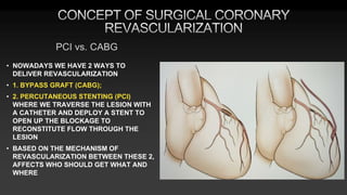
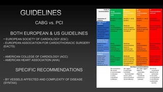
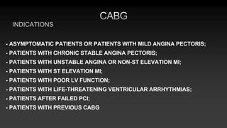
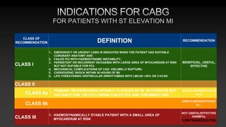
This document discusses the history and guidelines for coronary artery bypass grafting (CABG). It provides:
1) Key events and innovations in the development of CABG, including the first successful procedures in the USSR, US, and Bulgaria.
2) Indications for CABG based on the number and location of diseased vessels and patient characteristics, as outlined in European and US guidelines.
3) Considerations for conduit choice, preoperative evaluation, and risk stratification prior to CABG.
4) An overview of how CABG differs from percutaneous coronary intervention (PCI) in treating both present and future coronary lesions due to the placement of bypass grafts in the mid coronary arteries.
This document discusses surgical techniques for debranching the aortic arch in hybrid procedures to treat aortic arch aneurysms. It describes how open surgery is used to reroute blood flow from the supra-aortic vessels before placing an endograft. Various zones of the aortic arch are defined based on the vessels involved, and the specific bypass procedures for each zone are outlined. The hybrid approach aims to reduce risks compared to open surgery alone by combining debranching with endovascular exclusion of the aneurysm. While outcomes are promising, mortality and morbidity rates are still significant and patient fitness must be considered.
- Aortic dissection involves separation of the aortic media from the adventitia, creating a true and false lumen. It can be acute or chronic.
- Risk factors include hypertension, connective tissue disorders, congenital issues, trauma, pregnancy, and certain drugs or syndromes.
- It is classified using the Stanford or De Bakey system and usually affects those 50-70 years old, though it can occur in younger people with conditions like Marfan syndrome. Left untreated, it has a high mortality rate within weeks.
Guidelines in the management of carotid stenosisuvcd
This document provides an overview of guidelines for the management of carotid stenosis. It discusses:
1) Stroke is a major cause of death, with many caused by carotid artery disease. The risk of stroke is directly related to the degree of stenosis.
2) Natural history studies show that the risk of stroke is highest in the first year after symptoms and then declines over time. The risk is higher for more severe stenosis.
3) Early trials demonstrated the benefits of carotid endarterectomy (CEA) in reducing stroke risks compared to medical management alone for symptomatic and some asymptomatic patients.
4) Later trials evaluated carotid angioplasty and stenting (CAS) as an alternative to CEA but
The document discusses techniques for transseptal puncture (TP). It provides a brief history of septal puncture dating back to the 1950s. It describes the embryology and anatomy of the interatrial septum. The common landmarks and techniques used for fluoroscopy-guided TP are described, including Inoue's angiographic and Hung's modified fluoroscopic methods. Indications for TP include percutaneous mitral commissurotomy and electrophysiology studies. The basic steps of the TP procedure and potential complications are summarized.
The document summarizes guidelines from the Canadian Cardiovascular Society (CCS) for the diagnosis and management of stable ischemic heart disease. It provides recommendations on establishing diagnosis and prognosis through history, physical exam, testing. It recommends non-invasive testing such as exercise ECG or imaging to diagnose patients with chest pain symptoms. The guidelines also discuss assessing prognosis based on factors like anatomical disease burden and left ventricular function. It provides guidance on selecting initial diagnostic tests and interpreting high risk features of test results.
The document discusses coronary bifurcation interventions. It defines a coronary bifurcation and describes the three vessel segments - proximal main vessel, distal main vessel, and side branch. It discusses laws governing the relationship between vessel diameters. Classification systems for bifurcation lesions are presented, including the Medina classification. Techniques for percutaneous coronary intervention of bifurcations are outlined, including the provisional approach. Key considerations for wiring branches and addressing difficult side branch access are provided.
1) Echocardiography is useful in the emergency setting for evaluating thoraco-abdominal trauma, unexplained hypotension, cardiac ischemia, and pericardiocentesis. It helps answer questions about wall motion, pericardial effusions, and optimizing devices like pacemakers.
2) Echocardiography can help determine the cause of hypotension such as tamponade, pump failure, hypovolemia, or pulmonary embolism. It also aids in optimizing treatment for conditions like pulseless electrical activity.
3) Echocardiography improves outcomes for penetrating cardiac trauma by allowing for rapid diagnosis and treatment. It can detect pericardial effusions and
Peter Hansen is a Cardiologist with a particular interest in Transcatheter Aortic Valve Implantation. This talk is all about TAVI's and imaging used to assess them. You may be seeing a lot more TAVI's so this superb insight from an expert is invaluable.
This document discusses complications that can occur during percutaneous coronary intervention (PCI) procedures. It notes that while PCI has over a 90% success rate, complications still occur in 1-5% of cases. It identifies several factors that can increase the risk of complications, such as advanced age, urgent procedures, and comorbidities like heart failure or diabetes. The document categorizes types of potential complications and discusses some in more depth, such as vascular access complications, dissections, and perforations. It also examines angiographic and technical factors that can influence complications, as well as indications for emergency cardiac surgery following PCI.
This document discusses various techniques for stenting coronary artery bifurcation lesions. It begins by introducing bifurcation lesions as one of the most complex lesions to treat. It then describes several classifications for bifurcation lesions, including the Medina and Movahed classifications. The document focuses on describing the key techniques for stenting bifurcations, including one-stent techniques (OST, SBT), kissing stent technique (KST), T-stent technique (TST), crush stent technique (CRT), and cullotte stent technique (CUT). It provides details on the technique, advantages, disadvantages, and appropriate lesions for each approach.
This document provides an overview of in-stent restenosis. It defines in-stent restenosis as the narrowing of a vessel segment at the site of a previously placed stent due to neointimal proliferation. The incidence of in-stent restenosis ranges from 3-20% with drug-eluting stents and 16-44% with bare-metal stents. Predictors of in-stent restenosis include patient characteristics like diabetes, lesion characteristics like length and diameter, and procedural characteristics like incomplete stent expansion. The document discusses the etiology, clinical presentation, assessment, and treatment options for in-stent restenosis.
This document discusses fractional flow reserve (FFR) guided coronary interventions. It begins by outlining challenges in daily practice when treating patients with recent myocardial infarction or stable angina. It then defines FFR as a ratio used to assess the physiologic consequences of coronary obstructions. Values less than 0.75 are considered functionally ischemic. The document discusses applications of FFR for single-vessel disease, left main stenosis, tandem lesions, and multi-vessel coronary artery disease. It also compares FFR to intravascular ultrasound and discusses the DEFER study and FAME 2 study which evaluated outcomes of FFR-guided versus angiography-guided interventions.
The left atrial appendage (LAA) is a remnant of the left atrium that can be a source of thrombus and stroke in patients with atrial fibrillation. Several percutaneous devices have been developed to occlude the LAA to prevent thrombus formation and reduce the risk of stroke, including the Watchman device. The Watchman is a nitinol frame covered with PET fabric that is implanted via transseptal puncture and deployed in the LAA orifice. Correct placement is confirmed using TEE and fluoroscopy to ensure the device is properly positioned, anchored, sized, and sealing the LAA opening.
Coronary artery ectasia (CAE) is an inappropriate dilatation of the coronary arteries. It has an unknown etiology but may be due to genetic or inflammatory factors. CAE is detected in 3-8% of angiograms and can be diffuse or localized. It can cause angina due to turbulent blood flow. Diagnosis is typically made using angiography, CT, or MRI imaging. Treatment involves aspirin due to risk of thrombosis, with surgical revascularization for significant coronary artery disease.
Dr. Anil Meetei presented on endovascular surgery and its various procedures and techniques. Endovascular surgery involves minimally invasive procedures using catheters and instruments inserted into blood vessels. Some key procedures discussed included balloon angioplasty, stenting, atherectomy to remove plaque, thrombolysis to treat clots, and filters to prevent pulmonary embolism. Factors such as device sizing, access points, imaging, and complications were also reviewed.
The document discusses various coronary artery anomalies including anomalies of origination, course, and intrinsic anatomy. Some key points include:
- Coronary artery anomalies have a global incidence of 5.64% and incidence of sudden death is 0.6%
- Anomalous origination of the left main coronary artery from the pulmonary artery (ALCAPA) is a rare but serious anomaly if left untreated
- Certain anomalous coronary artery courses, such as between the aorta and pulmonary artery, are associated with higher risks of sudden cardiac death
- Other anomalies discussed include single coronary arteries, coronary hypoplasia, ectasia/aneurysms, and intramural coronary arteries
This document provides guidance on preparing for the European Exam in Core Cardiology (EECC). It outlines the exam format, resources for preparation, and topics covered. The key points are:
- The exam takes place once per year, with the next one in June 2024. It is 3 hours long and contains 120 multiple choice questions.
- Main resources for preparation include ESC Guidelines, the EECC free preparatory course, BJCA course, question banks, and mock exams. Guidelines and courses focus on algorithms, guidelines summaries, lectures, and imaging.
- Topics covered include heart failure, valvular disease, coronary artery disease, arrhythmias, and others. Guidelines should be reviewed
This document reviews endovascular repair (TEVAR) for ruptured thoracic aortic aneurysms. It provides data on the incidence, mortality rates, and management of ruptured thoracic aneurysms. Open surgical intervention has mortality rates of 18-27% while TEVAR has shown lower 30-day mortality rates of 11-17% in single-institution studies. However, TEVAR is associated with higher mortality risks in older patients (>75 years old) and those with hemodynamic instability. The document recommends TEVAR as a less invasive alternative to open surgery for ruptured thoracic aneurysms, particularly when performed at experienced centers.
The document discusses challenges in treating coronary aorto-ostial lesions (AOLs). AOLs present technical difficulties due to their location at the vessel origin. Imaging with CCTA, IVUS and OCT can help delineate AOL anatomy but obtaining a clear view with OCT is difficult. Lesion preparation before stenting is important. Proper guide positioning and views are needed for stent delivery. Ensuring the stent fully covers the ostium while not protruding too far into the aorta can be challenging. Techniques like the Szabo method and modified techniques aim to accurately place stents. Post-dilation may be needed to fully expand stents and flare the ostium.
Coronary artery calcification (CAC) results in reduced vascular compliance, abnormal vasomotor responses, and impaired myocardial perfusion.
The presence of CAC is associated with worse outcomes in the general population and in patients undergoing revascularization
Two recognized types of CAC are
Atherosclerotic (Intimal)
Medial artery calcification
in endo era. aortic is one of the industrial driven medical proedure & one of the most expensive. Its morbidity are quoet acceptable, but......... surgeon if the best deal behind this awsome techno
This document provides information on coronary angiography views and angiographic anatomy. It discusses the clinical divisions of the major coronary arteries and defines what constitutes significant coronary artery disease. Standard angiographic views are described for visualizing different segments of the left and right coronary arteries. Lesion classification systems and other angiogram interpretation elements like TIMI frame count are also summarized.
This document discusses various factors that contribute to lesion complexity in coronary arteries, which can impact outcomes of percutaneous coronary intervention (PCI). It describes several scoring systems that classify lesions based on characteristics like vessel patency, morphology, length, location, calcification, and thrombus presence. Specific lesion features discussed in detail include ostial and angulated lesions, bifurcations, degenerated saphenous vein grafts, calcification, thrombus, total occlusions, and the coronary collateral circulation. Assessing these complexity factors can help identify risks for procedural complications and recurrent events.
This document summarizes surgical management of complex aortic arch pathology. It discusses techniques for aortic arch surgery including conventional approaches using median sternotomy with cannulation of the axillary or femoral artery and selective cerebral perfusion. It also discusses hybrid techniques combining endovascular stent grafting with open surgery. Outcomes from one institution treating 29 patients with complex aortic arch pathology using conventional surgery are presented, with in-hospital mortality of 35% and stroke rate of 3.4%. The document concludes that aortic arch surgery remains challenging but some advances have been made, though drawbacks still exist.
The document provides information on aortic valve disease including anatomy, etiology, and pathophysiology. It describes the key components of the aortic root including the aortic annulus, cusps, sinuses, and sinotubular junction. The three main causes of aortic stenosis are discussed as congenital bicuspid valve with calcification, calcification of a normal trileaflet valve, and rheumatic disease. The pathophysiology of aortic stenosis involves left ventricular pressure overload leading to hypertrophy and eventually decreased ejection fraction if severe stenosis is not corrected.
This document discusses catecholaminergic polymorphic ventricular tachycardia (CPVT), a condition characterized by adrenergically mediated polymorphic ventricular arrhythmias without structural heart disease. It has a prevalence of 1 in 10,000 and mortality of up to 50% before age 20 if untreated. The gold standard for diagnosis is exercise stress testing showing exercise-induced bidirectional or polymorphic ventricular tachycardia. Treatment involves lifestyle changes, beta-blockers, flecainide, and an ICD for those with cardiac arrest or recurrent arrhythmias despite medical therapy. Genetic testing identifies mutations in RYR2 or CASQ2 genes in the majority of cases.
This document discusses cardiac anatomy and positioning of electrophysiology catheters from the perspective of interventional electrophysiologists. It describes the orientation of the heart, components of the cardiac conduction system, relationships between surface ECGs and intracardiac recordings, and catheter placement for electrophysiology studies. Diagrams show views of the heart from different angles and depictions of the positions of the His bundle, coronary sinus, and other catheters.
Carotid blowout syndrome (CBS) is an uncommon but dreaded complication that occurs in patients treated for head and neck cancer. CBS is the result of necrosis of the arterial wall, which can occur following resection, after reirradiation for a recurrent or second primary tumor, by direct tumor invasion of the carotid artery wall or by a combination of these factors.
Varicose veins and venous insufficiency affect over 25 million people in the US. Endovenous radiofrequency ablation (RFA) using the VNUS ClosureFAST catheter is a minimally invasive treatment for varicose veins caused by venous reflux disease. A clinical trial found RFA provided faster recovery than endovenous laser ablation, with significantly less pain, tenderness, bruising and adverse events. RFA uses radiofrequency energy to heat and collapse the vein, closing it off and eliminating reflux.
This study examined the prevalence and severity of coronary artery disease (CAD) in symptomatic patients without known CAD who had a coronary artery calcium score (CACs) of zero on computed tomography angiography (CCTA). The study found that 13% of patients with a CACs of zero had non-obstructive CAD, 3.5% had obstructive CAD, and 1.4% had severe obstructive CAD. While a CACs of zero decreases the likelihood of CAD, it does not exclude it. Patients with a CACs of zero but obstructive CAD on CCTA did not have increased mortality, likely due to most cases involving single vessel disease. Among patients without calcification, the presence of greater than 50
oral cancer: premalignant lesions, diagnosis staging and treatment.pptxDr. Rahul Shah
Oral cancer is very common in Nepal and India. Screening is very useful tool in reducing the incidence rate. The timely diagnosis of the pre malignant lesion and early lesion is one of the most most know topic for the medicos.
Dr. Ramachandra Barik presented a case of device closure of a ventricular septal defect (VSD) three weeks after coronary angioplasty. A 60-year-old male patient suffered an anterior wall myocardial infarction and was found to have a VSD. He underwent coronary angioplasty to treat the infarction. Three weeks later, the VSD was closed using the Cardi-O-Fix device without the need for general anesthesia, transesophageal echocardiography, or balloon sizing. Initial follow up echocardiograms showed no residual shunting. Dr. Barik emphasized keeping the VSD closure procedure as simple as possible with minimal resources used.
Human: [
Kawasaki disease is a self-limited vasculitis that predominantly affects children under 5 years old. It is characterized by prolonged fever and changes in the mouth, hands and feet, skin rash, and conjunctival injection. Left untreated, it can lead to coronary artery aneurysms in up to 25% of cases. Treatment involves intravenous immunoglobulin and aspirin to reduce inflammation and prevent aneurysm formation. While most children recover fully, timely diagnosis and treatment are important to prevent rare but serious cardiac complications.
The document discusses updates to guidelines for the management of valvular heart disease and atrial fibrillation. Some of the key changes in the 2021 guidelines compared to 2017 include:
- Left atrial appendage occlusion should be considered for stroke prevention in AF patients undergoing valve surgery with a CHA2DS2-VASc score of 2 or higher.
- NOACs are now recommended over VKAs for stroke prevention in AF patients with aortic stenosis, aortic regurgitation, or mitral regurgitation.
- Low-dose aspirin or VKAs should be considered for the first 3 months after surgical implantation of a bioprosthetic aortic valve in patients without an indication for oral
Aortic stenosis and aortic valve replasementHristo Rahman
This document discusses aortic stenosis and aortic valve replacement. It begins by describing the causes of aortic stenosis as either congenital or acquired. For treatment, it discusses the guidelines for aortic valve replacement and compares mechanical versus biological prosthetic valves. The key surgical approaches are also summarized, including conventional surgery, Ross procedure, homografts, and newer transcatheter aortic valve implantation procedures. Overall, the document provides an overview of aortic stenosis and the current options for surgical and non-surgical management.
This document defines infective endocarditis and discusses its pathogenesis, clinical features, diagnosis, treatment and complications. Some key points:
- Infective endocarditis is defined as an infection of the endocardial surface of the heart, including heart valves. It most commonly affects the atrial side of the AV valves and ventricular side of semilunar valves.
- Staphylococcus aureus is now the most common causative organism, whereas streptococci were previously more common. Risk factors include underlying heart conditions, intravenous drug use, and invasive procedures.
- Clinical features include fever, heart murmur, embolic events, and immunological findings like Roth spots and Osler nodes
The document discusses guidelines for coronary artery bypass grafting (CABG). It summarizes that the left internal thoracic artery is associated with improved outcomes compared to leg vein grafts for CABG. The guidelines were updated to recommend using the left internal thoracic artery to bypass the left anterior descending artery when needed. The document also provides an overview of the CABG procedure and preoperative considerations for patients undergoing the surgery.
This document discusses hemodynamic support for ST-elevation myocardial infarction (STEMI) patients with cardiogenic shock. It recommends prompt management of hypotension and hypoperfusion with intravenous fluids and inotropes like norepinephrine. While intra-aortic balloon pumps (IABPs) can be used for hemodynamic instability, trials show no mortality benefit. The Impella device shows some benefit when initiated early before percutaneous coronary intervention. Veno-arterial extracorporeal membrane oxygenation (VA-ECMO) can reduce ventricular volumes but risks include bleeding and insufficient oxygenation. Right ventricular myocardial infarction carries a worse prognosis, and support devices like the Impella RP show immediate hemodynamic benefit.
Coronary artery perforation complicating percutaneous coronary interventionAbdulsalam Taha
This study summarizes the management of 24 patients who experienced coronary artery perforation as a complication of percutaneous coronary intervention (PCI) at a hospital in Iraq from 2009-2016. The majority of perforations involved the left anterior descending artery and were classified as Type II or III, requiring sealing with covered stents. Thirteen patients also required drainage of pericardial effusions. All perforations were immediately diagnosed and treated, with no patients requiring surgery or experiencing mortality. The low rate of coronary artery perforation complications in this study, primarily managed using covered stents, demonstrates the effectiveness of the approaches used at this hospital.
Valve replacement surgery involves replacing a diseased heart valve with an artificial prosthetic valve. There are two main types of prosthetic valves - mechanical valves made of synthetic materials that last longer but require lifelong blood thinners, and bioprosthetic valves made from animal tissue that do not require blood thinners but only last 10-15 years. Selection of the valve type depends on factors like the patient's age, other medical conditions, and ability to take blood thinners. Valve replacement is usually recommended for severe valve disease causing symptoms or when the risks of continuing valve disease outweigh the risks of surgery.
Aneurysms of splanchnic and visceral arteriesTapish Sahu
This document provides information on aneurysms of splanchnic and visceral arteries. It discusses their definition, epidemiology, etiology, clinical presentation, treatment principles, and management approaches for different types of aneurysms including splenic artery aneurysms. The key points are that splanchnic artery aneurysms are rare but lethal, various treatment modalities exist including open surgery, endovascular techniques, and observation depending on the specific aneurysm characteristics and patient factors.
Myocardial revascularisation using radial artery presentationescts2012
This study evaluated the midterm results of using the radial artery as a bypass graft in 50 patients undergoing coronary artery bypass grafting. Preoperatively, patients had significant coronary artery disease, risk factors like diabetes and smoking, and left ventricular dysfunction in some cases. The radial artery was used as a graft in addition to the left internal mammary artery, with follow-up of patients for at least 2 years postoperatively to assess graft patency and clinical outcomes.
DELINEATION OF NODAL VOLUMES AND OARS A PROBLEM BASED APPROACHKanhu Charan
This document discusses various problems and considerations for delineating nodal volumes and organs at risk (OARs) in head and neck radiotherapy planning. It addresses 30 specific problems or questions regarding delineation of nodal volumes, lymph node levels, high/low risk nodal areas, OARs like parotid glands and dysphagia structures, and other challenges like extracapsular extension and unknown primary tumors. The document provides detailed guidelines and proposed solutions for each delineation problem.
Carotid artery disease is a major cause of stroke. Left untreated, carotid stenosis over 75% carries a risk of stroke of 2-5% per year. Carotid endarterectomy has been shown in clinical trials such as NASCET and ACAS to significantly reduce stroke risk compared to medical management alone, with perioperative stroke or death rates of less than 6% for symptomatic patients and 3% for asymptomatic patients. Carotid artery stenting is an alternative treatment that utilizes embolic protection devices and stent placement to treat carotid stenosis, but requires technical expertise to achieve outcomes comparable to surgery.
The document discusses endovenous radiofrequency ablation (RFA) for treating varicose veins caused by venous reflux disease. It notes that over 25 million Americans suffer from venous reflux disease, which often leads to varicose veins. RFA uses a catheter-based approach to deliver radiofrequency energy to heat and collapse the vein, providing an alternative treatment to surgery. Studies show RFA results in high occlusion rates with less pain and bruising than laser ablation treatment.
This document discusses a non-invasive technique using superparamagnetic iron oxide (SPIO) enhanced MRI to detect vulnerable plaque. SPIO particles are phagocytosed by macrophages and other phagocytic cells, accumulating in areas of inflammation. The document summarizes research showing SPIO uptake in atherosclerotic plaques in animal models using MRI and histology. It proposes that SPIO-enhanced MRI could be used to detect vulnerable human plaque characteristics like macrophage infiltration, thin fibrous caps, and leaky angiogenesis non-invasively. This may allow vulnerable plaque to be identified before acute events like rupture and thrombosis occur.
This document discusses a non-invasive technique using superparamagnetic iron oxide (SPIO) enhanced MRI to detect vulnerable plaque. SPIO particles are phagocytosed by macrophages and can thus be used to image inflammation. Studies in mice showed SPIO accumulation in atherosclerotic plaques after intravenous injection, indicating macrophage infiltration. MRI of abdominal aortas of mice after SPIO injection demonstrated higher signal in plaques compared to normal vessel walls, corresponding to greater iron uptake in inflamed plaques. This suggests SPIO enhanced MRI may non-invasively detect vulnerable plaque by imaging macrophage-mediated inflammation.
Acute aortic syndrome (AAS) refers to life-threatening pathologies of the thoracic aorta including aortic dissection, penetrating aortic ulcer, intramural hematoma, and leaking aortic aneurysm. Management of AAS involves surgery for ascending aorta pathologies and medical management or potential intervention for descending aorta issues depending on complications. Investigation options include TEE, CTA, MRI, and aortography but have limitations. Surgical and endovascular options aim to prevent complications like rupture but have significant mortality and morbidity risks.
Surgery has a long history, with the earliest known surgeries dating back over 7,000 years to trepanation procedures in Ukraine. Significant developments include ancient Egyptian brain surgery, Sushruta's pioneering of plastic surgery techniques in India in 600 BC, and advances made by Greek physicians like Hippocrates and Galen. In medieval times, surgery declined but was practiced by barbers and monks. Key historical figures helped establish modern surgical principles like controlling bleeding (Pare), understanding anatomy (Vesalius), anesthesia (Morton), antisepsis (Lister), and advances in multiple surgical specialties in the late 19th/early 20th centuries. Major 20th century developments include antibiotics, trans
Transplantology ( Basic terms & common drug regimens )Hristo Rahman
This document provides an overview of transplant surgery principles and history. It discusses how in ancient times, limb replacement was performed. In the 1950s, the first successful kidney transplant occurred between identical twins without immunosuppression. In the following decades, drugs like azathioprine and cyclosporine were discovered that allowed transplantation of other organs by preventing rejection. The document also describes the types of graft rejection, principles of immunosuppression therapy, complications of infection and malignancy, and definitions relevant to transplantation.
Malignant tumors can be classified based on their cell of origin and include carcinomas, sarcomas, and germ cell tumors. They are characterized by uncontrolled growth, invasion of surrounding tissues, and spread through lymphatics or bloodstream. Diagnosis is confirmed through biopsy and histopathological examination showing features of malignancy. While some genetic and environmental factors are known to increase cancer risk, the exact causes remain unclear. Staging systems help determine prognosis and appropriate treatment.
This document provides information on benign tumors, including their definition, differences between benign and malignant tumors, and descriptions of specific benign tumor types. Some key points:
- Benign tumors are slow-growing and do not invade other structures or spread to other parts of the body, while malignant tumors are rapidly growing and can invade nearby tissues and spread via lymphatics or bloodstream.
- Common benign tumor types described include lipomas, fibromas, papillomas, neurofibromas, and pigmented nevi. Specific features and classifications of lipomas and neurofibromas are outlined.
- Treatment options for benign tumors typically involve surgical excision to address cosmetic concerns or prevent complications from
The document discusses tricuspid valve disease, including causes of tricuspid regurgitation and stenosis. It describes how tricuspid regurgitation and stenosis are quantified by echocardiography. Surgical options for tricuspid valve annular dilatation are presented, including bicuspidization/plication, De Vega annuloplasty, and ring annuloplasty techniques. Risks of tricuspid valve surgery include damaging structures located in the triangle of Koch.
1. Cardiopulmonary bypass (CPB) was first successfully used by John Gibbon in 1953 and has since revolutionized cardiac surgery by allowing temporary replacement of heart and lung function.
2. CPB involves cannulating major vessels to establish bypass between the cardiopulmonary bypass machine and the patient, allowing the heart to be isolated from circulation.
3. Myocardial protection techniques like cardioplegic arrest and hypothermia are used during CPB to protect the heart from ischemic damage while it is stopped.
This document discusses several types of cyanotic congenital heart disease, including tetralogy of Fallot, transposition of the great arteries, total anomalous pulmonary venous connection, and Eisenmenger syndrome. It provides details on the pathophysiology, clinical presentation, diagnostic workup, and surgical treatment options for each condition. The standard of care has shifted from palliative procedures to complete anatomical repairs performed earlier in life to improve long-term outcomes.
This document discusses congenital heart disease (CHD), which are abnormalities in heart structure present from birth. CHDs arise during gestation and are the most common birth defect. While many CHDs cause increased blood flow to the lungs, some cause obstruction of blood flow. CHDs are classified as cyanotic if they involve deoxygenated blood in the arteries, or acyanotic. Common acyanotic defects include patent ductus arteriosus, atrial septal defect, and ventricular septal defect. Tetralogy of Fallot is a common cyanotic defect involving a right-to-left shunt.
1. Coronary artery disease is caused by atherosclerosis developing in three stages, culminating in plaque rupture and thrombosis.
2. Risk factors for atherosclerosis and CAD include smoking, hyperlipidemia, hypertension, diabetes, male gender, increasing age, and family history.
3. Acute coronary syndromes include unstable angina, NSTEMI, and STEMI, differentiated by cardiac enzymes and ECG changes. High-risk patients with ongoing symptoms should receive urgent angiography and revascularization.
The document discusses mitral regurgitation (MR), including its natural history, pathophysiology, classification, causes, symptoms, signs, quantification via echocardiography, and indications for mitral valve surgery. It notes that asymptomatic patients with MR can have a long latent period before symptoms develop. Severe MR is classified based on criteria such as jet area, regurgitant volume, and effective regurgitant orifice area. Etiologies include myxomatous degeneration, rheumatic fever, and ischemic cardiomyopathy. Evaluation involves assessing left atrial and ventricular size and function along with MR severity. Surgery is indicated for severe, symptomatic MR or asymptomatic patients with good ventricular function and a high likelihood of
This document discusses aneurysms and dissections of the thoracic aorta. It begins by describing the anatomical layers of the thoracic aorta and then defines different types of aneurysms including true aneurysms, false aneurysms, fusiform aneurysms, saccular aneurysms, and dissecting aneurysms. It discusses causes, symptoms, natural history, risk factors, and investigations for thoracic aortic aneurysms. It also describes specific conditions like Marfan syndrome, Ehlers-Danlos syndrome, and Loeys-Dietz syndrome that are associated with aortic aneurysms. The principles and indications for surgery to treat thoracic aortic aneurysms are outlined.
This document discusses mechanical circulatory support devices (MCSDs) and artificial hearts. It begins by explaining heart failure and its stages. It then describes various types of temporary and permanent MCSDs, including their goals, energy sources, blood flow characteristics, and implantation methods. Examples of specific MCSDs are provided like the IABP, ECMO, Impella, TandemHeart, HeartMate XVE, HeartMate II, HeartWare, Jarvik 2000, and total artificial hearts from SynCardia and Carmat. Major complications of MCSDs and artificial hearts discussed are bleeding, infection, and thrombosis.
This document provides information on congenital heart disease. It discusses various types of acyanotic heart defects including atrial septal defects (ASD), ventricular septal defects (VSD), patent ductus arteriosus (PDA), and coarctation of the aorta (CoA). For each condition, it describes the pathophysiology, clinical presentation, diagnostic evaluation, and treatment options including surgical repair. Common anatomical variations are defined for different subtypes of each condition.
This document discusses varicose veins and their treatment. It begins by describing the anatomy and physiology of the venous system, including the roles of superficial, perforator, and deep veins as well as venous valves and the venous pump. It then discusses varicose veins specifically, including risk factors, classifications, and complications. Clinical features of varicose veins are outlined. The pathophysiology of varicose veins involves valve incompetence and chronic venous hypertension. Investigative tests for varicose veins are described. Surgical and non-surgical treatment options are presented.
Cardiac surgery departments and providers face significant challenges in treating patients during the COVID-19 pandemic. Patients undergoing cardiac surgery are often older with pre-existing health conditions, putting them at high risk for severe COVID-19 infections before and after procedures. While emergency cardiac surgeries and interventions should continue following strict safety protocols, elective procedures may need to be postponed to avoid risks to patients and conserve critical care resources that may be needed to treat COVID-19 patients. Cardiac surgery teams also need to take precautions to avoid exposure given the risk of working with intubated COVID-19 patients. The long term impacts of the pandemic on cardiac surgery capabilities and the healthcare system remain uncertain.
This document discusses several mechanical complications that can arise from coronary artery disease, including left ventricular aneurysm, ventricular septal defect, left ventricular free wall rupture, and ischemic mitral regurgitation. It provides details on the causes, presentations, diagnoses, and surgical treatments for each complication. Surgical techniques like linear repair, patch repair, and infarct exclusion are described for repairing left ventricular aneurysms and ventricular septal defects. The importance of addressing these complications through early surgical intervention is emphasized.
This document discusses cardiac trauma, including traumatic aortic rupture and penetrating cardiac injuries. It provides details on:
- The pathophysiology, clinical features, diagnosis and treatment options for traumatic aortic rupture, including endovascular stenting or open surgical repair.
- The indications and techniques for emergency resuscitative thoracotomy to address penetrating cardiac injuries.
- The principles of managing blunt cardiac trauma and penetrating injuries, including decompressing cardiac tamponade, repairing lacerations, and addressing injuries to the coronary arteries.
Emergency surgical access methods like left anterolateral thoracotomy or clamshell thoracotomy are described for rapidly addressing life-threatening cardiac injuries.
The skin is the largest organ and its health plays a vital role among the other sense organs. The skin concerns like acne breakout, psoriasis, or anything similar along the lines, finding a qualified and experienced dermatologist becomes paramount.
8 Surprising Reasons To Meditate 40 Minutes A Day That Can Change Your Life.pptxHolistified Wellness
We’re talking about Vedic Meditation, a form of meditation that has been around for at least 5,000 years. Back then, the people who lived in the Indus Valley, now known as India and Pakistan, practised meditation as a fundamental part of daily life. This knowledge that has given us yoga and Ayurveda, was known as Veda, hence the name Vedic. And though there are some written records, the practice has been passed down verbally from generation to generation.
- Video recording of this lecture in English language: https://youtu.be/Pt1nA32sdHQ
- Video recording of this lecture in Arabic language: https://youtu.be/uFdc9F0rlP0
- Link to download the book free: https://nephrotube.blogspot.com/p/nephrotube-nephrology-books.html
- Link to NephroTube website: www.NephroTube.com
- Link to NephroTube social media accounts: https://nephrotube.blogspot.com/p/join-nephrotube-on-social-media.html
Histololgy of Female Reproductive System.pptxAyeshaZaid1
Dive into an in-depth exploration of the histological structure of female reproductive system with this comprehensive lecture. Presented by Dr. Ayesha Irfan, Assistant Professor of Anatomy, this presentation covers the Gross anatomy and functional histology of the female reproductive organs. Ideal for students, educators, and anyone interested in medical science, this lecture provides clear explanations, detailed diagrams, and valuable insights into female reproductive system. Enhance your knowledge and understanding of this essential aspect of human biology.
Adhd Medication Shortage Uk - trinexpharmacy.comreignlana06
The UK is currently facing a Adhd Medication Shortage Uk, which has left many patients and their families grappling with uncertainty and frustration. ADHD, or Attention Deficit Hyperactivity Disorder, is a chronic condition that requires consistent medication to manage effectively. This shortage has highlighted the critical role these medications play in the daily lives of those affected by ADHD. Contact : +1 (747) 209 – 3649 E-mail : sales@trinexpharmacy.com
Osteoporosis - Definition , Evaluation and Management .pdfJim Jacob Roy
Osteoporosis is an increasing cause of morbidity among the elderly.
In this document , a brief outline of osteoporosis is given , including the risk factors of osteoporosis fractures , the indications for testing bone mineral density and the management of osteoporosis
5-hydroxytryptamine or 5-HT or Serotonin is a neurotransmitter that serves a range of roles in the human body. It is sometimes referred to as the happy chemical since it promotes overall well-being and happiness.
It is mostly found in the brain, intestines, and blood platelets.
5-HT is utilised to transport messages between nerve cells, is known to be involved in smooth muscle contraction, and adds to overall well-being and pleasure, among other benefits. 5-HT regulates the body's sleep-wake cycles and internal clock by acting as a precursor to melatonin.
It is hypothesised to regulate hunger, emotions, motor, cognitive, and autonomic processes.
Clinic ^%[+27633867063*Abortion Pills For Sale In Tembisa Central19various
Clinic ^%[+27633867063*Abortion Pills For Sale In Tembisa Central Clinic ^%[+27633867063*Abortion Pills For Sale In Tembisa CentralClinic ^%[+27633867063*Abortion Pills For Sale In Tembisa CentralClinic ^%[+27633867063*Abortion Pills For Sale In Tembisa CentralClinic ^%[+27633867063*Abortion Pills For Sale In Tembisa Central
5. • STENOSES IN CORONARY ARTERIES CAUSE IMPAIRED BLOOD FLOW AND CAN RESULT
IN:
• MYOCARDIAL ISCHAEMIA,
• ANGINA,
• ARRHYTHMIAS,
• MYOCARDIAL DEATH,
• DEATH OF THE PATIENT
• THE STENOSES OCCUR IN THE PROXIMAL CORONARY ARTERIES
• IF BYPASS GRAFTS CAN BE CONSTRUCTED THAT ROUTE BLOOD AROUND & BEYOND THE
STENOSES, SYMPTOMS WILL BE LESS, QALITY OF LIFE - BETTER, AND SURVIVAL WILL BE
PROLONGED
6. • NOWADAYS WE HAVE 2 WAYS TO
DELIVER REVASCULARIZATION
• 1. BYPASS GRAFT (CABG);
• 2. PERCUTANEOUS STENTING (PCI)
WHERE WE TRAVERSE THE LESION WITH
A CATHETER AND DEPLOY A STENT TO
OPEN UP THE BLOCKAGE TO
RECONSTITUTE FLOW THROUGH THE
LESION
• BASED ON THE MECHANISM OF
REVASCULARIZATION BETWEEN THESE 2,
AFFECTS WHO SHOULD GET WHAT AND
WHERE
PCI vs. CABG
7. • LEFT MAIN >50%
• OTHER >70%
• IF IN DOUBT ON ANGIOGRAPHY,
ADDITIONAL MEANS - FFR
• - FRACTIONAL FLOW RESERVE (FFR);
• - DURING CATHETERIZATION, PRESSURE
DIFFERENCES ACROSS STENOSIS ARE
MEASURED TO DETERMINE LIKELIHOOD
THE STENOSIS IMPEDES OXYGEN
DELIVERY TO THE MYOCARDIUM
• - SIGNIFICANT WHEN <0.8
SIGNIFICANT STENOSES
8. • ANGIOGRAPHIC GRADING TOOL TO
DETERMINE THE COMPLEXITY OF
CAD
• INCORPORATES LESION
COMPLEXITY, LOCATION AND
NUMBER
• LOW SCORE = LOW COMPLEXITY
• LOW SCORE: 0-22
• INTERMEDIATE: 23-32
• HIGH >/= 33
SYNTAX SCORE
9.
10.
11. BROAD INDICATIONS
• LEFT MAIN DESEASE >50%
• LEFT MAIN EQUIVALENT (LAD % LCX STENOSED)
• THREE VESSEL DISEASE
• MULTIVESSEL DISEASE WITH LEFT VENTRICULAR DYSFUNCTION
• LIFESTYLE LIMITING ANGINAUNRESPONSIVE TO MAXIMUM MEDICAL
THERAPY OR PERCUTANEOUS STENTING (PCI)
12. BROAD INDICATIONS
• ONGOING ISCHAEMIA IN SETTING OF NSTEMI UNRESPONSIVE TO
MEDICAL THERAPY
• IN STEMI WHEN NOT POSSIBLE TO PCI LESION OR WHERE PCI HAS
FAILED
• IN COMBINED WITH VALVE, AORTIC, OTHER CARDIAC SURGICAL
PROCEDURE
14. BASED ON ALL AVAILABLE EVIDENCE
A. BASED ON STRONG EVIDENCE;
B. BASED ON MODERATE EVIDENCE;
C. BASED ON EXPERT OPINION
GREEN: STRONG RECOMMENDATION
YELLOW: MODERATE
RECOMMENDATION
ORANGE: WEAK RECOMMENDATION
RED: NO BENEFIT, POTENTIAL HARM
15. CABG vs. PCI
BOTH EUROPEAN & US GUIDELINES
- EUROPEAN SOCIETY OF CARDIOLOGY (ESC)
- EUROPEAN ASSOCIATION FOR CARDIOTHORACIC SURGERY
(EACTS)
-
- AMERICAN COLLEGE OF CARDIOLOGY (ACC)
- AMERICAN HEART ASSOCIATION (AHA)
SPECIFIC RECOMMENDATIONS
- BY VESSELS AFFECTED AND COMPLEXITY OF DISEASE
(SYNTAX)
16. INDICATIONS
- ASYMPTOMATIC PATIENTS OR PATIENTS WITH MILD ANGINA PECTORIS;
- PATIENTS WITH CHRONIC STABLE ANGINA PECTORIS;
- PATIENTS WITH UNSTABLE ANGINA OR NON-ST ELEVATION MI;
- PATIENTS WITH ST ELEVATION MI;
- PATIENTS WITH POOR LV FUNCTION;
- PATIENTS WITH LIFE-THREATENING VENTRICULAR ARRHYTHMIAS;
- PATIENTS AFTER FAILED PCI;
- PATIENTS WITH PREVIOUS CABG
17. FOR ASYMPTOMATIC PATIENTS OR PATIENTS WITH MILD ANGINA
PECTORIS
CLASS OF
RECOMMENDATION DEFINITION RECOMMENDATION
CLASS I
1. SIGNIFICANT LMCA DISEASE (>50%);
2. LMCA DISEASE EQUIVALENT (LADp & LCXp >70%);
3. 3-VCAD WITH LVEF <50%;
4. 1-VCAD / 2-CAD (incl. LADp) WITH LVEF <5O%
BENEFICIAL, USEFUL,
EFFECTIVE
CLASS II
CLASS IIa 1.1-VCAD / 2-VCAD (incl. LADp) WITH LVEF >50%
USEFULNESS/EFFICASY
++/-
CLASS IIb
1.1-VCAD / 2-VCAD (without LADp) BUT WITH LARGE AREA
OF MYOCARDIUM AT RISK, DEMONTRATED ON NON-
INVASIVE TESTING
USEFULNESS/EFFICASY
+/- -
CLASS III
NOT USEFUL/EFFECTIVE
HARMFUL
18. FOR PATIENTS WITH CHRONIC STABLE ANGINA PECTORIS
CLASS OF
RECOMMENDATION DEFINITION RECOMMENDATION
CLASS I
1. SIGNIFICANT LMCA DISEASE (>50%);
2. LMCA DISEASE EQUIVALENT (LADp & LCXp >70%);
3. 3-VCAD WITH LVEF <50%;
4. 1-VCAD / 2-CAD (incl. LADp) WITH LVEF <5O%;
5. 1-VCAD / 2-VCAD (without LADp) BUT WITH LARGE AREA OF MYOCARDIUM
AT RISK, DEMONTRATED ON NON-INVASIVE TESTING
BENEFICIAL, USEFUL,
EFFECTIVE
CLASS II
CLASS IIa
1. 1-VCAD / 2-CAD (incl. LADp) WITH LVEF <5O%;
2. 1-VCAD / 2-VCAD (without LADp) BUT WITH MODERATE AREA OF
MYOCARDIUM AT RISK, DEMONSTRATED ON NON-INVASIVE TESTING
USEFULNESS/EFFICASY
++/-
CLASS IIb
USEFULNESS/EFFICASY
+/- -
CLASS III
1. 1-VCAD / 2-VCAD (without LADp) WITH NO MYOCARDIUM AT RISK;
2. BORDERLINE STENOSIS 50-60% WITH NO MYOCARDIUM AT RISK;
3. INSIGNIFICANT STENOSIS (<50%)
NOT USEFUL/EFFECTIVE
HARMFUL
CONTRAINDICATED
19. • Broad agreement in guidelines
between Europe and US - though
Europe a bit more “PCI friendly”
• GREEN- DO
• RED-DON’T
INDICATIONS IN CHRONIC STABLE ANGINA
EUROPEAN AND US GUIDELINES
HEART TEAM
CABG when three vessels
involved
PCI when non LAD
disease one or 2 vessels
20. • LEFT MAIN DISEASE +
MULTIPLE VESSELS
• CABG
• EU, US, BG
INDICATIONS IN LEFT MAIN CAD
EUROPEAN AND US GUIDELINES
21. FOR PATIENTS WITH UNSTABLE ANGINA OR NON-ST ELEVATION
MI
CLASS OF
RECOMMENDATION DEFINITION RECOMMENDATION
CLASS I
1. SIGNIFICANT LMCA DISEASE (>50%);
2. LMCA DISEASE EQUIVALENT (LADp & LCXp >70%);
3. OTHER CAD WITH ONGOING ISCHAEMIA UNRESPONSIVE
TO MAXIMAL NON-SURGICAL TREATMENT
BENEFICIAL, USEFUL,
EFFECTIVE
CLASS II
CLASS IIa 1. 1-VCAD / 2-CAD (incl. LADp) WITH LVEF <5O%;
USEFULNESS/EFFICASY
++/-
CLASS IIb
1. 1-VCAD / 2-VCAD (without LADp) WHEN PCI IS NOT AN
OPTION BUT A LARGE AREA OF MYOCARDIUM IS AT RISK
USEFULNESS/EFFICASY
+/- -
CLASS III
NOT USEFUL/EFFECTIVE
HARMFUL
CONTRAINDICATED
22. FOR PATIENTS WITH ST ELEVATION MI
CLASS OF
RECOMMENDATION DEFINITION RECOMMENDATION
CLASS I
1. EMERGENCY OR URGENT CABG IS INDICATED WHEN THE PATIENT HAS SUITABLE
CORONARY ANATOMY AND:
2. FAILED PCI WITH HAEMODYNAMIC INSTABILITY;
3. PERSISTENT OR RECURRENT ISCHAEMIA WITH LARGE AREA OF MYOCARDIUM AT RISK
BUT NOT SUITABLE FOR PCI;
4. MECHANICAL COMPLICATIONS OF CAD: VSD,IMR,LV RUPTURE;
5. CARDIOGENIC SHOCK WITHIN 36 HOURS OF MI;
6. LIFE-THREATENING VENTRICULAR ARRHYTHMIAS WITH LMCAD >50% OR 3-VCAD
BENEFICIAL, USEFUL,
EFFECTIVE
CLASS II
CLASS IIa
1. PRIMARY RE-PERFUSION WITHIN 6-12 HOURS OF MI, IN PATIENTS NOT
SUITABLE FOR, OR FOLLOWING FAILED PCI AND THROMBOLYSIS
USEFULNESS/EFFICASY
++/-
CLASS IIb
USEFULNESS/EFFICASY
+/- -
CLASS III
1. HAEMODYNAMICALLY STABLE PATIENT WITH A SMALL AREA OF
MYOCARDIUM AT RISK
NOT USEFUL/EFFECTIVE
HARMFUL
CONTRAINDICATED
23. FOR PATIENTS WITH POOR LV FUNCTION
CLASS OF
RECOMMENDATION DEFINITION RECOMMENDATION
CLASS I
1. SIGNIFICANT LMCA DISEASE (>50%);
2. LMCA DISEASE EQUIVALENT (LADp & LCXp >70%);
3. 3-VCAD WITH LVEF <50%;
4. 1-VCAD / 2-CAD (incl. LADp)
BENEFICIAL, USEFUL,
EFFECTIVE
CLASS II
CLASS IIa
1. SIGNIFICANT VIABLE NON-CONTRACTING REVASCULARISABLE
MYOCARDIUM (WITHOUT ABOVE ANATOMY)
USEFULNESS/EFFICASY
++/-
CLASS IIb
USEFULNESS/EFFICASY
+/- -
CLASS III
1. NO EVIDENCE OF INTERMITTENT ISCHAEMIA OR VIABLE NON-CONTRACTING
REVASCULARISABLE MYOCARDIUM
NOT USEFUL/EFFECTIVE
HARMFUL
CONTRAINDICATED
24. FOR PATIENTS WITH LIFE-THREATENING VENTRICULAR
ARRHYTHMIAS
CLASS OF
RECOMMENDATION DEFINITION RECOMMENDATION
CLASS I
1. LIFE-THREATENING VENTRICULAR ARRHYTHMIA CAUSED
BY LMCAD OR 3-VCAD;
2. RESUSCITATED SCD OR SUSTAINED VENTRICULAR
TACHYCARDIA IN PATIENTS WITH 1-VCAD / 2-VCAD
BENEFICIAL, USEFUL,
EFFECTIVE
CLASS II
CLASS IIa
1. LIFE-THREATENING VENTRICULAR ARRHYTHMIA CAUSED BY 1-VCAD /
2-VCAD
USEFULNESS/EFFICASY
++/-
CLASS IIb
USEFULNESS/EFFICASY
+/- -
CLASS III
1. VENTRICULAR TACHYCARDIA WITH MYOCARDIAL SCAR AND NO EVIDENCE
OF ISCHAEMIA
NOT USEFUL/EFFECTIVE
HARMFUL
CONTRAINDICATED
25. FOR PATIENTS AFTER FAILED PCI
CLASS OF
RECOMMENDATION DEFINITION RECOMMENDATION
CLASS I
1. ONGOING ISCHAEMIA;
2. THREATENED OCCLUSION;
3. HAEMODYNAMIC COMPROMISE
BENEFICIAL, USEFUL,
EFFECTIVE
CLASS II
CLASS IIa
1. RETAINED FOREIGN BODY;
2. HAEMODYNAMIC COMPROMISE WITH IMPAIRED CLOTTING AND NO
PREVIOUS MEDIAN STERNOTOMY
USEFULNESS/EFFICASY
++/-
CLASS IIb
1. HAEMODYNAMIC COMPROMISE WITH IMPAIRED CLOTTING AND
PREVIOUS MEDIAN STERNOTOMY
USEFULNESS/EFFICASY
+/- -
CLASS III
1. NO EVIDENCE OF ISCHAEMIA;
2. NO SUITABLE TARGETS FOR GRAFTING
NOT USEFUL/EFFECTIVE
HARMFUL
CONTRAINDICATED
26. FOR PATIENTS WITH PREVIOUS CABG
CLASS OF
RECOMMENDATION DEFINITION RECOMMENDATION
CLASS I
1. DISABLING ANGINA PECTORIS DESPITE MAXIMAL NON-
SURGICAL THERAPY;
2. IF 0 GRAFTS ARE PATENT, INDICATIONS ARE SIMILAR TO
PRIMARY CABG ( LMCAD, 3-VCAD )
BENEFICIAL, USEFUL,
EFFECTIVE
CLASS II
CLASS IIa
1. THREATENED MYOCARDIUM, DEMONSTRATED BY NON-INVASIVE
STUDIES;
2. ATHEROSCLEROTIC VEIN GRAFTS WITH >50% STENOSIS SUPPLYING A
LARGE AREA OF MYOCARDIUM
USEFULNESS/EFFICASY
++/-
CLASS IIb
USEFULNESS/EFFICASY
+/- -
CLASS III
NOT USEFUL/EFFECTIVE
HARMFUL
CONTRAINDICATED
27. PCI treats present lesion:
stenosis that is visible or the
plaque that has just ruptured
but the rest of the vessels are
unprotected,
Whereas:
Bypass graft (CABG)- usually
placed in the mid/distal vessel
so anything proximal or the
coronary stenoses usually
develop, we may have
ongoing disease there, but we
still have blood flow around it,
and more complete
revascularization is achieved
that way.
CABG treats present and
future lesions
28. • CAD IS LOCATED MAILY IN PROXIMAL CORONARY ARTERIES
• PLACING A BYPASS IN THE MID CORONARY ARTERY PROTECTS
AGAINST FUTURE LESIONS
• CAD IS A PROGRESSIVE DISEASE - CONTINUES TO DEVELOP
• PCI ONLY TREATS ONE LESION, DOES NOT TREAT FUTURE LESIONS
31. CHEST WALL DEFECT WITH INFECTION IN FEMALE PATIENT AFTER LIMA
HARVESTING FOR CABGx3 WITH PREVIOUS HISTORY OF LEFT RADICAL
MASTECTOMY FOLLOWED BY RADIOTHERAPY
CAUTION IN FEMALE PATIENTS
WITH MASTECTOMY
32. PREOPERATIVE REVIEW
• MEDICATIONS:
• - DISCONTINUE ANTIPLATELET DRUGS 7 DAYS PRIOR SURGERY
• - BUT CONTINUE WITH HEPARIN/LMWH UP TO SURGERY (LMCAD)
• -NORMALIZE INR (DISCONTINUE ANTICOAGULATION 7 DAYS PRIOR SURGERY)
• -PLAVIX - AVOID IF POSSIBLE PRIOR SURGERY OR REPLACE WITH HEPARIN
• ECHOCARDIOGRAPHY:
• - FUNCTIONAL SIGNIFICANCE OF CAD (LVEF)
• - VALVES
• - LV SYSTOLIC WALL MOTION, RV FUNCTION
• CORONARY ANGIOGRAPHY:
• - IDENTIFY TARGET VESSELS
46. BITA
• SURVIVAL ADVANTAGE AND LOWER REINTERVENTION INTO 3-RD DECADE
• SKELETONIZE LITA & RITA
• RITA:
• - IN SITU VIA TRANSVERSE SINUS TO HIGH OM BRANCHES
• - IN SITU TO LAD ( NOT FAVORED)
• - FREE GRAFT VIA HOOD OF SVG OR Y- GRAFT FROM IN SITU LITA
• AVOID IN :
• OBESITY +/- COPD ON STEROIDS;
• EMERGENCY CABG;
• INSULIN DEPENDENT DIABETES ( check HgA1C)