This document provides information on hemorrhage control techniques for use in tactical environments. It discusses the leading causes of preventable death on the battlefield being hemorrhage and the importance of hemorrhage control to save lives. New hemostatic agents are available to help with hemorrhage control, such as chitosan bandages and QuikClot powder. Proper use of tourniquets, such as the Combat Application Tourniquet, and direct pressure are emphasized as primary methods for controlling hemorrhage from external wounds, while internal signs of hemorrhage are also reviewed.
This document provides guidance on managing hypovolemic shock and fluid resuscitation for combat medics. It discusses the importance of proper airway management and ventilation techniques. It recommends starting an IV with saline lock for casualties with significant injuries, or encouraging oral fluids for those without. The document reviews using a saline lock kit and performing intraosseous access, particularly a sternal IO. It discusses fluid types, flow rates, administering blood, and problems to watch for. The focus is on choosing the right equipment and fluids and effectively applying ventilation and IV techniques in tactical environments.
This document discusses various aspects of bleeding control, including the importance of homeostasis, types and causes of bleeding, and methods for controlling bleeding. It covers the body's natural barriers against hemorrhage as well as its response to injury, including local vasoconstriction, platelet aggregation, and coagulation. It describes different types of hemorrhage and the signs of bleeding. Finally, it outlines various surgical and medical methods for achieving hemostasis, such as mechanical techniques like ligation and suturing, thermal methods like electrocautery and laser surgery, and chemical agents that promote coagulation.
This document discusses diagnosis and management of hemorrhage in oral surgery. It defines hemorrhage as prolonged or uncontrolled bleeding. Hemorrhage can occur during surgery and depends on a patient's hematological status. In healthy patients, postoperative bleeding is usually from local causes like arteries, veins, or bone in the surgery site. For patients with bleeding disorders or those taking anticoagulants, preoperative testing and correction of any deficiencies is important. Proper use of hemostatic agents, sutures, and other local measures can manage hemorrhage from different causes.
This document discusses hemorrhage, or abnormal blood loss. It describes external hemorrhage from soft tissue injuries and internal hemorrhage that can result from trauma or medical illnesses in body cavities like the chest, abdomen, pelvis or retroperitoneum. Signs of internal hemorrhage include blood from orifices or vomit. The body's response to hemorrhage is hemostasis to stop bleeding. Stages of hemorrhage are described based on percentage of circulating blood volume lost. Assessment of hemorrhage includes mental status, vital signs and interventions to control bleeding, provide oxygen and treat for shock.
This document discusses methods of hemorrhage control in the pre-hospital setting. Hemorrhage is the leading cause of preventable death in trauma. The document outlines signs of blood loss and sources of external and internal bleeding. It describes techniques for hemorrhage control including applying direct pressure, pressure dressings, and tourniquets. The goals of pre-hospital care are continuing hemorrhage control and rapid transport to a surgical facility to prevent patients from bleeding to death.
The document is a training course on how to stop bleeding from injuries. It teaches the ABCs of bleeding control: A) Alert emergency services, B) Find the source of bleeding, and C) Apply pressure, packing, or a tourniquet to compress the wound and stop blood loss. Personal safety is the top priority, and treatment involves directly pressing on wounds or using gauze and approved tourniquets until emergency help arrives. The goal is to save lives by empowering more people with the skills to stop life-threatening bleeding in emergency situations.
The STOP THE BLEED course teaches individuals how to control life-threatening bleeding through the ABCs of bleeding control: A) Alert 911, B) Find the source of bleeding, and C) Apply pressure, packing, or a tourniquet to compress the wound and stop blood loss. Personal safety is the top priority, and help should only be provided if it can be done safely. Direct pressure, wound packing, or a tourniquet above the wound can help control bleeding until emergency responders arrive. The course aims to provide bystanders with skills that could save a life in an emergency situation.
The document discusses the initial assessment and management of trauma patients. It outlines the ABCDE approach to prioritize airway, breathing, circulation, disability, and exposure. Specific interventions are described for addressing life-threatening injuries associated with each category. The goal is to rapidly identify and treat issues that pose the greatest risk to life, such as airway obstruction, tension pneumothorax, and hemorrhagic shock from blood loss.
This document provides information on controlling bleeding in a tactical field care setting. It discusses the types of bleeding, signs of shock, methods of bleeding control including tourniquets and hemostatic agents. Key points covered include that the leading preventable cause of death on the battlefield is bleeding from an extremity. Tourniquets should only be used to control arterial bleeding or amputation of a limb. New hemostatic agents like Combat Gauze have been shown to be more effective at stopping bleeding than previous agents.
Passive closure devices such as hemostasis pads, Chito-Seal, Clo-Sur PAD, and SyvekPatch rely on accelerating the body's natural clotting process without leaving any materials in the body. Compression devices provide mechanical compression to allow hemostasis but do not shorten time to ambulation compared to manual compression. Active closure devices such as Angio-Seal, VasoSeal, and Mynx deploy plugs or sealants outside the artery, while some like Cardiva Catalyst provide temporary intravascular tamponade. Overall, closure devices aim to improve patient comfort over manual compression but have varying effects on time to hemostasis and ambulation or risks of complications.
Intraoperative monitoring involves monitoring key patient vital signs throughout surgery to ensure patient safety and well-being. The four basic monitors are ECG to monitor heart rate and rhythm, pulse oximetry (SpO2) to monitor oxygen saturation and perfusion, and blood pressure (either non-invasive or invasive). Modern monitors make monitoring easier but clinical judgement is still most important. Any monitor readings require correlation with the patient's clinical condition.
CPCR Basic Life Support by Midland HealthcareAbhishek Singh
1. James Elam -first to experimentally demonstrate CPR
2. Dr. Peter Safar- brought to light effective procedures putting them together into what he called “the ABCs”
3. Claude Beck- Internal defibrillator
4. Paul Zoll- AC External defibrillator
5. Bernard Lown- DC external defibrillator
6. Foundation of successful ACLS is good BLS
For Help Visit: https://midlandhealthcare.org/
This document discusses principles of casualty triage and evacuation categories. It describes how triage is used to categorize casualties according to severity of injury and available resources. Casualties are sorted into immediate, delayed, minimal or expectant categories. The document then outlines evacuation categories including urgent, urgent surgical, priority, routine and convenience based on the time frame in which evacuation is required to save life, limb or eyesight.
The document is a training course on how to stop bleeding from injuries. It teaches the ABCs of bleeding control: A is for Alert by calling 911, B is for finding Bleeding by locating wounds, and C is for Compressing bleeding through direct pressure, wound packing, or using a tourniquet. Personal safety is emphasized, and appropriate ways to apply pressure, pack wounds, and use tourniquets are demonstrated for arms, legs, neck, and other body parts in adults and children. Questions about special situations are addressed. The overall goal is to recognize life-threatening bleeding and take steps to stop it until emergency help arrives.
Parálisis Cerebral Infantil: Tratamiento ortopédico de las alteraciones de ca...Teletón Paraguay
This document discusses orthopedic care for cerebral palsy, focusing on treatments for hip issues. It describes stages of muscle contractures and treatments like botulinum toxin, casting, and surgery. Surgical options for hip subluxation/dislocation are discussed, including adductor releases, femoral and pelvic osteotomies. Dega osteotomy and the San Diego procedure are described for acetabular dysplasia. Post-operative management involves splinting and rehabilitation. The authors' experience with over 70 hip surgeries in 4 years is presented, showing low rates of reoperation.
The document provides guidelines for diagnosing and managing cardiac arrest in cardiac surgical patients, noting they require rapid diagnosis and treatment such as immediate defibrillation if needed. It outlines the cardiac arrest protocol including defibrillation, medications, pacing, basic life support, and performing emergency resternotomy to access the heart if initial resuscitative efforts are unsuccessful. The guidelines emphasize the importance of teamwork and defined roles to efficiently manage the cardiac arrest according to the protocol.
This document provides information on Bernadette Speiser's credentials and experience as well as techniques and considerations for transradial cardiac catheterization. It details steps for patient preparation including arm positioning, anesthesia administration and hemostasis device protocols. Assessment points are outlined to determine patient eligibility for radial access. Intraprocedural monitoring including pulse checks and oximetry readings are described. Post-procedure care instructions are also provided.
Endovascular repair is a safe and effective treatment for traumatic aortic transections with lower mortality and paraplegia rates compared to open surgical repair. However, endovascular repair of transections poses critical issues including appropriate timing, managing small aortic diameters, preventing endograft collapse, and avoiding left subclavian artery occlusion. Newer endografts aim to address some of these issues through features like enhanced control during deployment and ability to treat a broader range of anatomies. Overall endovascular repair shows promise as the preferred treatment but requires close follow-up and further technical improvements to devices.
This document discusses chest trauma, specifically mechanisms of injury to the chest, frequency of injuries from motor vehicle accidents, and types of thoracic organ injuries. It provides details on evaluating patients with chest trauma through imaging and tests. Treatment of pneumothorax, hemothorax, and flail chest is covered. Hemothorax is diagnosed using diminished breath sounds, muffled percussion, and chest X-ray showing clouding of the thorax. Treatment involves early, aggressive drainage of blood from the chest cavity through thoracostomy or thoracotomy.
This document discusses the application of tourniquets in orthopaedics. It provides a history of tourniquet development and describes the types of tourniquets. Pneumatic tourniquets are now most commonly used and can be non-automated or automated. Guidelines are presented for the safe use of tourniquets, including appropriate application sites, padding, pressures, and time limits. Potential complications from ischemia, pressure in the cuff, and failure to remove the tourniquet are outlined. Proper exsanguination techniques using an Esmarch bandage are also described.
The document provides instruction on advanced airway techniques for combat medics, including how to insert a nasopharyngeal airway, use a Combitube for ventilation, and perform an emergency cricothyrotomy when other airway methods have failed or are contraindicated due to trauma. It reviews airway anatomy and physiology, indications and contraindications for various airway devices, and step-by-step procedures for proper insertion and use of nasopharyngeal airways, Combitubes, and emergency cricothyrotomies.
Emergency thoracotomy is a procedure performed in the emergency department or operating room to treat penetrating or blunt chest trauma with signs of life. It involves making an incision in the chest wall to access the heart, lungs, and great vessels to control bleeding, release pericardial tamponade, or perform open cardiac massage. Factors associated with increased survival include signs of life on arrival, penetrating rather than blunt trauma, shorter duration of CPR, and certain cardiac rhythms. Proper patient preparation, equipment, and a trained team are required to perform the procedure. Complications can include bleeding, infection, and injury to surrounding structures.
The document summarizes the systemic effects of using a tourniquet. It discusses how tourniquets work by compressing blood vessels to occlude blood flow in a limb. Key effects include tissue hypoxia, acidosis, and physiological changes in the limb as well as cardiovascular, respiratory, temperature and drug kinetic alterations. Prolonged tourniquet use can also cause nerve paralysis or damage and muscle injury due to ischemia. Proper tourniquet size, pressure levels and duration are important to prevent complications.
The document discusses the systemic effects of using a tourniquet. It notes that a tourniquet occludes blood vessels in the limb to create a bloodless field for surgery. While useful for reducing blood loss, prolonged tourniquet times can cause hypoxia, acidosis, and cellular damage due to lack of blood flow. This can impact the cardiovascular, hematological and central nervous systems. The summary recommends carefully monitoring tourniquet pressure and time based on the individual patient to minimize potential complications.
An increased pressure within enclosed
osteofascial space that reduces capillary per-
fusion below level necessary for tissue
viability; the underlying mechanism is:
- increased volume within space
- decreased space for contents
- combination of both
This document provides guidelines for using different types of antenna devices to extend the range of a wireless network. It describes range extender antennas, broadband amplifiers, wireless range extenders, and outdoor wireless access points/client bridges. The document also lists 15 guidelines for maximizing wireless signal range, such as keeping the number of walls between devices low, placing devices high and away from metal, and ensuring antenna cables are short. Building materials, trees, and other wireless networks can weaken signals.
Fast1 training ppt 2006 - mil - compressAKsentinel
The document provides instructions for using the FAST1 Sternal Intraosseous device to access the central vasculature through the sternum bone. The device can be used when peripheral IV access has failed or is inadequate. It inserts a needle cluster perpendicular to the sternum surface to penetrate the bone and provide equivalent access to a central line. Precautions are given for patient size and bone fractures. Instructions detail preparing the device, insertion at a perpendicular angle to the sternum, securing the device, initiating fluid flow, and addressing potential problems.
The document discusses the initial assessment and management of trauma patients. It outlines the ABCDE approach to prioritize airway, breathing, circulation, disability, and exposure. Specific interventions are described for addressing life-threatening injuries associated with each category. The goal is to rapidly identify and treat issues that pose the greatest risk to life, such as airway obstruction, tension pneumothorax, and hemorrhagic shock from blood loss.
This document provides information on controlling bleeding in a tactical field care setting. It discusses the types of bleeding, signs of shock, methods of bleeding control including tourniquets and hemostatic agents. Key points covered include that the leading preventable cause of death on the battlefield is bleeding from an extremity. Tourniquets should only be used to control arterial bleeding or amputation of a limb. New hemostatic agents like Combat Gauze have been shown to be more effective at stopping bleeding than previous agents.
Passive closure devices such as hemostasis pads, Chito-Seal, Clo-Sur PAD, and SyvekPatch rely on accelerating the body's natural clotting process without leaving any materials in the body. Compression devices provide mechanical compression to allow hemostasis but do not shorten time to ambulation compared to manual compression. Active closure devices such as Angio-Seal, VasoSeal, and Mynx deploy plugs or sealants outside the artery, while some like Cardiva Catalyst provide temporary intravascular tamponade. Overall, closure devices aim to improve patient comfort over manual compression but have varying effects on time to hemostasis and ambulation or risks of complications.
Intraoperative monitoring involves monitoring key patient vital signs throughout surgery to ensure patient safety and well-being. The four basic monitors are ECG to monitor heart rate and rhythm, pulse oximetry (SpO2) to monitor oxygen saturation and perfusion, and blood pressure (either non-invasive or invasive). Modern monitors make monitoring easier but clinical judgement is still most important. Any monitor readings require correlation with the patient's clinical condition.
CPCR Basic Life Support by Midland HealthcareAbhishek Singh
1. James Elam -first to experimentally demonstrate CPR
2. Dr. Peter Safar- brought to light effective procedures putting them together into what he called “the ABCs”
3. Claude Beck- Internal defibrillator
4. Paul Zoll- AC External defibrillator
5. Bernard Lown- DC external defibrillator
6. Foundation of successful ACLS is good BLS
For Help Visit: https://midlandhealthcare.org/
This document discusses principles of casualty triage and evacuation categories. It describes how triage is used to categorize casualties according to severity of injury and available resources. Casualties are sorted into immediate, delayed, minimal or expectant categories. The document then outlines evacuation categories including urgent, urgent surgical, priority, routine and convenience based on the time frame in which evacuation is required to save life, limb or eyesight.
The document is a training course on how to stop bleeding from injuries. It teaches the ABCs of bleeding control: A is for Alert by calling 911, B is for finding Bleeding by locating wounds, and C is for Compressing bleeding through direct pressure, wound packing, or using a tourniquet. Personal safety is emphasized, and appropriate ways to apply pressure, pack wounds, and use tourniquets are demonstrated for arms, legs, neck, and other body parts in adults and children. Questions about special situations are addressed. The overall goal is to recognize life-threatening bleeding and take steps to stop it until emergency help arrives.
Parálisis Cerebral Infantil: Tratamiento ortopédico de las alteraciones de ca...Teletón Paraguay
This document discusses orthopedic care for cerebral palsy, focusing on treatments for hip issues. It describes stages of muscle contractures and treatments like botulinum toxin, casting, and surgery. Surgical options for hip subluxation/dislocation are discussed, including adductor releases, femoral and pelvic osteotomies. Dega osteotomy and the San Diego procedure are described for acetabular dysplasia. Post-operative management involves splinting and rehabilitation. The authors' experience with over 70 hip surgeries in 4 years is presented, showing low rates of reoperation.
The document provides guidelines for diagnosing and managing cardiac arrest in cardiac surgical patients, noting they require rapid diagnosis and treatment such as immediate defibrillation if needed. It outlines the cardiac arrest protocol including defibrillation, medications, pacing, basic life support, and performing emergency resternotomy to access the heart if initial resuscitative efforts are unsuccessful. The guidelines emphasize the importance of teamwork and defined roles to efficiently manage the cardiac arrest according to the protocol.
This document provides information on Bernadette Speiser's credentials and experience as well as techniques and considerations for transradial cardiac catheterization. It details steps for patient preparation including arm positioning, anesthesia administration and hemostasis device protocols. Assessment points are outlined to determine patient eligibility for radial access. Intraprocedural monitoring including pulse checks and oximetry readings are described. Post-procedure care instructions are also provided.
Endovascular repair is a safe and effective treatment for traumatic aortic transections with lower mortality and paraplegia rates compared to open surgical repair. However, endovascular repair of transections poses critical issues including appropriate timing, managing small aortic diameters, preventing endograft collapse, and avoiding left subclavian artery occlusion. Newer endografts aim to address some of these issues through features like enhanced control during deployment and ability to treat a broader range of anatomies. Overall endovascular repair shows promise as the preferred treatment but requires close follow-up and further technical improvements to devices.
This document discusses chest trauma, specifically mechanisms of injury to the chest, frequency of injuries from motor vehicle accidents, and types of thoracic organ injuries. It provides details on evaluating patients with chest trauma through imaging and tests. Treatment of pneumothorax, hemothorax, and flail chest is covered. Hemothorax is diagnosed using diminished breath sounds, muffled percussion, and chest X-ray showing clouding of the thorax. Treatment involves early, aggressive drainage of blood from the chest cavity through thoracostomy or thoracotomy.
This document discusses the application of tourniquets in orthopaedics. It provides a history of tourniquet development and describes the types of tourniquets. Pneumatic tourniquets are now most commonly used and can be non-automated or automated. Guidelines are presented for the safe use of tourniquets, including appropriate application sites, padding, pressures, and time limits. Potential complications from ischemia, pressure in the cuff, and failure to remove the tourniquet are outlined. Proper exsanguination techniques using an Esmarch bandage are also described.
The document provides instruction on advanced airway techniques for combat medics, including how to insert a nasopharyngeal airway, use a Combitube for ventilation, and perform an emergency cricothyrotomy when other airway methods have failed or are contraindicated due to trauma. It reviews airway anatomy and physiology, indications and contraindications for various airway devices, and step-by-step procedures for proper insertion and use of nasopharyngeal airways, Combitubes, and emergency cricothyrotomies.
Emergency thoracotomy is a procedure performed in the emergency department or operating room to treat penetrating or blunt chest trauma with signs of life. It involves making an incision in the chest wall to access the heart, lungs, and great vessels to control bleeding, release pericardial tamponade, or perform open cardiac massage. Factors associated with increased survival include signs of life on arrival, penetrating rather than blunt trauma, shorter duration of CPR, and certain cardiac rhythms. Proper patient preparation, equipment, and a trained team are required to perform the procedure. Complications can include bleeding, infection, and injury to surrounding structures.
The document summarizes the systemic effects of using a tourniquet. It discusses how tourniquets work by compressing blood vessels to occlude blood flow in a limb. Key effects include tissue hypoxia, acidosis, and physiological changes in the limb as well as cardiovascular, respiratory, temperature and drug kinetic alterations. Prolonged tourniquet use can also cause nerve paralysis or damage and muscle injury due to ischemia. Proper tourniquet size, pressure levels and duration are important to prevent complications.
The document discusses the systemic effects of using a tourniquet. It notes that a tourniquet occludes blood vessels in the limb to create a bloodless field for surgery. While useful for reducing blood loss, prolonged tourniquet times can cause hypoxia, acidosis, and cellular damage due to lack of blood flow. This can impact the cardiovascular, hematological and central nervous systems. The summary recommends carefully monitoring tourniquet pressure and time based on the individual patient to minimize potential complications.
An increased pressure within enclosed
osteofascial space that reduces capillary per-
fusion below level necessary for tissue
viability; the underlying mechanism is:
- increased volume within space
- decreased space for contents
- combination of both
Similar to C191 w5tc cmast hemorrhage control (20)
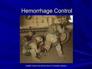
This document provides guidelines for using different types of antenna devices to extend the range of a wireless network. It describes range extender antennas, broadband amplifiers, wireless range extenders, and outdoor wireless access points/client bridges. The document also lists 15 guidelines for maximizing wireless signal range, such as keeping the number of walls between devices low, placing devices high and away from metal, and ensuring antenna cables are short. Building materials, trees, and other wireless networks can weaken signals.
Fast1 training ppt 2006 - mil - compressAKsentinel
The document provides instructions for using the FAST1 Sternal Intraosseous device to access the central vasculature through the sternum bone. The device can be used when peripheral IV access has failed or is inadequate. It inserts a needle cluster perpendicular to the sternum surface to penetrate the bone and provide equivalent access to a central line. Precautions are given for patient size and bone fractures. Instructions detail preparing the device, insertion at a perpendicular angle to the sternum, securing the device, initiating fluid flow, and addressing potential problems.
C191 w9tc cmast int humanitarian law and geneva conventionsAKsentinel
The document discusses the key principles of International Humanitarian Law (IHL) and the Geneva Conventions, which regulate the conduct of hostilities and aim to protect civilians and those who are wounded, sick, shipwrecked or detained. It outlines that IHL protects people who are not participating in fighting, such as civilians, medical personnel, chaplains and aid workers. It also protects those who can no longer fight, like wounded or sick soldiers and prisoners of war. The Geneva Conventions establish humanitarian protections that must be observed by governments and their armed forces during war.
This document discusses management of chest trauma. It covers common causes of chest injuries including gunshots, explosions, and crashes. It describes the organs in the chest cavity and how to assess a casualty for chest injuries by examining their breathing, neck, chest wall, and lung sounds. Open and tension pneumothoraces are discussed as well as how to perform a needle decompression in the field to relieve pressure in the chest from a tension pneumothorax. Proper management of chest wounds and decompression of tension pneumothoraces are essential for stabilizing chest trauma patients.
The document discusses Tactical Combat Casualty Care (TC-3) training for combat medics, which focuses on providing medical care on the battlefield in 3 stages - Care Under Fire, Tactical Field Care, and Combat Casualty Evacuation Care - with an emphasis on controlling hemorrhage, maintaining airways, and fluid resuscitation when possible given limited medical resources and ongoing tactical threats.
The document discusses point of wounding care for combat casualties. It notes that 90% of battlefield casualties die before reaching definitive care, so self-aid/buddy-aid is critical. The primary causes of preventable death are hemorrhage from extremity wounds, tension pneumothorax, and airway problems. Soldiers must be trained and equipped to control bleeding, treat chest wounds, and maintain airways. This includes improved individual first aid kits and training of Combat Lifesavers in key lifesaving skills like tourniquet application and needle decompression.
Fast1 training ppt 2006 - mil - compressAKsentinel
The FAST1 sternal intraosseous device provides rapid central vascular access by inserting a needle cluster perpendicular to the sternum. It is recommended for patients with inadequate peripheral IV access who are over 40-50kg and do not have sternum fractures or damage. The device can infuse fluids at 15-80ml/min by gravity or 125ml/min with pressure. Blood must be infused under pressure due to its higher viscosity. Proper insertion requires cleaning the site, placing the device perpendicular to the sternum midline, and connecting it to an IV line.
This document provides guidance for sustaining critical medical skills through annual training and validation testing for Soldiers with an MOS of 68W (Health Care Specialist). It outlines seven training tables focused on trauma assessment, airway management, intravenous access and more. It also includes an annual skills validation test to demonstrate competency. Completing this training satisfies requirements to maintain National Registry of Emergency Medical Technicians certification and provides continuing education credits for recertification. The core focus is on key combat casualty care skills to ensure Soldier Medics are prepared to provide lifesaving care on the battlefield.
One health condition that is becoming more common day by day is diabetes.
According to research conducted by the National Family Health Survey of India, diabetic cases show a projection which might increase to 10.4% by 2030.
8 Surprising Reasons To Meditate 40 Minutes A Day That Can Change Your Life.pptxHolistified Wellness
We’re talking about Vedic Meditation, a form of meditation that has been around for at least 5,000 years. Back then, the people who lived in the Indus Valley, now known as India and Pakistan, practised meditation as a fundamental part of daily life. This knowledge that has given us yoga and Ayurveda, was known as Veda, hence the name Vedic. And though there are some written records, the practice has been passed down verbally from generation to generation.
Clinic ^%[+27633867063*Abortion Pills For Sale In Tembisa Central19various
Clinic ^%[+27633867063*Abortion Pills For Sale In Tembisa Central Clinic ^%[+27633867063*Abortion Pills For Sale In Tembisa CentralClinic ^%[+27633867063*Abortion Pills For Sale In Tembisa CentralClinic ^%[+27633867063*Abortion Pills For Sale In Tembisa CentralClinic ^%[+27633867063*Abortion Pills For Sale In Tembisa Central
TEST BANK For Community Health Nursing A Canadian Perspective, 5th Edition by...Donc Test
TEST BANK For Community Health Nursing A Canadian Perspective, 5th Edition by Stamler, Verified Chapters 1 - 33, Complete Newest Version Community Health Nursing A Canadian Perspective, 5th Edition by Stamler, Verified Chapters 1 - 33, Complete Newest Version Community Health Nursing A Canadian Perspective, 5th Edition by Stamler Community Health Nursing A Canadian Perspective, 5th Edition TEST BANK by Stamler Test Bank For Community Health Nursing A Canadian Perspective, 5th Edition Pdf Chapters Download Test Bank For Community Health Nursing A Canadian Perspective, 5th Edition Pdf Download Stuvia Test Bank For Community Health Nursing A Canadian Perspective, 5th Edition Study Guide Test Bank For Community Health Nursing A Canadian Perspective, 5th Edition Ebook Download Stuvia Test Bank For Community Health Nursing A Canadian Perspective, 5th Edition Questions and Answers Quizlet Test Bank For Community Health Nursing A Canadian Perspective, 5th Edition Studocu Test Bank For Community Health Nursing A Canadian Perspective, 5th Edition Quizlet Test Bank For Community Health Nursing A Canadian Perspective, 5th Edition Stuvia Community Health Nursing A Canadian Perspective, 5th Edition Pdf Chapters Download Community Health Nursing A Canadian Perspective, 5th Edition Pdf Download Course Hero Community Health Nursing A Canadian Perspective, 5th Edition Answers Quizlet Community Health Nursing A Canadian Perspective, 5th Edition Ebook Download Course hero Community Health Nursing A Canadian Perspective, 5th Edition Questions and Answers Community Health Nursing A Canadian Perspective, 5th Edition Studocu Community Health Nursing A Canadian Perspective, 5th Edition Quizlet Community Health Nursing A Canadian Perspective, 5th Edition Stuvia Community Health Nursing A Canadian Perspective, 5th Edition Test Bank Pdf Chapters Download Community Health Nursing A Canadian Perspective, 5th Edition Test Bank Pdf Download Stuvia Community Health Nursing A Canadian Perspective, 5th Edition Test Bank Study Guide Questions and Answers Community Health Nursing A Canadian Perspective, 5th Edition Test Bank Ebook Download Stuvia Community Health Nursing A Canadian Perspective, 5th Edition Test Bank Questions Quizlet Community Health Nursing A Canadian Perspective, 5th Edition Test Bank Studocu Community Health Nursing A Canadian Perspective, 5th Edition Test Bank Quizlet Community Health Nursing A Canadian Perspective, 5th Edition Test Bank Stuvia
Our backs are like superheroes, holding us up and helping us move around. But sometimes, even superheroes can get hurt. That’s where slip discs come in.
Osteoporosis - Definition , Evaluation and Management .pdfJim Jacob Roy
Osteoporosis is an increasing cause of morbidity among the elderly.
In this document , a brief outline of osteoporosis is given , including the risk factors of osteoporosis fractures , the indications for testing bone mineral density and the management of osteoporosis
These lecture slides, by Dr Sidra Arshad, offer a simplified look into the mechanisms involved in the regulation of respiration:
Learning objectives:
1. Describe the organisation of respiratory center
2. Describe the nervous control of inspiration and respiratory rhythm
3. Describe the functions of the dorsal and respiratory groups of neurons
4. Describe the influences of the Pneumotaxic and Apneustic centers
5. Explain the role of Hering-Breur inflation reflex in regulation of inspiration
6. Explain the role of central chemoreceptors in regulation of respiration
7. Explain the role of peripheral chemoreceptors in regulation of respiration
8. Explain the regulation of respiration during exercise
9. Integrate the respiratory regulatory mechanisms
10. Describe the Cheyne-Stokes breathing
Study Resources:
1. Chapter 42, Guyton and Hall Textbook of Medical Physiology, 14th edition
2. Chapter 36, Ganong’s Review of Medical Physiology, 26th edition
3. Chapter 13, Human Physiology by Lauralee Sherwood, 9th edition
2. CMAST 2
IntroductionIntroduction
Review methods of hemorrhage control inReview methods of hemorrhage control in
a tactical environment.a tactical environment.
Hemorrhage is the leading cause ofHemorrhage is the leading cause of
preventable death on the battlefield.preventable death on the battlefield.
Hemorrhage control save lives.Hemorrhage control save lives.
New Hemostatic agents available.New Hemostatic agents available.
6. CMAST 6
BloodBlood
Adult body:Adult body:
– Contains approximately 5 to 6 liters of bloodContains approximately 5 to 6 liters of blood
– Loss of 1 pint of blood without harmful effectsLoss of 1 pint of blood without harmful effects
– Loss of 2 pints may cause shockLoss of 2 pints may cause shock
Three phases ofThree phases of
hemostasis:hemostasis:
– Vascular spasmVascular spasm
– Platelet plug formationPlatelet plug formation
– Blood clottingBlood clotting
(coagulation cascade)(coagulation cascade)
7. CMAST 7
HemorrhageHemorrhage
Pulse vs. Blood Pressure.Pulse vs. Blood Pressure.
How long until there are changes?How long until there are changes?
Young healthy adults compensate for longYoung healthy adults compensate for long
periods, then decompensate rapidly.periods, then decompensate rapidly.
At what blood pressure do casualties loseAt what blood pressure do casualties lose
consciousness?consciousness?
─ @ 50 mm Hg@ 50 mm Hg
8. CMAST 8
Clinical Signs of Acute HemorrhageClinical Signs of Acute Hemorrhage
ClassClass % Blood% Blood
LossLoss
Clinical SignsClinical Signs
II Up to 750 mlUp to 750 ml
(15%)(15%)
Slight increase in HR; no change in BPSlight increase in HR; no change in BP
or respirationsor respirations
IIII 750-1500 ml750-1500 ml
(15-30%)(15-30%)
Increased HR and respirations;Increased HR and respirations;
increased diastolic BP; anxiety, fright orincreased diastolic BP; anxiety, fright or
hostilityhostility
IIIIII 1500-20001500-2000
ml (30-40%)ml (30-40%)
Increased HR and respirations; fall inIncreased HR and respirations; fall in
systolic BP; significantsystolic BP; significant AMSAMS
IVIV >2000>2000
(>40%)(>40%)
Severe tachycardia; severe lowering ofSevere tachycardia; severe lowering of
BP; cold, pale skin; severe AMSBP; cold, pale skin; severe AMS
9. CMAST 9
Sources of HemorrhageSources of Hemorrhage
External:External:
– Visible blood is hard to estimateVisible blood is hard to estimate
Internal:Internal:
– May be hidden within the torso or even inMay be hidden within the torso or even in
the extremities secondary to fracturesthe extremities secondary to fractures
10. CMAST 10
Sources of External BleedingSources of External Bleeding
Arterial:Arterial:
─Rapid, profuse and pulsatingRapid, profuse and pulsating
─Bright red in colorBright red in color
Venous:Venous:
─Steady flowSteady flow
─Dark red or maroon in colorDark red or maroon in color
Capillary:Capillary:
─Slow and oozingSlow and oozing
─Often clots spontaneouslyOften clots spontaneously
14. CMAST 14
Hemorrhage ControlHemorrhage Control
Assess the tactical situation.Assess the tactical situation.
Expose the wound.Expose the wound.
Attempt to control theAttempt to control the
bleeding with directbleeding with direct
pressure or a pressurepressure or a pressure
dressing.dressing.
17. CMAST 17
Hemorrhage ControlHemorrhage Control
Life-threatening arterial bleedingLife-threatening arterial bleeding
(amputation) may require early use of a(amputation) may require early use of a
tourniquet.tourniquet.
If under enemy fire or in a dangerousIf under enemy fire or in a dangerous
position rapidly apply a tourniquet andposition rapidly apply a tourniquet and
move casualty to cover.move casualty to cover.
18. CMAST 18
TourniquetsTourniquets
Several new tourniquets have beenSeveral new tourniquets have been
selected as primary means to controlselected as primary means to control
hemorrhage in combat.hemorrhage in combat.
19. CMAST 19
Combat Application TourniquetCombat Application Tourniquet
WINDLASS
SELF ADHERING BAND
WINDLASS STRAP
The C-A-T was selected as the primary tourniquetThe C-A-T was selected as the primary tourniquet
for every soldier.for every soldier.
20. C-A-T Step 1C-A-T Step 1
Place the
wounded
extremity
through the loop
of the Self-
adhering Band
29. C-A-T TourniquetC-A-T Tourniquet
NOTE:
The friction adaptor
buckle is not
necessary for
proper C-A-T
application to an
arm. It MUST be
used with two
hands when
applying to a leg.
30. C-A-T: Lower ExtremityC-A-T: Lower Extremity
To use, wrap
the Self-
adhering Band
through the
friction
adaptor
buckle.
31. C-A-T: Lower ExtremityC-A-T: Lower Extremity
This prevents
the Self-
adhering Band
from loosening
during
transport.
35. CMAST 35
SOFTT ApplicationSOFTT Application
Similar to the CAT:Similar to the CAT:
─ Slide loop over extremitySlide loop over extremity
─ Pull strap tightPull strap tight
─ Twist windlass untilTwist windlass until
bleeding stopsbleeding stops
─ Latch the windlassLatch the windlass
with one of thewith one of the
tri-ringstri-rings
─ Tighten the safetyTighten the safety
screwscrew
37. CMAST 37
Improvised TourniquetImprovised Tourniquet
Place cravat between heart and wound.Place cravat between heart and wound.
Tie a half-knot on upper surface.Tie a half-knot on upper surface.
Place a short stick on half-knot.Place a short stick on half-knot.
Tie a square knot on top ofTie a square knot on top of
stick.stick.
Twist stick (windlass) toTwist stick (windlass) to
tighten.tighten.
UNTIL BLEEDING STOPS.UNTIL BLEEDING STOPS.
Secure windlass to prevent unwinding.Secure windlass to prevent unwinding.
39. CMAST 39
Tourniquet PrinciplesTourniquet Principles
Never cover a tourniquet.Never cover a tourniquet.
Mark a “T” on the casualty's forehead orMark a “T” on the casualty's forehead or
somewhere obvious (sharpie pen).somewhere obvious (sharpie pen).
In combat when the tactical situationIn combat when the tactical situation
allows, loosening a tourniquet isallows, loosening a tourniquet is
appropriate.appropriate.
40. CMAST 40
Tourniquet RemovalTourniquet Removal
Once the tactical situation allows,Once the tactical situation allows,
tourniquets should be loosened and othertourniquets should be loosened and other
methods to stop bleeding applied.methods to stop bleeding applied.
─Direct pressure - pressure dressingDirect pressure - pressure dressing
─HemCon Chitosan BandageHemCon Chitosan Bandage
─QuikClot powderQuikClot powder
41. CMAST 41
Tourniquet RemovalTourniquet Removal
When loosening a tourniquet, do notWhen loosening a tourniquet, do not
remove it from the limb.remove it from the limb.
If the tourniquet has been in place forIf the tourniquet has been in place for
> 6 hours, do not remove.> 6 hours, do not remove.
If fluid resuscitation is required, it shouldIf fluid resuscitation is required, it should
be accomplished before the tourniquetbe accomplished before the tourniquet
is removed.is removed.
Tourniquets are very painful, provideTourniquets are very painful, provide
pain medications as needed.pain medications as needed.
42. CMAST 42
Tourniquet RemovalTourniquet Removal
If tourniquet has been in place for onlyIf tourniquet has been in place for only
1-2 hours, loosening and using other1-2 hours, loosening and using other
methods to control hemorrhage canmethods to control hemorrhage can
salvage limbs.salvage limbs.
Remember: if unable to controlRemember: if unable to control
hemorrhage by other means, re-tightenhemorrhage by other means, re-tighten
the tourniquet.the tourniquet.
It is better to sacrifice the limb than toIt is better to sacrifice the limb than to
lose a life to hemorrhage.lose a life to hemorrhage.
43. CMAST 43
AmputationAmputation
Apply a pressure dressing to cover theApply a pressure dressing to cover the
end of the stump.end of the stump.
Kerlix and 6” Ace wrap for effectiveKerlix and 6” Ace wrap for effective
pressure dressing.pressure dressing.
Rinse amputated part free of debris.Rinse amputated part free of debris.
Wrap loosely in saline-moistened sterileWrap loosely in saline-moistened sterile
gauze.gauze.
44. CMAST 44
Preservation of Amputation PartsPreservation of Amputation Parts
Seal amputated part in a plastic bag orSeal amputated part in a plastic bag or
cravat.cravat.
Place in a cool container; do not allow toPlace in a cool container; do not allow to
freeze.freeze.
Never place an amputated part in water.Never place an amputated part in water.
Never place amputated part directly on ice.Never place amputated part directly on ice.
Never use dry ice to cool an amputatedNever use dry ice to cool an amputated
part.part.
46. CMAST 46
Chitosan Hemostatic DressingChitosan Hemostatic Dressing
Hold the foil over-pouch so that instructions canHold the foil over-pouch so that instructions can
be read. Identify unsealed edges at the top of thebe read. Identify unsealed edges at the top of the
over-pouch.over-pouch.
47. CMAST 47
Chitosan Hemostatic DressingChitosan Hemostatic Dressing
Peel open over-pouch by pulling the unsealedPeel open over-pouch by pulling the unsealed
edges apart.edges apart.
48. CMAST 48
Chitosan Hemostatic DressingChitosan Hemostatic Dressing
Trap dressing between bottom foil and non-Trap dressing between bottom foil and non-
absorbable green/black polyester backing withabsorbable green/black polyester backing with
your hand and thumb.your hand and thumb.
49. CMAST 49
Chitosan Hemostatic DressingChitosan Hemostatic Dressing
Hold dressing by the non-absorbable polyester backingHold dressing by the non-absorbable polyester backing
and discard the foil over-pouch. Hands must be dry toand discard the foil over-pouch. Hands must be dry to
prevent dressing from sticking to them.prevent dressing from sticking to them.
51. CMAST 51
Chitosan Hemostatic DressingChitosan Hemostatic Dressing
Place the light colored sponge portion of thePlace the light colored sponge portion of the
dressing directly to the wound area with thedressing directly to the wound area with the
most severe bleeding. Apply pressure for 2most severe bleeding. Apply pressure for 2
minutes or until the dressing adheres andminutes or until the dressing adheres and
bleeding stops. Once applied and in contactbleeding stops. Once applied and in contact
with the blood and other fluids, the dressingwith the blood and other fluids, the dressing
cannot be repositioned.cannot be repositioned.
A new dressing should be applied to otherA new dressing should be applied to other
exposed bleeding sites; each new dressingexposed bleeding sites; each new dressing
must be in contact with tissue where bleedingmust be in contact with tissue where bleeding
is heaviest. Care must be taken to avoidis heaviest. Care must be taken to avoid
contact with the casualty’s eyes.contact with the casualty’s eyes.
52. CMAST 52
Chitosan Hemostatic DressingChitosan Hemostatic Dressing
If dressing is not effective in stoppingIf dressing is not effective in stopping
bleeding after 4 minutes, remove the originalbleeding after 4 minutes, remove the original
and apply a new dressing. Additionaland apply a new dressing. Additional
dressings cannot be applied over ineffectivedressings cannot be applied over ineffective
dressings.dressings.
Apply a battle dressing/bandage to secure aApply a battle dressing/bandage to secure a
hemostatic dressing in place.hemostatic dressing in place.
Hemostatic dressings should only beHemostatic dressings should only be
removed by responsible persons afterremoved by responsible persons after
evacuation to the next level of care.evacuation to the next level of care.
56. CMAST 56
QuikClotQuikClot
Warning: Avoid contact with wet skin;Warning: Avoid contact with wet skin;
product reacts with small amounts ofproduct reacts with small amounts of
water and can cause burning.water and can cause burning.
Stop burning by brushing away granulesStop burning by brushing away granules
and flooding area with large volume ofand flooding area with large volume of
water.water.
If ingested, immediately drink two or moreIf ingested, immediately drink two or more
glasses of water.glasses of water.
57. CMAST 57
QuikClotQuikClot
Directions:Directions:
1-Apply direct firm pressure to wound using sterile1-Apply direct firm pressure to wound using sterile
dressing or best available substitutedressing or best available substitute
2-If bleeding is stopped or nearly stopped after2-If bleeding is stopped or nearly stopped after
approximately 1 minute of pressure, wrap and tieapproximately 1 minute of pressure, wrap and tie
bandage to maintain pressure on woundbandage to maintain pressure on wound
3-If moderate to severe bleeding continues, hold3-If moderate to severe bleeding continues, hold
pack away from face and tear open at tabspack away from face and tear open at tabs
58. CMAST 58
QuikClotQuikClot
4-Use wiping motion to remove gauze and4-Use wiping motion to remove gauze and
excess blood – immediately start a slowexcess blood – immediately start a slow
pouring of one QuikClot packet directly ontopouring of one QuikClot packet directly onto
the wound. Stop pouring as soon as drythe wound. Stop pouring as soon as dry
granules cover the wound areagranules cover the wound area
5-Use only enough QuikClot to stop bleeding. If5-Use only enough QuikClot to stop bleeding. If
bleeding continues open a second packet ofbleeding continues open a second packet of
QuikClot and continue to use as directedQuikClot and continue to use as directed
59. CMAST 59
QuikClotQuikClot
6-Reapply firm pressure to QuikClot covered6-Reapply firm pressure to QuikClot covered
wound using sterile gauze. Wrap and tiewound using sterile gauze. Wrap and tie
bandage to maintain pressurebandage to maintain pressure
61. CMAST 61
Treatment GoalsTreatment Goals
Hemorrhage control continues to be theHemorrhage control continues to be the
priority in battlefield care.priority in battlefield care.
Hemorrhage is the leading cause ofHemorrhage is the leading cause of
preventable death on the battlefield.preventable death on the battlefield.
Our focus must be on stopping soldiersOur focus must be on stopping soldiers
from bleeding to death on the battlefield.from bleeding to death on the battlefield.