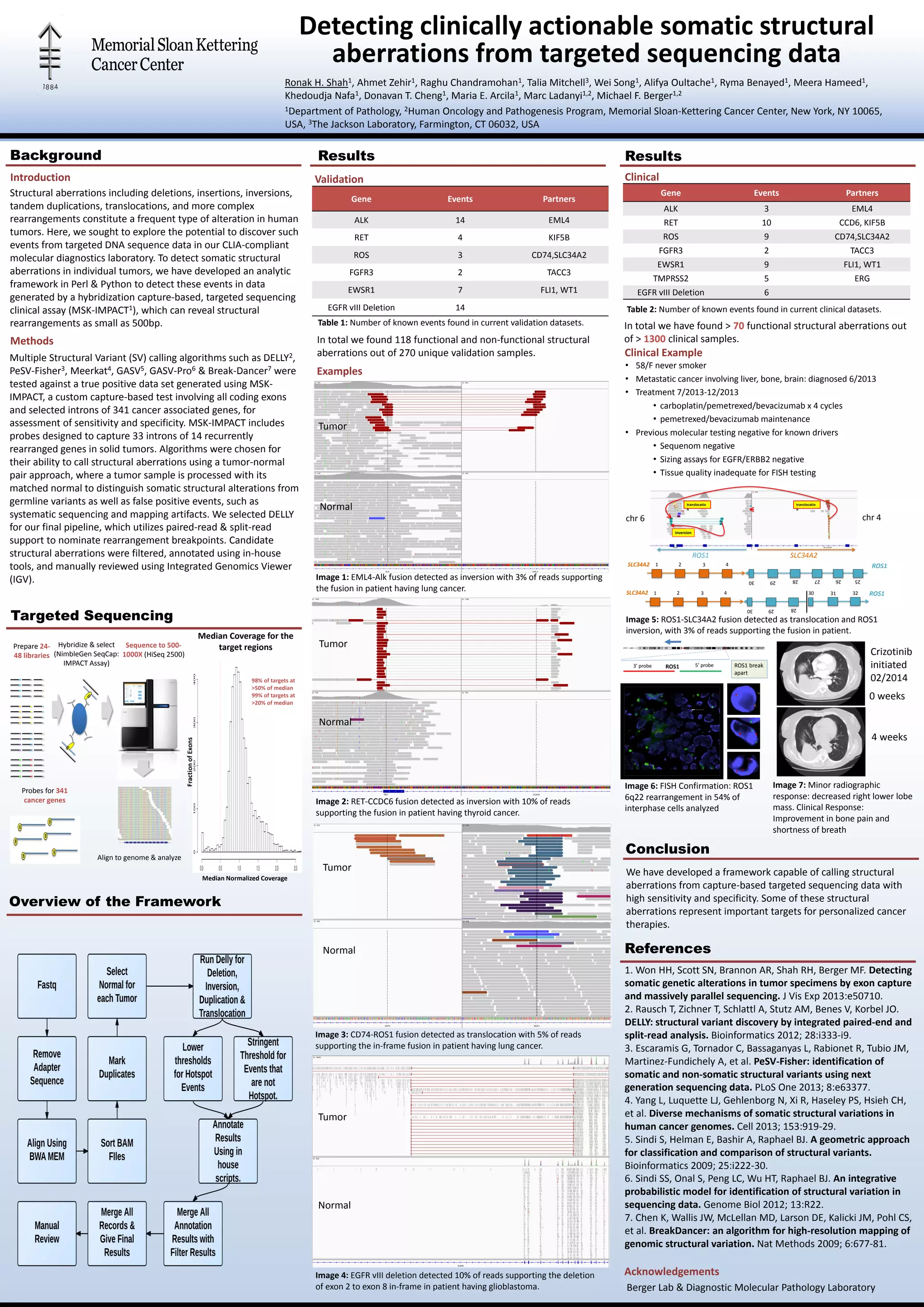
Detecting clinically actionable somatic structural 
aberrations from targeted sequencing data 
Ronak H. Shah1, Ahmet Zehir1, Raghu Chandramohan1, Talia Mitchell3, Wei Song1, Alifya Oultache1, Ryma Benayed1, Meera Hameed1, 
Khedoudja Nafa1, Donavan T. Cheng1, Maria E. Arcila1, Marc Ladanyi1,2, Michael F. Berger1,2 
1Department of Pathology, 2Human Oncology and Pathogenesis Program, Memorial Sloan-Kettering Cancer Center, New York, NY 10065, 
USA, 3The Jackson Laboratory, Farmington, CT 06032, USA 
Background Results 
Structural aberrations including deletions, insertions, inversions, 
tandem duplications, translocations, and more complex 
rearrangements constitute a frequent type of alteration in human 
tumors. Here, we sought to explore the potential to discover such 
events from targeted DNA sequence data in our CLIA-compliant 
molecular diagnostics laboratory. To detect somatic structural 
aberrations in individual tumors, we have developed an analytic 
framework in Perl & Python to detect these events in data 
generated by a hybridization capture-based, targeted sequencing 
clinical assay (MSK-IMPACT1), which can reveal structural 
rearrangements as small as 500bp. 
Multiple Structural Variant (SV) calling algorithms such as DELLY2, 
PeSV-Fisher3, Meerkat4, GASV5, GASV-Pro6 & Break-Dancer7 were 
tested against a true positive data set generated using MSK-IMPACT, 
a custom capture-based test involving all coding exons 
and selected introns of 341 cancer associated genes, for 
assessment of sensitivity and specificity. MSK-IMPACT includes 
probes designed to capture 33 introns of 14 recurrently 
rearranged genes in solid tumors. Algorithms were chosen for 
their ability to call structural aberrations using a tumor-normal 
pair approach, where a tumor sample is processed with its 
matched normal to distinguish somatic structural alterations from 
germline variants as well as false positive events, such as 
systematic sequencing and mapping artifacts. We selected DELLY 
for our final pipeline, which utilizes paired-read & split-read 
support to nominate rearrangement breakpoints. Candidate 
structural aberrations were filtered, annotated using in-house 
tools, and manually reviewed using Integrated Genomics Viewer 
(IGV). 
Targeted Sequencing 
Results 
translocatio 
n 
translocatio 
n 
chr 6 chr 4 
SLC34A2 ROS1 
SLC34A2 30 31 32 ROS1 
Image 5: ROS1-SLC34A2 fusion detected as translocation and ROS1 
inversion, with 3% of reads supporting the fusion in patient. 
Conclusion 
Hybridize & select 
(NimbleGen SeqCap: 
IMPACT Assay) 
Overview of the Framework 
Crizotinib 
initiated 
02/2014 
We have developed a framework capable of calling structural 
aberrations from capture-based targeted sequencing data with 
high sensitivity and specificity. Some of these structural 
aberrations represent important targets for personalized cancer 
therapies. 
1. Won HH, Scott SN, Brannon AR, Shah RH, Berger MF. Detecting 
somatic genetic alterations in tumor specimens by exon capture 
and massively parallel sequencing. J Vis Exp 2013:e50710. 
2. Rausch T, Zichner T, Schlattl A, Stutz AM, Benes V, Korbel JO. 
DELLY: structural variant discovery by integrated paired-end and 
split-read analysis. Bioinformatics 2012; 28:i333-i9. 
3. Escaramis G, Tornador C, Bassaganyas L, Rabionet R, Tubio JM, 
Martinez-Fundichely A, et al. PeSV-Fisher: identification of 
somatic and non-somatic structural variants using next 
generation sequencing data. PLoS One 2013; 8:e63377. 
4. Yang L, Luquette LJ, Gehlenborg N, Xi R, Haseley PS, Hsieh CH, 
et al. Diverse mechanisms of somatic structural variations in 
human cancer genomes. Cell 2013; 153:919-29. 
5. Sindi S, Helman E, Bashir A, Raphael BJ. A geometric approach 
for classification and comparison of structural variants. 
Bioinformatics 2009; 25:i222-30. 
6. Sindi SS, Onal S, Peng LC, Wu HT, Raphael BJ. An integrative 
probabilistic model for identification of structural variation in 
sequencing data. Genome Biol 2012; 13:R22. 
7. Chen K, Wallis JW, McLellan MD, Larson DE, Kalicki JM, Pohl CS, 
et al. BreakDancer: an algorithm for high-resolution mapping of 
genomic structural variation. Nat Methods 2009; 6:677-81. 
Acknowledgements 
Introduction 
Methods 
Prepare 24- 
48 libraries 
Probes for 341 
cancer genes 
Sequence to 500- 
1000X (HiSeq 2500) 
Align to genome & analyze 
98% of targets at 
>50% of median 
99% of targets at 
>20% of median 
References 
Berger Lab & Diagnostic Molecular Pathology Laboratory 
Validation 
Gene Events Partners 
ALK 14 EML4 
RET 4 KIF5B 
ROS 3 CD74,SLC34A2 
FGFR3 2 TACC3 
EWSR1 7 FLI1, WT1 
EGFR vIII Deletion 14 
In total we found 118 functional and non-functional structural 
aberrations out of 270 unique validation samples. 
Examples 
Tumor 
Normal 
Image 1: EML4-Alk fusion detected as inversion with 3% of reads supporting 
the fusion in patient having lung cancer. 
Tumor 
Normal 
Image 2: RET-CCDC6 fusion detected as inversion with 10% of reads 
supporting the fusion in patient having thyroid cancer. 
Tumor 
Normal 
Image 3: CD74-ROS1 fusion detected as translocation with 5% of reads 
supporting the in-frame fusion in patient having lung cancer. 
Tumor 
Normal 
Image 4: EGFR vIII deletion detected 10% of reads supporting the deletion 
of exon 2 to exon 8 in-frame in patient having glioblastoma. 
Clinical 
Gene Events Partners 
ALK 3 EML4 
RET 10 CCD6, KIF5B 
ROS 9 CD74,SLC34A2 
FGFR3 2 TACC3 
EWSR1 9 FLI1, WT1 
TMPRSS2 5 ERG 
EGFR vIII Deletion 6 
In total we have found > 70 functional structural aberrations out 
of > 1300 clinical samples. 
Clinical Example 
• 58/F never smoker 
• Metastatic cancer involving liver, bone, brain: diagnosed 6/2013 
• Treatment 7/2013-12/2013 
• carboplatin/pemetrexed/bevacizumab x 4 cycles 
• pemetrexed/bevacizumab maintenance 
• Previous molecular testing negative for known drivers 
• Sequenom negative 
• Sizing assays for EGFR/ERBB2 negative 
• Tissue quality inadequate for FISH testing 
inversion 
ROS1 SLC34A2 
1 2 3 4 
28 29 30 
1 2 3 4 
25 26 27 28 29 30 
3’ probe ROS1 5’ probe ROS1 break 
apart 
Image 6: FISH Confirmation: ROS1 
6q22 rearrangement in 54% of 
interphase cells analyzed 
0 weeks 
4 weeks 
Image 7: Minor radiographic 
response: decreased right lower lobe 
mass. Clinical Response: 
Improvement in bone pain and 
shortness of breath 
Table 1: Number of known events found in current validation datasets. 
Table 2: Number of known events found in current clinical datasets. 
Median Coverage for the 
target regions 
Median Normalized Coverage 
Fraction of Exons

Detecting clinically actionable somatic structural aberrations from targeted sequencing data

  • 1.
    Detecting clinically actionablesomatic structural aberrations from targeted sequencing data Ronak H. Shah1, Ahmet Zehir1, Raghu Chandramohan1, Talia Mitchell3, Wei Song1, Alifya Oultache1, Ryma Benayed1, Meera Hameed1, Khedoudja Nafa1, Donavan T. Cheng1, Maria E. Arcila1, Marc Ladanyi1,2, Michael F. Berger1,2 1Department of Pathology, 2Human Oncology and Pathogenesis Program, Memorial Sloan-Kettering Cancer Center, New York, NY 10065, USA, 3The Jackson Laboratory, Farmington, CT 06032, USA Background Results Structural aberrations including deletions, insertions, inversions, tandem duplications, translocations, and more complex rearrangements constitute a frequent type of alteration in human tumors. Here, we sought to explore the potential to discover such events from targeted DNA sequence data in our CLIA-compliant molecular diagnostics laboratory. To detect somatic structural aberrations in individual tumors, we have developed an analytic framework in Perl & Python to detect these events in data generated by a hybridization capture-based, targeted sequencing clinical assay (MSK-IMPACT1), which can reveal structural rearrangements as small as 500bp. Multiple Structural Variant (SV) calling algorithms such as DELLY2, PeSV-Fisher3, Meerkat4, GASV5, GASV-Pro6 & Break-Dancer7 were tested against a true positive data set generated using MSK-IMPACT, a custom capture-based test involving all coding exons and selected introns of 341 cancer associated genes, for assessment of sensitivity and specificity. MSK-IMPACT includes probes designed to capture 33 introns of 14 recurrently rearranged genes in solid tumors. Algorithms were chosen for their ability to call structural aberrations using a tumor-normal pair approach, where a tumor sample is processed with its matched normal to distinguish somatic structural alterations from germline variants as well as false positive events, such as systematic sequencing and mapping artifacts. We selected DELLY for our final pipeline, which utilizes paired-read & split-read support to nominate rearrangement breakpoints. Candidate structural aberrations were filtered, annotated using in-house tools, and manually reviewed using Integrated Genomics Viewer (IGV). Targeted Sequencing Results translocatio n translocatio n chr 6 chr 4 SLC34A2 ROS1 SLC34A2 30 31 32 ROS1 Image 5: ROS1-SLC34A2 fusion detected as translocation and ROS1 inversion, with 3% of reads supporting the fusion in patient. Conclusion Hybridize & select (NimbleGen SeqCap: IMPACT Assay) Overview of the Framework Crizotinib initiated 02/2014 We have developed a framework capable of calling structural aberrations from capture-based targeted sequencing data with high sensitivity and specificity. Some of these structural aberrations represent important targets for personalized cancer therapies. 1. Won HH, Scott SN, Brannon AR, Shah RH, Berger MF. Detecting somatic genetic alterations in tumor specimens by exon capture and massively parallel sequencing. J Vis Exp 2013:e50710. 2. Rausch T, Zichner T, Schlattl A, Stutz AM, Benes V, Korbel JO. DELLY: structural variant discovery by integrated paired-end and split-read analysis. Bioinformatics 2012; 28:i333-i9. 3. Escaramis G, Tornador C, Bassaganyas L, Rabionet R, Tubio JM, Martinez-Fundichely A, et al. PeSV-Fisher: identification of somatic and non-somatic structural variants using next generation sequencing data. PLoS One 2013; 8:e63377. 4. Yang L, Luquette LJ, Gehlenborg N, Xi R, Haseley PS, Hsieh CH, et al. Diverse mechanisms of somatic structural variations in human cancer genomes. Cell 2013; 153:919-29. 5. Sindi S, Helman E, Bashir A, Raphael BJ. A geometric approach for classification and comparison of structural variants. Bioinformatics 2009; 25:i222-30. 6. Sindi SS, Onal S, Peng LC, Wu HT, Raphael BJ. An integrative probabilistic model for identification of structural variation in sequencing data. Genome Biol 2012; 13:R22. 7. Chen K, Wallis JW, McLellan MD, Larson DE, Kalicki JM, Pohl CS, et al. BreakDancer: an algorithm for high-resolution mapping of genomic structural variation. Nat Methods 2009; 6:677-81. Acknowledgements Introduction Methods Prepare 24- 48 libraries Probes for 341 cancer genes Sequence to 500- 1000X (HiSeq 2500) Align to genome & analyze 98% of targets at >50% of median 99% of targets at >20% of median References Berger Lab & Diagnostic Molecular Pathology Laboratory Validation Gene Events Partners ALK 14 EML4 RET 4 KIF5B ROS 3 CD74,SLC34A2 FGFR3 2 TACC3 EWSR1 7 FLI1, WT1 EGFR vIII Deletion 14 In total we found 118 functional and non-functional structural aberrations out of 270 unique validation samples. Examples Tumor Normal Image 1: EML4-Alk fusion detected as inversion with 3% of reads supporting the fusion in patient having lung cancer. Tumor Normal Image 2: RET-CCDC6 fusion detected as inversion with 10% of reads supporting the fusion in patient having thyroid cancer. Tumor Normal Image 3: CD74-ROS1 fusion detected as translocation with 5% of reads supporting the in-frame fusion in patient having lung cancer. Tumor Normal Image 4: EGFR vIII deletion detected 10% of reads supporting the deletion of exon 2 to exon 8 in-frame in patient having glioblastoma. Clinical Gene Events Partners ALK 3 EML4 RET 10 CCD6, KIF5B ROS 9 CD74,SLC34A2 FGFR3 2 TACC3 EWSR1 9 FLI1, WT1 TMPRSS2 5 ERG EGFR vIII Deletion 6 In total we have found > 70 functional structural aberrations out of > 1300 clinical samples. Clinical Example • 58/F never smoker • Metastatic cancer involving liver, bone, brain: diagnosed 6/2013 • Treatment 7/2013-12/2013 • carboplatin/pemetrexed/bevacizumab x 4 cycles • pemetrexed/bevacizumab maintenance • Previous molecular testing negative for known drivers • Sequenom negative • Sizing assays for EGFR/ERBB2 negative • Tissue quality inadequate for FISH testing inversion ROS1 SLC34A2 1 2 3 4 28 29 30 1 2 3 4 25 26 27 28 29 30 3’ probe ROS1 5’ probe ROS1 break apart Image 6: FISH Confirmation: ROS1 6q22 rearrangement in 54% of interphase cells analyzed 0 weeks 4 weeks Image 7: Minor radiographic response: decreased right lower lobe mass. Clinical Response: Improvement in bone pain and shortness of breath Table 1: Number of known events found in current validation datasets. Table 2: Number of known events found in current clinical datasets. Median Coverage for the target regions Median Normalized Coverage Fraction of Exons