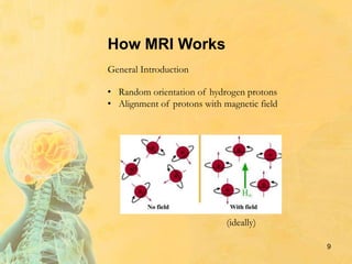
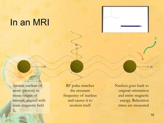
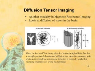
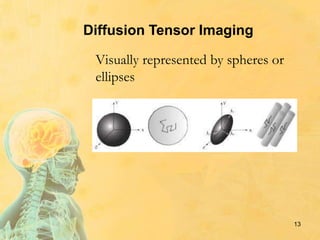
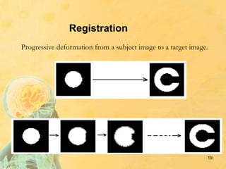
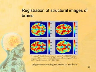
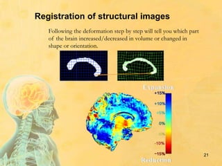
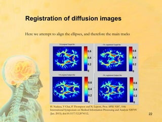
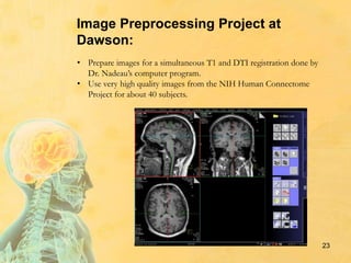
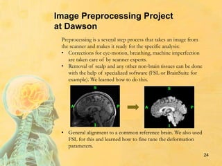
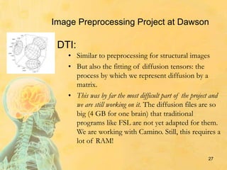
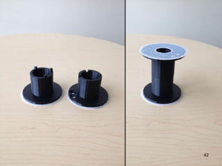
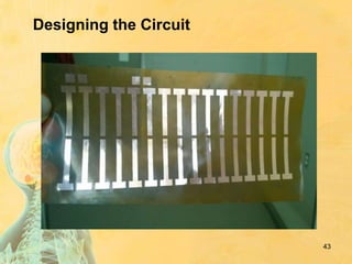
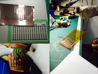
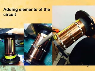
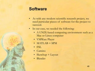
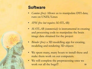
Group Summer Internship
on
Brain Imaging
Supervised by Dr. Hélène Nadeau
Dawson College
Summer 2015
Introduction
2
Group internship:
• Experience a research environment under
the supervision of experts.
• Learn the background as a group (8-10
students), then specialise to a specific
project (2 students per project) while
continuing to meet twice a week as a group.
• Multidisciplinary the whole summer. (Math,
Physics, Anatomy, Computer Science, etc..)
• Very flexible to accommodate complex
schedules. Some of the work is done from
home.
Introduction
3
Experts:
• Dr. Hélène Nadeau, Dawson College
• Dr. Jamie Near, Centre d’imagerie cérébrale, Douglas Mental
Health Institute
• Dr. Franco Leporé and Dr. Latifa Lazzouni, Département de
psychologie, Université de Montréal
Introduction
Students:
• Amro Abdrabo
• Yassmine Abdrabo
• Zahra Altalibi
• Julia Cohen
• Jessica Di Bartolomeo
• Myriam Dimanche
• Nafisa Husein
• Avigayil Sorokine
4
A variety of skills and interests
5
Background
• Anatomy of the brain
• Physics behind Magnetic Resonance Imaging and
spectroscopy
• Image registration
• Matrices, Fourier transforms, complex numbers
• Specialized software
Projects
• Preprocessing for non-linear registration (H.N.)
• Myriam, Jessica & Julia
• Analysis of functional MRI (L.L.)
• Avi & Myriam
• Phase correction of MR spectra (J.N.)
• Amro & Yassmine
• Conception and realisation of a bird cage coil
(J.N.)
• Zahra & Nafisa
6
Anatomy of the Brain
• We began the summer by learning about the
different parts of the brain and their functions.
• Below is a link to a study tool one can use to
learn the anatomy of the brain:
• http://headneckbrainspine.com/web_flash/
newmodules/Brain%20MRI.swf
• These images are MRI slices in three planes,
the sagittal, coronal, and axial.
7
Introduction to MRI
8
• Magnetic Resonance Imaging scans
• Used for diagnosing diseases and disorders
• Used to study development of the brain
and to learn about various brain functions
• Structural, functional, diffusion
Geraint Rees, Ewa Wojciulik, Karen Clarke, Masud Husain, Chris Frith, Jon Driver
Brain Aug 2000, 123 (8) 1624-1633; DOI: 10.1093/brain/123.8.1624
How MRI Works
• Random orientation of hydrogen protons
• Alignment of protons with magnetic field
General Introduction
(ideally)
9
In an MRI
Atomic nucleus of
atom (proton) in
tissue/organ of
interest, aligned with
main magnetic field
β0
RF pulse matches
the resonant
frequency of nucleus
and causes it to
reorient itself
Nucleus goes back to
original orientation
and emits magnetic
energy. Relaxation
times are measured
N N
N N
E
10
Magnetic Resonance Imaging
11
• β0 magnetic field
• Radiofrequency pulse
• Net magnetization vector
• Time to return to z-axis
T1 Relaxation
T2 Relaxation
• Dephasing in x and y axes
• Spin interaction
T2* Relaxation
• Decrease in signal strength
Different tissues have different
relaxation time, so this translates in a
distinction between white matter, grey
matter and cerebral fluid on an image
12
Diffusion Tensor Imaging
• Another modality in Magnetic Resonance Imaging
• Looks at diffusion of water in the brain
Water is free to diffuse in any direction in cerebrospinal fluid, but has
a strongly preferred direction of diffusion in a tube-like structure, as in
white matter. Studying anisotropic diffusion is especially useful for
mapping orientation of white-matter tracks.
Diffusion Tensor Imaging
13
Visually represented by spheres or
ellipses
14
x
z
y
λ1
λ2
λ3
x
z
y
λ3
θ
𝐿′
= 𝑅𝐿𝑅 𝑇
=
𝜆1 𝑐𝑜𝑠𝜃2 + 𝜆2 𝑠𝑖𝑛𝜃2 (𝜆1 − 𝜆2)𝑠𝑖𝑛𝜃𝑐𝑜𝑠𝜃 0
(𝜆1 − 𝜆2)𝑠𝑖𝑛𝜃𝑐𝑜𝑠𝜃 𝜆1 𝑠𝑖𝑛𝜃2 + 𝜆2 𝑐𝑜𝑠𝜃2 0
0 0 𝜆3
𝐿 =
𝜆1 0 0
0 𝜆2 0
0 0 𝜆3
Diffusion Tensor Imaging
Mathematically represented by matrices
(tensors).
If λ1=λ2=λ3 we
have a sphere
What a diffusion scan looks like
15
Colors give main direction of diffusion
Functional MRI
16
Functional magnetic resonance imaging or functional MRI
(fMRI) is a functional neuroimaging procedure using MRI
technology that measures brain activity by detecting
associated changes in blood flow. When an area of the brain
is in use, blood flow to that region also increases.
These fMRI images are from a study showing parts of the brain lighting up
on seeing houses and other parts on seeing faces. The 'r' values are
correlations, with higher positive or negative values indicating a better match.
Source:
"Haxby2001" by
National Institute of
Mental Health - US
Department of
Health and Human
Services: National
Institute of Mental
Health. Licensed
under Public
Domain via
Commons
17
Functional MRI at the Department of
Psychology of Université de Montréal
• Under the supervision of Dr. Lazzouni
• Visualization of the sections of the auditory cortex
activated during certain tasks
• Eventually to study the effect of cochlear implants
Statistical parametric mapping (SPM)
18
Registration
• Two populations (blind with seeing people,
active 70-year-old with sedentary 70-year-old,
musicians with non-musicians, schizophrenic
with healthy subjects, etc..)
• Images from the same individual at different
times (development, results of treatment, etc…)
Used to compare:
For the project on preprocessing of structural and diffusion
images we had to familiarize ourselves with the concept of
registration.
Registration
Progressive deformation from a subject image to a target image.
19
u
20
Registration of structural images of
brains
Align corresponding structures of the brain
H. Nadeau, Y Chai, P. Thompson and N. Leporé, Proc. SPIE 9287, 10th
International Symposium on Medical Information Processing and Analysis
92870V (Jan. 2015); doi:10.1117/12.2074112.
21
Registration of structural images
Following the deformation step by step will tell you which part
of the brain increased/decreased in volume or changed in
shape or orientation.
22
Registration of diffusion images
Here we attempt to align the ellipses, and therefore the main tracks
H. Nadeau, Y Chai, P. Thompson and N. Leporé, Proc. SPIE 9287, 10th
International Symposium on Medical Information Processing and Analysis 92870V
(Jan. 2015); doi:10.1117/12.2074112.
23
• Prepare images for a simultaneous T1 and DTI registration done by
Dr. Nadeau’s computer program.
• Use very high quality images from the NIH Human Connectome
Project for about 40 subjects.
Image Preprocessing Project at
Dawson:
24
Preprocessing is a several step process that takes an image from
the scanner and makes it ready for the specific analysis:
• Corrections for eye-motion, breathing, machine imperfection
are taken care of by scanner experts.
• Removal of scalp and any other non-brain tissues can be done
with the help of specialized software (FSL or BrainSuite for
example). We learned how to do this.
• General alignment to a common reference brain. We also used
FSL for this and learned how to fine tune the deformation
parameters.
Image Preprocessing Project
at Dawson
Various types of transformations
of images
Rotation
Shearing
Translation
Scaling
25
Transformations
are performed with
the help of
matrices
26
• Similar to preprocessing for structural images
• But also the fitting of diffusion tensors: the
process by which we represent diffusion by a
matrix.
• This was by far the most difficult part of the project and
we are still working on it. The diffusion files are so
big (4 GB for one brain) that traditional
programs like FSL are not yet adapted for them.
We are working with Camino. Still, this requires a
lot of RAM!
27
DTI:
Image Preprocessing Project at Dawson
Radio wave Frequency Spectroscopy
E = hf
28
RF Spectroscopy
Just as a neon tube emits visible light in different colors or frequencies when
stimulated by electric power, a brain stimulated by a radio frequency transmitter
will emit radio frequencies depending on the properties of the chemical
compounds founds in different locations of the brain. Thus, RF spectroscopy
probes the chemistry or micro-biology of the brain.
29
RF spectroscopy
enables us to identify
the metabolites of a
brain, which include
GABA, N-
acetylaspartate
(NAA), glutamate, etc.
Signal Processing: The Signal
• The signal is created from the oscillating
magnetization vector of the proton.
• Since the magnetization vector is moving , an
alternating voltage will be induced in the receiver
coil. (Lenz’s Law).
• This voltage supplies info about the proton’s
magnetization vector whose magnitude is
proportional to the signal intensity:
• The real part:
• The imaginary part:
30
Project under
the supervision
of Dr. Near
FID Signal Collection
• Time Domain Signal varies with time
• Frequency Domain Intensity vs. Frequency
Vs.
Signal from a Single Proton Signal from Many Protons
31
Fourier Transform
32
Fourier Transform
• Takes a scalar real value function as input.
• This function, s(t), has time as its domain, and
S(f) has frequency as the domain.
• This transition is represented by:
33
Post FT: Phase Correction
In order for the signal peaks to look positive and
upright, phase correction must be applied.
34
Phase Correction
• In order to get upright (absorptive) peaks for the
x component of the signal, the signal (which has
the same direction as the magnetization vector)
must be rotated by theta.
• Specifically,
• Ideally, the protons start their precession at the
positive x axis (after being rotated 90 degrees).
However, flip angles vary.
• There is also a time delay between excitation and
detection.
35
Phase Correction
Before Correction After Correction
36
Phase Correction
• The algorithm for phase correction maximizes
the area under the real component of the signal
plotted versus the frequency.
37
Our Project
• Supervisor: Dr. Jamie Near
• Goal: Designing an MRI machine to scan rat brains
38
Birdcage Coil
• Transmitter
• Receiver
39
RF Coils
• Transmitters • Receivers
• Both
N
N
E
40
41
Designing the Casing
Sketchup: a 3D modeling software
42
Designing the Circuit
43
44
Adding elements of the
circuit
45
46
Tuning & Matching
47
Software
• As with any modern scientific research project, we
need particular pieces of software for the project to
succeed.
• In our case, we needed the following:
• A UNIX-based computing environment such as a
Mac or Linux computer
• VMWare Player
• MATLAB + SPM
• FSL
• Camino
• Sketchup + Layout
• Blender
48
Software
• A UNIX-based computing environment: Why?
Because the scientific community predominately
uses this type of environment for software
development
• VMWare Player (free): Allows a Windows user to
install another type of computing environment
within a wrapped window on their PC; we used
this to install Linux CentOS
• FSL (free): This app is one of the standard
programs for manipulating and managing MRI
images; runs on UNIX/Linux
49
Software
• Camino (free): Allows us to manipulate DTI data;
runs on UNIX/Linux
• SPM (free but requires MATLAB)
• MATLAB (commercial): is instrumental in creating
and processing code to manipulate the brain
image data obtained for the project
• Blender (free): a 3D modelling app for creating,
modeling and rendering 3D objects
• We spent many, many hours to install these and
make them work on our computers.
• We will complete the preprocessing once we
work out all the bugs!!! 50
Conclusion
• Researching a very unfamiliar topic and
presenting it to the group helped develop
independent learning skills.
• Working with peers proved to be highly
beneficial, and boosted the confidence of our
beginner researchers.
• Experiencing a real research environment was
very stimulating.
• Learning the basics with the group and then
getting specific training with an established
researcher meant that the training in the lab
could really focus on the project.
51
52
Looking forward
We would like to pursue our projects and
expand our group:
• extend to more disciplines: include
students from a wider variety of
programs;
• involve more Dawson faculty
members as mentors;
• recruit additional supervisors from
Montreal universities and research
centers.
Many thanks to:
Dr. Leporé
Dr. Lazzouni
Dr. Near
53

BrainImaging_2015-10-06_22h29

  • 1.
    Group Summer Internship on BrainImaging Supervised by Dr. Hélène Nadeau Dawson College Summer 2015
  • 2.
    Introduction 2 Group internship: • Experiencea research environment under the supervision of experts. • Learn the background as a group (8-10 students), then specialise to a specific project (2 students per project) while continuing to meet twice a week as a group. • Multidisciplinary the whole summer. (Math, Physics, Anatomy, Computer Science, etc..) • Very flexible to accommodate complex schedules. Some of the work is done from home.
  • 3.
    Introduction 3 Experts: • Dr. HélèneNadeau, Dawson College • Dr. Jamie Near, Centre d’imagerie cérébrale, Douglas Mental Health Institute • Dr. Franco Leporé and Dr. Latifa Lazzouni, Département de psychologie, Université de Montréal
  • 4.
    Introduction Students: • Amro Abdrabo •Yassmine Abdrabo • Zahra Altalibi • Julia Cohen • Jessica Di Bartolomeo • Myriam Dimanche • Nafisa Husein • Avigayil Sorokine 4 A variety of skills and interests
  • 5.
    5 Background • Anatomy ofthe brain • Physics behind Magnetic Resonance Imaging and spectroscopy • Image registration • Matrices, Fourier transforms, complex numbers • Specialized software
  • 6.
    Projects • Preprocessing fornon-linear registration (H.N.) • Myriam, Jessica & Julia • Analysis of functional MRI (L.L.) • Avi & Myriam • Phase correction of MR spectra (J.N.) • Amro & Yassmine • Conception and realisation of a bird cage coil (J.N.) • Zahra & Nafisa 6
  • 7.
    Anatomy of theBrain • We began the summer by learning about the different parts of the brain and their functions. • Below is a link to a study tool one can use to learn the anatomy of the brain: • http://headneckbrainspine.com/web_flash/ newmodules/Brain%20MRI.swf • These images are MRI slices in three planes, the sagittal, coronal, and axial. 7
  • 8.
    Introduction to MRI 8 •Magnetic Resonance Imaging scans • Used for diagnosing diseases and disorders • Used to study development of the brain and to learn about various brain functions • Structural, functional, diffusion Geraint Rees, Ewa Wojciulik, Karen Clarke, Masud Husain, Chris Frith, Jon Driver Brain Aug 2000, 123 (8) 1624-1633; DOI: 10.1093/brain/123.8.1624
  • 9.
    How MRI Works •Random orientation of hydrogen protons • Alignment of protons with magnetic field General Introduction (ideally) 9
  • 10.
    In an MRI Atomicnucleus of atom (proton) in tissue/organ of interest, aligned with main magnetic field β0 RF pulse matches the resonant frequency of nucleus and causes it to reorient itself Nucleus goes back to original orientation and emits magnetic energy. Relaxation times are measured N N N N E 10
  • 11.
    Magnetic Resonance Imaging 11 •β0 magnetic field • Radiofrequency pulse • Net magnetization vector • Time to return to z-axis T1 Relaxation T2 Relaxation • Dephasing in x and y axes • Spin interaction T2* Relaxation • Decrease in signal strength Different tissues have different relaxation time, so this translates in a distinction between white matter, grey matter and cerebral fluid on an image
  • 12.
    12 Diffusion Tensor Imaging •Another modality in Magnetic Resonance Imaging • Looks at diffusion of water in the brain Water is free to diffuse in any direction in cerebrospinal fluid, but has a strongly preferred direction of diffusion in a tube-like structure, as in white matter. Studying anisotropic diffusion is especially useful for mapping orientation of white-matter tracks.
  • 13.
    Diffusion Tensor Imaging 13 Visuallyrepresented by spheres or ellipses
  • 14.
    14 x z y λ1 λ2 λ3 x z y λ3 θ 𝐿′ = 𝑅𝐿𝑅 𝑇 = 𝜆1𝑐𝑜𝑠𝜃2 + 𝜆2 𝑠𝑖𝑛𝜃2 (𝜆1 − 𝜆2)𝑠𝑖𝑛𝜃𝑐𝑜𝑠𝜃 0 (𝜆1 − 𝜆2)𝑠𝑖𝑛𝜃𝑐𝑜𝑠𝜃 𝜆1 𝑠𝑖𝑛𝜃2 + 𝜆2 𝑐𝑜𝑠𝜃2 0 0 0 𝜆3 𝐿 = 𝜆1 0 0 0 𝜆2 0 0 0 𝜆3 Diffusion Tensor Imaging Mathematically represented by matrices (tensors). If λ1=λ2=λ3 we have a sphere
  • 15.
    What a diffusionscan looks like 15 Colors give main direction of diffusion
  • 16.
    Functional MRI 16 Functional magneticresonance imaging or functional MRI (fMRI) is a functional neuroimaging procedure using MRI technology that measures brain activity by detecting associated changes in blood flow. When an area of the brain is in use, blood flow to that region also increases. These fMRI images are from a study showing parts of the brain lighting up on seeing houses and other parts on seeing faces. The 'r' values are correlations, with higher positive or negative values indicating a better match. Source: "Haxby2001" by National Institute of Mental Health - US Department of Health and Human Services: National Institute of Mental Health. Licensed under Public Domain via Commons
  • 17.
    17 Functional MRI atthe Department of Psychology of Université de Montréal • Under the supervision of Dr. Lazzouni • Visualization of the sections of the auditory cortex activated during certain tasks • Eventually to study the effect of cochlear implants Statistical parametric mapping (SPM)
  • 18.
    18 Registration • Two populations(blind with seeing people, active 70-year-old with sedentary 70-year-old, musicians with non-musicians, schizophrenic with healthy subjects, etc..) • Images from the same individual at different times (development, results of treatment, etc…) Used to compare: For the project on preprocessing of structural and diffusion images we had to familiarize ourselves with the concept of registration.
  • 19.
    Registration Progressive deformation froma subject image to a target image. 19 u
  • 20.
    20 Registration of structuralimages of brains Align corresponding structures of the brain H. Nadeau, Y Chai, P. Thompson and N. Leporé, Proc. SPIE 9287, 10th International Symposium on Medical Information Processing and Analysis 92870V (Jan. 2015); doi:10.1117/12.2074112.
  • 21.
    21 Registration of structuralimages Following the deformation step by step will tell you which part of the brain increased/decreased in volume or changed in shape or orientation.
  • 22.
    22 Registration of diffusionimages Here we attempt to align the ellipses, and therefore the main tracks H. Nadeau, Y Chai, P. Thompson and N. Leporé, Proc. SPIE 9287, 10th International Symposium on Medical Information Processing and Analysis 92870V (Jan. 2015); doi:10.1117/12.2074112.
  • 23.
    23 • Prepare imagesfor a simultaneous T1 and DTI registration done by Dr. Nadeau’s computer program. • Use very high quality images from the NIH Human Connectome Project for about 40 subjects. Image Preprocessing Project at Dawson:
  • 24.
    24 Preprocessing is aseveral step process that takes an image from the scanner and makes it ready for the specific analysis: • Corrections for eye-motion, breathing, machine imperfection are taken care of by scanner experts. • Removal of scalp and any other non-brain tissues can be done with the help of specialized software (FSL or BrainSuite for example). We learned how to do this. • General alignment to a common reference brain. We also used FSL for this and learned how to fine tune the deformation parameters. Image Preprocessing Project at Dawson
  • 25.
    Various types oftransformations of images Rotation Shearing Translation Scaling 25
  • 26.
  • 27.
    • Similar topreprocessing for structural images • But also the fitting of diffusion tensors: the process by which we represent diffusion by a matrix. • This was by far the most difficult part of the project and we are still working on it. The diffusion files are so big (4 GB for one brain) that traditional programs like FSL are not yet adapted for them. We are working with Camino. Still, this requires a lot of RAM! 27 DTI: Image Preprocessing Project at Dawson
  • 28.
    Radio wave FrequencySpectroscopy E = hf 28
  • 29.
    RF Spectroscopy Just asa neon tube emits visible light in different colors or frequencies when stimulated by electric power, a brain stimulated by a radio frequency transmitter will emit radio frequencies depending on the properties of the chemical compounds founds in different locations of the brain. Thus, RF spectroscopy probes the chemistry or micro-biology of the brain. 29 RF spectroscopy enables us to identify the metabolites of a brain, which include GABA, N- acetylaspartate (NAA), glutamate, etc.
  • 30.
    Signal Processing: TheSignal • The signal is created from the oscillating magnetization vector of the proton. • Since the magnetization vector is moving , an alternating voltage will be induced in the receiver coil. (Lenz’s Law). • This voltage supplies info about the proton’s magnetization vector whose magnitude is proportional to the signal intensity: • The real part: • The imaginary part: 30 Project under the supervision of Dr. Near
  • 31.
    FID Signal Collection •Time Domain Signal varies with time • Frequency Domain Intensity vs. Frequency Vs. Signal from a Single Proton Signal from Many Protons 31
  • 32.
  • 33.
    Fourier Transform • Takesa scalar real value function as input. • This function, s(t), has time as its domain, and S(f) has frequency as the domain. • This transition is represented by: 33
  • 34.
    Post FT: PhaseCorrection In order for the signal peaks to look positive and upright, phase correction must be applied. 34
  • 35.
    Phase Correction • Inorder to get upright (absorptive) peaks for the x component of the signal, the signal (which has the same direction as the magnetization vector) must be rotated by theta. • Specifically, • Ideally, the protons start their precession at the positive x axis (after being rotated 90 degrees). However, flip angles vary. • There is also a time delay between excitation and detection. 35
  • 36.
  • 37.
    Phase Correction • Thealgorithm for phase correction maximizes the area under the real component of the signal plotted versus the frequency. 37
  • 38.
    Our Project • Supervisor:Dr. Jamie Near • Goal: Designing an MRI machine to scan rat brains 38
  • 39.
  • 40.
    RF Coils • Transmitters• Receivers • Both N N E 40
  • 41.
    41 Designing the Casing Sketchup:a 3D modeling software
  • 42.
  • 43.
  • 44.
  • 45.
    Adding elements ofthe circuit 45
  • 46.
  • 47.
  • 48.
    Software • As withany modern scientific research project, we need particular pieces of software for the project to succeed. • In our case, we needed the following: • A UNIX-based computing environment such as a Mac or Linux computer • VMWare Player • MATLAB + SPM • FSL • Camino • Sketchup + Layout • Blender 48
  • 49.
    Software • A UNIX-basedcomputing environment: Why? Because the scientific community predominately uses this type of environment for software development • VMWare Player (free): Allows a Windows user to install another type of computing environment within a wrapped window on their PC; we used this to install Linux CentOS • FSL (free): This app is one of the standard programs for manipulating and managing MRI images; runs on UNIX/Linux 49
  • 50.
    Software • Camino (free):Allows us to manipulate DTI data; runs on UNIX/Linux • SPM (free but requires MATLAB) • MATLAB (commercial): is instrumental in creating and processing code to manipulate the brain image data obtained for the project • Blender (free): a 3D modelling app for creating, modeling and rendering 3D objects • We spent many, many hours to install these and make them work on our computers. • We will complete the preprocessing once we work out all the bugs!!! 50
  • 51.
    Conclusion • Researching avery unfamiliar topic and presenting it to the group helped develop independent learning skills. • Working with peers proved to be highly beneficial, and boosted the confidence of our beginner researchers. • Experiencing a real research environment was very stimulating. • Learning the basics with the group and then getting specific training with an established researcher meant that the training in the lab could really focus on the project. 51
  • 52.
    52 Looking forward We wouldlike to pursue our projects and expand our group: • extend to more disciplines: include students from a wider variety of programs; • involve more Dawson faculty members as mentors; • recruit additional supervisors from Montreal universities and research centers.
  • 53.
    Many thanks to: Dr.Leporé Dr. Lazzouni Dr. Near 53

Editor's Notes