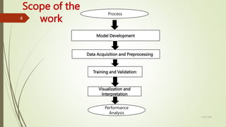
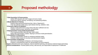
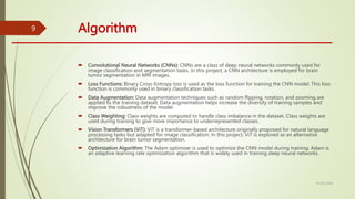
Medical Image Segmentation
By
SACHIN S
1
Project Objectives
 To develop a deep learning model to accurately segment brain tumors in MRI images.
 To ensure the model's reliability and performance across diverse datasets and imaging conditions.
 To demonstrate the model's practical utility in assisting medical professionals with tumor detection and
treatment planning.
 To compare the model's performance against established segmentation methods to validate its
effectiveness and potential clinical impact.
10-02-2024
2
Need of the project
 Improved Diagnosis: Automating brain tumor segmentation in MRI images
streamlines the diagnostic process, aiding healthcare professionals in detecting
tumors earlier and more accurately.
 Time Efficiency: Manual segmentation is time-consuming and requires specialized
skills. Automated segmentation models save time and resources, allowing medical
staff to focus on patient care.
 Enhanced Treatment Planning: Accurate segmentation helps in precise treatment
planning, including surgery, radiation therapy, and chemotherapy, leading to better
outcomes for patients with brain tumors.
 Access to Healthcare: By developing accessible and reliable segmentation tools, the
project aims to improve healthcare accessibility, especially in regions with limited
medical resources or expertise, ultimately benefiting a larger population of
patients.
10-02-2024
Design and Implementation of Fractional Order IMC Controller for Nonlinear Process
3
Data Acquisition and Preprocessing
Model Development
Training and Validation:
Process
Visualization and
Interpretation
Scope of the
work
Performance
Analysis
10-02-2024
4
Work Progress
Project Work completed
First review Model Model Development: Explored different deep learning architectures.
Conducted initial model experiments.
Data Preprocessing: Collected MRI datasets. Started preprocessing tasks like
resizing and normalization.
Training Preparation: Set up initial training pipeline. Defined basic data
augmentation techniques.
Second review Model Training: Completed initial model training. Monitored training progress and
performance.
Evaluation: Evaluated models using standard metrics. Analyzed model accuracy and
performance.
Visualization: Visualized segmentation results. Examined model outputs
forinterpretation.
Third review Model Refinement:
Made adjustments based on training insights.
Fine-tuned model hyperparameters.
Documentation:
Documented model architecture and training procedures.
Prepared initial project documentation.
Next Steps:
Discussed future research directions.
Identified areas for improvement and collaboration
10-02-2024
5
10-02-2024
6
 Challenge: Manual segmentation of brain tumors in MRI images is time-
consuming and prone to errors.
 Objective: Develop a deep learning model for accurate and efficient
automated segmentation.
 Purpose: Assist medical professionals in early diagnosis and treatment
planning, enhancing patient outcomes.
 Approach: Leveraging deep learning techniques to analyze MRI data and
identify tumor regions.
 Impact: Revolutionize brain tumor detection, streamline healthcare
workflows, and improve patient care.
 Ethical Considerations: Prioritize patient privacy, data security, and
responsible deployment of AI technology in healthcare.
INTRODUCTION
Block diagram
10-02-2024
7
Proposed metholodgy
1.Data Acquisition & Preprocessing:
•Obtain MRI datasets with brain images and tumor masks.
•Preprocess data by resizing, normalizing, and addressing artifacts.
2.Model Selection & Training:
•Explore deep learning architectures like U-Net or DeepLabv3+.
•Train the selected model using a split dataset (training, validation, test).
3.Evaluation Metrics & Validation:
•Assess model performance using metrics like Dice coefficient and IoU.
•Validate model accuracy, sensitivity, and specificity.
4.Hyperparameter Tuning & Data Augmentation:
•Tune hyperparameters (learning rates, batch sizes).
•Apply data augmentation (rotation, flipping) to enhance model generalization.
5.Visualization & Interpretation:
•Visualize segmentation results by overlaying predicted masks.
•Interpret model outputs for accuracy and improvement insights.
6.Documentation & Reporting:
•Document methodology, architecture, and training process.
•Prepare a comprehensive report for reproducibility and future research.
Impact: Streamline brain tumor diagnosis, improve treatment planning, and advance medical imaging technology.
Ethical Considerations: Prioritize patient privacy, data security, and responsible AI deployment in healthcare.
10-02-2024
8
Algorithm
 Convolutional Neural Networks (CNNs): CNNs are a class of deep neural networks commonly used for
image classification and segmentation tasks. In this project, a CNN architecture is employed for brain
tumor segmentation in MRI images.
 Loss Functions: Binary Cross-Entropy loss is used as the loss function for training the CNN model. This loss
function is commonly used in binary classification tasks.
 Data Augmentation: Data augmentation techniques such as random flipping, rotation, and zooming are
applied to the training dataset. Data augmentation helps increase the diversity of training samples and
improve the robustness of the model.
 Class Weighting: Class weights are computed to handle class imbalance in the dataset. Class weights are
used during training to give more importance to underrepresented classes.
 Vision Transformers (ViT): ViT is a transformer-based architecture originally proposed for natural language
processing tasks but adapted for image classification. In this project, ViT is explored as an alternative
architecture for brain tumor segmentation.
 Optimization Algorithm: The Adam optimizer is used to optimize the CNN model during training. Adam is
an adaptive learning rate optimization algorithm that is widely used in training deep neural networks.
10-02-2024
9
Pseudocode
10
Here are the headings for each section of the simplified pseudocode:
Medical Image Segmentation for Brain Tumor
Detection
1.Import Libraries
2.Define Parameters
3.Data Preprocessing
4.Model Architecture
5.Compile Model
6.Model Training
7.Model Evaluation
8.Fine-tuning (Optional)
9.Documentation
10.Conclusion
Result Analysis
 Result Analysis Techniques
 Accuracy & Loss Curves
 Track model performance over epochs.
 Identify overfitting or underfitting.
 Confusion Matrix
 Evaluate classification model performance.
 Summarize correct/incorrect predictions by class.
 Classification Report
 Provide precision, recall, F1-score metrics.
 Assess model performance comprehensively.
 Intersection over Union (IoU)
 Measure segmentation mask overlap.
 Evaluate accuracy of segmentation.
 Dice Coefficient
 Assess similarity between samples.
 Useful for binary segmentation tasks.
 F1-Score
 Harmonic mean of precision and recall.
 Balanced measure of model performance.
 Visual Inspection
 Overlay predicted masks on MRI images.
 Validate segmentation accuracy visually
10-02-2024
11
SUMMARY
 Project Overview:
 Objective: Develop a deep learning model for automatic brain tumor segmentation in MRI images.
 Aim: Assist medical professionals in early diagnosis and treatment planning.
 Approach:
 Utilize Convolutional Neural Networks (CNNs) and Vision Transformers (ViT) for image segmentation.
 Train the model on MRI brain images with corresponding tumor segmentation masks.
 Implementation:
 Data preprocessing: Resize, normalize, and augment images.
 Model development: CNN with convolutional and dense layers, ViT with patch creation and encoding.
 Evaluation: Assess model accuracy and performance using appropriate metrics.
 Tools Used:
 Libraries: TensorFlow, OpenCV, NumPy, Matplotlib, Pandas, scikit-learn.
 Frameworks: Keras, TensorFlow-Addons.
 Outcome:
 Improved early detection and treatment planning for brain tumors.
 Potential to enhance patient outcomes and streamline medical diagnosis processes.
 Conclusion:
 Medical image segmentation with deep learning offers promising avenues for healthcare advancement.
 Collaboration between technology and medicine can revolutionize diagnostic practices.
10-02-2024
12
Acknowledgement
 Acknowledgements:
 We would like to express our gratitude to the following individuals, organizations, and sources for their contributions and support during the
development of this project:
 Kaggle: We acknowledge brain Tumor Dataset for providing the brain tumor detection dataset used in this project.
 - Libraries and Tools: We extend our appreciation to the developers and contributors of TensorFlow, OpenCV, NumPy, PIL, scikit-learn, and other
libraries and tools used in this project for their invaluable contributions to the field of deep learning and image processing.
 - Inspiration and References: We are thankful to the authors of [Reference Papers or Projects] for their pioneering work in medical image
segmentation and brain tumor detection, which served as inspiration and references during the development of our model.
 - Classmates, Mentors, or Advisors: We would like to thank for their support, guidance, and feedback during the course of this project.
 - Institution or Organization: This project was conducted as part of [Name of Institution or Organization]. We acknowledge Ramco Institute of
Technology for providing resources, facilities, and support for this research.
10-02-2024
13
Thank You
10-02-2024
14

Medical Image segmentation from dl .pptx

  • 1.
  • 2.
    Project Objectives  Todevelop a deep learning model to accurately segment brain tumors in MRI images.  To ensure the model's reliability and performance across diverse datasets and imaging conditions.  To demonstrate the model's practical utility in assisting medical professionals with tumor detection and treatment planning.  To compare the model's performance against established segmentation methods to validate its effectiveness and potential clinical impact. 10-02-2024 2
  • 3.
    Need of theproject  Improved Diagnosis: Automating brain tumor segmentation in MRI images streamlines the diagnostic process, aiding healthcare professionals in detecting tumors earlier and more accurately.  Time Efficiency: Manual segmentation is time-consuming and requires specialized skills. Automated segmentation models save time and resources, allowing medical staff to focus on patient care.  Enhanced Treatment Planning: Accurate segmentation helps in precise treatment planning, including surgery, radiation therapy, and chemotherapy, leading to better outcomes for patients with brain tumors.  Access to Healthcare: By developing accessible and reliable segmentation tools, the project aims to improve healthcare accessibility, especially in regions with limited medical resources or expertise, ultimately benefiting a larger population of patients. 10-02-2024 Design and Implementation of Fractional Order IMC Controller for Nonlinear Process 3
  • 4.
    Data Acquisition andPreprocessing Model Development Training and Validation: Process Visualization and Interpretation Scope of the work Performance Analysis 10-02-2024 4
  • 5.
    Work Progress Project Workcompleted First review Model Model Development: Explored different deep learning architectures. Conducted initial model experiments. Data Preprocessing: Collected MRI datasets. Started preprocessing tasks like resizing and normalization. Training Preparation: Set up initial training pipeline. Defined basic data augmentation techniques. Second review Model Training: Completed initial model training. Monitored training progress and performance. Evaluation: Evaluated models using standard metrics. Analyzed model accuracy and performance. Visualization: Visualized segmentation results. Examined model outputs forinterpretation. Third review Model Refinement: Made adjustments based on training insights. Fine-tuned model hyperparameters. Documentation: Documented model architecture and training procedures. Prepared initial project documentation. Next Steps: Discussed future research directions. Identified areas for improvement and collaboration 10-02-2024 5
  • 6.
    10-02-2024 6  Challenge: Manualsegmentation of brain tumors in MRI images is time- consuming and prone to errors.  Objective: Develop a deep learning model for accurate and efficient automated segmentation.  Purpose: Assist medical professionals in early diagnosis and treatment planning, enhancing patient outcomes.  Approach: Leveraging deep learning techniques to analyze MRI data and identify tumor regions.  Impact: Revolutionize brain tumor detection, streamline healthcare workflows, and improve patient care.  Ethical Considerations: Prioritize patient privacy, data security, and responsible deployment of AI technology in healthcare. INTRODUCTION
  • 7.
  • 8.
    Proposed metholodgy 1.Data Acquisition& Preprocessing: •Obtain MRI datasets with brain images and tumor masks. •Preprocess data by resizing, normalizing, and addressing artifacts. 2.Model Selection & Training: •Explore deep learning architectures like U-Net or DeepLabv3+. •Train the selected model using a split dataset (training, validation, test). 3.Evaluation Metrics & Validation: •Assess model performance using metrics like Dice coefficient and IoU. •Validate model accuracy, sensitivity, and specificity. 4.Hyperparameter Tuning & Data Augmentation: •Tune hyperparameters (learning rates, batch sizes). •Apply data augmentation (rotation, flipping) to enhance model generalization. 5.Visualization & Interpretation: •Visualize segmentation results by overlaying predicted masks. •Interpret model outputs for accuracy and improvement insights. 6.Documentation & Reporting: •Document methodology, architecture, and training process. •Prepare a comprehensive report for reproducibility and future research. Impact: Streamline brain tumor diagnosis, improve treatment planning, and advance medical imaging technology. Ethical Considerations: Prioritize patient privacy, data security, and responsible AI deployment in healthcare. 10-02-2024 8
  • 9.
    Algorithm  Convolutional NeuralNetworks (CNNs): CNNs are a class of deep neural networks commonly used for image classification and segmentation tasks. In this project, a CNN architecture is employed for brain tumor segmentation in MRI images.  Loss Functions: Binary Cross-Entropy loss is used as the loss function for training the CNN model. This loss function is commonly used in binary classification tasks.  Data Augmentation: Data augmentation techniques such as random flipping, rotation, and zooming are applied to the training dataset. Data augmentation helps increase the diversity of training samples and improve the robustness of the model.  Class Weighting: Class weights are computed to handle class imbalance in the dataset. Class weights are used during training to give more importance to underrepresented classes.  Vision Transformers (ViT): ViT is a transformer-based architecture originally proposed for natural language processing tasks but adapted for image classification. In this project, ViT is explored as an alternative architecture for brain tumor segmentation.  Optimization Algorithm: The Adam optimizer is used to optimize the CNN model during training. Adam is an adaptive learning rate optimization algorithm that is widely used in training deep neural networks. 10-02-2024 9
  • 10.
    Pseudocode 10 Here are theheadings for each section of the simplified pseudocode: Medical Image Segmentation for Brain Tumor Detection 1.Import Libraries 2.Define Parameters 3.Data Preprocessing 4.Model Architecture 5.Compile Model 6.Model Training 7.Model Evaluation 8.Fine-tuning (Optional) 9.Documentation 10.Conclusion
  • 11.
    Result Analysis  ResultAnalysis Techniques  Accuracy & Loss Curves  Track model performance over epochs.  Identify overfitting or underfitting.  Confusion Matrix  Evaluate classification model performance.  Summarize correct/incorrect predictions by class.  Classification Report  Provide precision, recall, F1-score metrics.  Assess model performance comprehensively.  Intersection over Union (IoU)  Measure segmentation mask overlap.  Evaluate accuracy of segmentation.  Dice Coefficient  Assess similarity between samples.  Useful for binary segmentation tasks.  F1-Score  Harmonic mean of precision and recall.  Balanced measure of model performance.  Visual Inspection  Overlay predicted masks on MRI images.  Validate segmentation accuracy visually 10-02-2024 11
  • 12.
    SUMMARY  Project Overview: Objective: Develop a deep learning model for automatic brain tumor segmentation in MRI images.  Aim: Assist medical professionals in early diagnosis and treatment planning.  Approach:  Utilize Convolutional Neural Networks (CNNs) and Vision Transformers (ViT) for image segmentation.  Train the model on MRI brain images with corresponding tumor segmentation masks.  Implementation:  Data preprocessing: Resize, normalize, and augment images.  Model development: CNN with convolutional and dense layers, ViT with patch creation and encoding.  Evaluation: Assess model accuracy and performance using appropriate metrics.  Tools Used:  Libraries: TensorFlow, OpenCV, NumPy, Matplotlib, Pandas, scikit-learn.  Frameworks: Keras, TensorFlow-Addons.  Outcome:  Improved early detection and treatment planning for brain tumors.  Potential to enhance patient outcomes and streamline medical diagnosis processes.  Conclusion:  Medical image segmentation with deep learning offers promising avenues for healthcare advancement.  Collaboration between technology and medicine can revolutionize diagnostic practices. 10-02-2024 12
  • 13.
    Acknowledgement  Acknowledgements:  Wewould like to express our gratitude to the following individuals, organizations, and sources for their contributions and support during the development of this project:  Kaggle: We acknowledge brain Tumor Dataset for providing the brain tumor detection dataset used in this project.  - Libraries and Tools: We extend our appreciation to the developers and contributors of TensorFlow, OpenCV, NumPy, PIL, scikit-learn, and other libraries and tools used in this project for their invaluable contributions to the field of deep learning and image processing.  - Inspiration and References: We are thankful to the authors of [Reference Papers or Projects] for their pioneering work in medical image segmentation and brain tumor detection, which served as inspiration and references during the development of our model.  - Classmates, Mentors, or Advisors: We would like to thank for their support, guidance, and feedback during the course of this project.  - Institution or Organization: This project was conducted as part of [Name of Institution or Organization]. We acknowledge Ramco Institute of Technology for providing resources, facilities, and support for this research. 10-02-2024 13
  • 14.