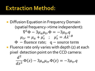
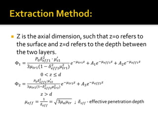
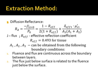
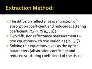
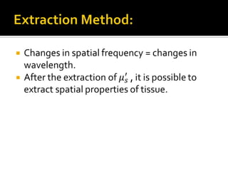
Brain Optical Properties Extraction Using Structured LightEdited by  Trakhtman  AnaAriel University Center of SamariaDepartment of Applied  Physics
Tissue Optics Tissue Optical PropertiesStructured lightContent:Extraction MethodOptical SystemSummery
Tissue Optics – What is it?Tissue optics is a branch in optical science, which describes EM radiation propagation in biological tissue. Determines optical properties for diagnostic applications allowing imaging of a tissue non-invasively and in vivo.
Optical Properties:Reduced Scattering Coefficient :𝜇𝑠′=(1−𝑔)𝜇𝑠for a wavelength of 600−1000 𝑛𝑚:𝜇𝑠′=𝐴𝜆−𝐵where A, B depend on spatial properties of 	a tissueAbsorption Coefficient - 𝜇𝑎Scattering Anisotropy – 𝑔=0.9 
Structured Light:Projecting the light in a specific pattern (plane, grid, horizontal bars or  a more complex shape) at a known angle onto an object.Very useful for imaging and acquiring dimensional information.
Extraction Method:Diffusion Equation in Frequency Domain(spatial frequency->time independent):𝛻2Φ−3𝜇𝑎𝜇𝑡𝑟Φ=−3𝜇𝑡𝑟𝑞𝜇𝑡𝑟=𝜇𝑎+𝜇𝑠′    ;    𝜇𝑠′=𝐴𝜆−𝐵Φ −fluence rate;   q−source termFluence rate only varies with depth (z) at each pixel  detection point on the CCD camera:𝑑2𝑑𝑧2Φ𝑧−3𝜇𝑎𝜇𝑡𝑟Φ(𝑧)=−3𝜇𝑡𝑟𝑞 
Extraction Method:Z is the axial dimension, such that z=0 refers to the surface and z=d refers to the depth between the two layers.Φ1=𝑃0𝛿𝑒𝑓𝑓12∙𝜇𝑠1′ 3𝜇𝑡𝑟1(1−𝛿𝑒𝑓𝑓12𝜇𝑡𝑟12)𝑒−𝜇𝑡𝑟1𝑧+𝐴1𝑒−𝜇𝑒𝑓𝑓1𝑧+𝐴2𝑒−𝜇𝑒𝑓𝑓1𝑧0<𝑧≤𝑑Φ2=𝑃0𝛿𝑒𝑓𝑓22∙𝜇𝑠2′ 3𝜇𝑡𝑟2(1−𝛿𝑒𝑓𝑓22𝜇𝑡𝑟22)𝑒−𝜇𝑡𝑟2𝑧+𝐴3𝑒−𝜇𝑒𝑓𝑓2𝑧𝑧>𝑑𝜇𝑒𝑓𝑓=1𝛿𝑒𝑓𝑓=3𝜇𝑎𝜇𝑡𝑟   ; 𝛿𝑒𝑓𝑓 - effective penetration depth 
Extraction Method:Diffusion Reflectance:𝑅𝑑=−𝐽|𝑧=0𝑃0=1−𝑅𝑒𝑓𝑓2(1+𝑅𝑒𝑓𝑓)×𝛿𝑒𝑓𝑓1∙𝜇𝑠1′𝐴3(𝐴1+𝐴2)J – flux   ; 𝑅𝑒𝑓𝑓 - effective reflection coefficient𝑅𝑒𝑓𝑓=0.493 for tissue𝐴1 , 𝐴2 , 𝐴3 −can be obtained from the following boundary conditions:Fluence and flux are continuous across the boundary between layers.The flux just below surface is related to the fluence just below the surface. 
Extraction Method:The diffusion reflectance is a function of absorption coefficient and reduced scattering coefficient: 𝑅𝑑=𝑅(𝜇𝑎 , 𝜇𝑠′)Two diffusion reflectance measurements = two equations with two variables (𝜇𝑎 , 𝜇𝑠′)Solving this equations gives us the optical parameters (absorption coefficient and reduced scattering coefficient) of the tissue. 
Extraction Method:Changes in spatial frequency = changes in wavelength.After the extraction of 𝜇𝑠′ , it is possible to extract spatial properties of tissue. 
Optical System:
Summery:At this point in research, illumination is done directly upon brain tissue.Although, the extraction method was given for two layered tissue, at this point we refer to one layer only (surface layer).This method gives us information about optical and spatial properties of tissue.The light we are using is in visible spectrum and does no harm.Optical System is very simple, and can be build at low costs.

Brain Optical Properties Extraction

  • 1.
    Brain Optical PropertiesExtraction Using Structured LightEdited by Trakhtman AnaAriel University Center of SamariaDepartment of Applied Physics
  • 2.
    Tissue Optics TissueOptical PropertiesStructured lightContent:Extraction MethodOptical SystemSummery
  • 3.
    Tissue Optics –What is it?Tissue optics is a branch in optical science, which describes EM radiation propagation in biological tissue. Determines optical properties for diagnostic applications allowing imaging of a tissue non-invasively and in vivo.
  • 4.
    Optical Properties:Reduced ScatteringCoefficient :𝜇𝑠′=(1−𝑔)𝜇𝑠for a wavelength of 600−1000 𝑛𝑚:𝜇𝑠′=𝐴𝜆−𝐵where A, B depend on spatial properties of a tissueAbsorption Coefficient - 𝜇𝑎Scattering Anisotropy – 𝑔=0.9 
  • 5.
    Structured Light:Projecting thelight in a specific pattern (plane, grid, horizontal bars or a more complex shape) at a known angle onto an object.Very useful for imaging and acquiring dimensional information.
  • 6.
    Extraction Method:Diffusion Equationin Frequency Domain(spatial frequency->time independent):𝛻2Φ−3𝜇𝑎𝜇𝑡𝑟Φ=−3𝜇𝑡𝑟𝑞𝜇𝑡𝑟=𝜇𝑎+𝜇𝑠′    ;    𝜇𝑠′=𝐴𝜆−𝐵Φ −fluence rate;   q−source termFluence rate only varies with depth (z) at each pixel detection point on the CCD camera:𝑑2𝑑𝑧2Φ𝑧−3𝜇𝑎𝜇𝑡𝑟Φ(𝑧)=−3𝜇𝑡𝑟𝑞 
  • 7.
    Extraction Method:Z isthe axial dimension, such that z=0 refers to the surface and z=d refers to the depth between the two layers.Φ1=𝑃0𝛿𝑒𝑓𝑓12∙𝜇𝑠1′ 3𝜇𝑡𝑟1(1−𝛿𝑒𝑓𝑓12𝜇𝑡𝑟12)𝑒−𝜇𝑡𝑟1𝑧+𝐴1𝑒−𝜇𝑒𝑓𝑓1𝑧+𝐴2𝑒−𝜇𝑒𝑓𝑓1𝑧0<𝑧≤𝑑Φ2=𝑃0𝛿𝑒𝑓𝑓22∙𝜇𝑠2′ 3𝜇𝑡𝑟2(1−𝛿𝑒𝑓𝑓22𝜇𝑡𝑟22)𝑒−𝜇𝑡𝑟2𝑧+𝐴3𝑒−𝜇𝑒𝑓𝑓2𝑧𝑧>𝑑𝜇𝑒𝑓𝑓=1𝛿𝑒𝑓𝑓=3𝜇𝑎𝜇𝑡𝑟 ; 𝛿𝑒𝑓𝑓 - effective penetration depth 
  • 8.
    Extraction Method:Diffusion Reflectance:𝑅𝑑=−𝐽|𝑧=0𝑃0=1−𝑅𝑒𝑓𝑓2(1+𝑅𝑒𝑓𝑓)×𝛿𝑒𝑓𝑓1∙𝜇𝑠1′𝐴3(𝐴1+𝐴2)J– flux ; 𝑅𝑒𝑓𝑓 - effective reflection coefficient𝑅𝑒𝑓𝑓=0.493 for tissue𝐴1 , 𝐴2 , 𝐴3 −can be obtained from the following boundary conditions:Fluence and flux are continuous across the boundary between layers.The flux just below surface is related to the fluence just below the surface. 
  • 9.
    Extraction Method:The diffusionreflectance is a function of absorption coefficient and reduced scattering coefficient: 𝑅𝑑=𝑅(𝜇𝑎 , 𝜇𝑠′)Two diffusion reflectance measurements = two equations with two variables (𝜇𝑎 , 𝜇𝑠′)Solving this equations gives us the optical parameters (absorption coefficient and reduced scattering coefficient) of the tissue. 
  • 10.
    Extraction Method:Changes inspatial frequency = changes in wavelength.After the extraction of 𝜇𝑠′ , it is possible to extract spatial properties of tissue. 
  • 11.
  • 12.
    Summery:At this pointin research, illumination is done directly upon brain tissue.Although, the extraction method was given for two layered tissue, at this point we refer to one layer only (surface layer).This method gives us information about optical and spatial properties of tissue.The light we are using is in visible spectrum and does no harm.Optical System is very simple, and can be build at low costs.