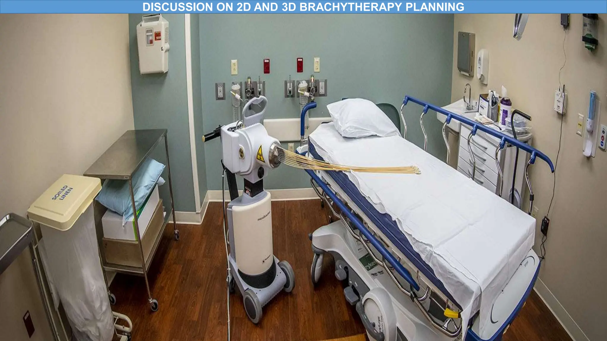
DISCUSSION ON 2D AND 3D BRACHYTHERAPY PLANNING
• INTRODUCTION
• ADVANTAGES AND DISADVANTAGES OF BRACHYTHERAPY
• BRACHYTHERAPY APPLICATIONS
• INDICATIONS FOR GYNAECOLOGICAL CANCERS
• 2D BRACHYTHERAPY
• 2D BRACHYTHERAPY PLANNING
• 3D BRACHYTHERAPY
• 3D BRACHYTHERAPY PLANNING
• ADVANTAGES AND DISADVANTAGES OF 2D & 3D BRACHYTHERAPY PLANNING
• CONCLUSION
• REFERENCES
OBJECTIVES
Figure 1:https://www.google.com/url?sa=i&url=https%3A%2F%2Fwww.healtheuropa.com%2Fpersonalising-brachytherapy-for-better-
results-against-cervical-cancer%2F120273%2F&psig=AOvVaw2OVs-
EYzE2E4KRp4h1DVfC&ust=1690286113755000&source=images&cd=vfe&opi=89978449&ved=0CBEQjRxqFwoTCNCEgM2kp4ADF
QAAAAAdAAAAABAP
• The term Brachytherapy is derived from the Greek words “brachy’’ meaning short, and ‘’therapy’’ meaning treatment (Skowronek,2017:581).
• Treatment at short distance, refers to radiotherapy in which sources of radiation are placed close to or directly in the target volume (Washington
Leaver & leaver,2016:265).
• Radiation sources are isotopes that may cause radiation exposure by emitting ionizing radiation or releasing radioactive substances e.g. Iodine
125,Cobalt 60, Iridium 195 etc.
• Sources are encapsulated by titanium or stainless steel in order to provide adequate shielding against the Alpha or Beta radiation emitted from the
source to prevent leakage of the radioactive material (Skowronek,2017:581).
• Brachytherapy allows high radiation dose to be delivered locally to the tumour with rapid dose fall-off in the surrounding normal tissues (Washington
Leaver & leaver,2016:265)
1. INTRODUCTION
Figure 2:https://www.google.com/url?sa=i&url=https%3A%2F%2Fwww.mayoclinic.org%2Ftests-
procedures%2Fbrachytherapy%2Fabout%2Fpac-
20385159&psig=AOvVaw3duqD_Zj8TJiHkQsW7Zctg&ust=1690113074776000&source=images&cd=vf
e&opi=89978449&ved=0CBEQjRxqFwoTCNDypv6fooADFQAAAAAdAAAAABAE
• High doses of radiation can be delivered locally to the tumor in a short time- ↑ cell killing
• Radiation does not penetrate through overlying tissues
• Surrounding tissues are spared from increased doses of radiation
• ↓ side effects
PRONS
CONS
• Invasive procedure
• Can only be used in localized small tumors
• Increase risk of high dose of radiation to staff and patients
• Costly procedure
Figure 3 :
https:://www.google.com/url?sa=i&url=https%3A%2F%2Fmountmiriam.com%2Fbrachytherapy%2F&psig=AOvVaw1
vGZrB_kFyBg3RMkBEMUQF&ust=1692185437375000&source=images&cd=vfe&opi=89978449&ved=0CBAQjRxq
FwoTCJDz0ZnI3oADFQAAAAAdAAAAABAK
2. ADVANTAGES AND DISADVANTAGES OF BRACHYTHERAPY
Brachytherapy applications Description
Interstitial brachytherapy
Placement of radioactive sources directly into a tumor or tumor bed: commonly used for head & neck,breast,prostate
and soft tissue sarcomas (Washington & Leaver,2016:268)
Intraluminal brachytherapy
Placement of sources of radiation within body such as Esophagus, trachea and bronchus (Washington &
Leaver,2016:268)
Intravascular brachytherapy
Widely used to reduce the rate of restenosis after angioplasty and the placement of stents in blood vessels
(Washington & Leaver,2016:268)
Surface or Topical brachytherapy
Places the radioactive sources on top of the area treated ,commonly used for skin cancer (Washington &
Leaver,2016:269)
Intracavitary brachytherapy Implants are placed within body cavity for treatment, its used for gynecological cancers
(Washington & Leaver,2016:268)
Figure 4 :https://www.google.com/url?sa=i&url=https%3A%2F%2Fwww.siemens-
healthineers.com%2Fen-
us%2Fradiotherapy%2Fbrachytherapy&psig=AOvVaw0wWNFXvMxY1WoiKcQIafRj&
ust=1692188949855000&source=images&cd=vfe&opi=89978449&ved=0CBAQjRxqF
woTCPDBvqTV3oADFQAAAAAdAAAAABAK
3. BRACHYTHERAPY APPLICATIONS
• Early stage tumors Banerjee,Kamrava,2014 :556)
• Tumors < 4-5cm (Banerjee,Kamrava,2014 :556)
• Lower vaginal involvement (Banerjee,Kamrava,2014 :556)
• Patients who have had a supracervical hysterectomy (Banerjee,Kamrava,2014 :556)
Figure 5:https://www.google.com/url?sa=i&url=https%3A%2F%2Fwww.iaea.org%2Fnewscenter%2Fnews%2Fcombining-external-
radiation-with-high-dose-rate-brachytherapy-effective-for-cervical-cancer-iaea-study-
confirms&psig=AOvVaw1vGZrB_kFyBg3RMkBEMUQF&ust=1692185437375000&source=images&cd=vfe&opi=89978449&ved=0CBAQj
RxqFwoTCJDz0ZnI3oADFQAAAAAdAAAAABAE
4. INDICATIONS FOR GYNAECOLOGICALCANCERS
• X-ray fluoroscopy or radiographs (Banerjee,Kamrava,2014 :559)
• Are used to verify the correct placement of the applicators in the patient (Banerjee,Kamrava,2014 :559)
• When the placement of the applicators is validated and at an adequate position (Banerjee,Kamrava,2014 :559)
• Dose is delivered to the anatomy around the applicators (Banerjee,Kamrava,2014 :559)
• Point doses are conventionally calculated for the rectum and bladder according to ICRU Report 38
recommendations (Banerjee,Kamrava,2014 :559)
5. 2D BRACHYTHERAPY
FIGURE 6:https://www.google.com/url?sa=i&url=https%3A%2F%2Fwww.fujifilm.com%2Fza%2Fen%2Fhealthcare%2Fx-ray%2Ffdx-
visionary-
mobile&psig=AOvVaw0r6hWViHktL5GS75XNPV2n&ust=1692198315484000&source=images&cd=vfe&opi=89978449&ved=0CBAQjRxqFwo
TCLiJspb43oADFQAAAAAdAAAAABAf
6. 2D BRACHYTHERAPY PLANNING
Insertion of applicators
• Patient is sedated for comfort, relaxation of muscles and easy insertion of applicators
• Oncologist clean the patient using cotton and safe disinfecting liquid
• Use cervical Os finder, speculum to view cervix and measure the size and lengths of the applicators
• Catheter is inserted into the bladder
• Applicators with rectal retractor and sterile gauze with contrast are inserted into cervix
Imaging
• A radiation therapist uses a C-arm for screening and exposure of Anterior posterior and lateral x ray images
• To validate the correct placement of applicators
Planning
• The prescription point A and B are used as reference points
• Prescription of 24Gy in 3#; delivering 8Gy daily is prescribed at point A
• Point A is 2cm lateral to uterine canal and 2cm superior to lateral fornice of vagina
Organs at risk dose
• Coordinates of prescription points in X (left and right), Y( sup and inf ) and Z (ant and post) from x ray images
• Coordinated points are entered onto the genie software and doses to bladder and rectum are recorded
• Tolerances for bladder and rectum are 85% and 75% respectively
Figure 7:
https://www.google.com/imgres?imgurl=https%3A%2F%2Fdoctorlib.info%2Ftherapy%2Fbrachytherapy%2Fbrachyther
apy.files%2Fimage216.jpg&tbnid=nXx1CZ-vd7EBaM&vet=10CAwQxiAoC2oXChMI-
NKHkv_tgAMVAAAAAB0AAAAAEBo..i&imgrefurl=https%3A%2F%2Fdoctorlib.info%2Ftherapy%2Fbrachytherapy%2F9
.html&docid=jkD2zvUvSDDEkM&w=491&h=521&itg=1&q=2d%20x%20ray%20brachytherapy%20cervix&ved=0CAwQx
iAoC2oXChMI-NKHkv_tgAMVAAAAAB0AAAAAEBo
7. 3D BRACHYTHERAPY
• Delivery of radiotherapy treatment plans based on 3-D image dataset (Weiner & Schwarz,2015 :370).
• Target , organs at risk localization, applicator reconstruction and dose calculations are based on 3D imaging modalities
(Weiner & Schwarz,2015 :370).
• CT, MRI and Ultrasound are commonly used for 3D brachytherapy planning (Weiner & Schwarz,2015 :370).
• Enables an anatomy-adapted implantation, treatment planning and optimization (Weiner & Schwarz,2015 :370).
Figure 8 :https://inis.iaea.org/collection/NCLCollectionStore/_Public/49/093/49093363.pdf
8. 3D BRACHYTHERAPY PLANNING
Localization
• The Applicators are scanned using the CT, MRI or Ultrasound
• Data set is imported to the Treatment planning system
• Organs at risk such as the bladder, rectum, sigmoid are contoured including Target volumes drawn
Planning
Target volumes include:
• GTV :Visible / palpable tumor
• High Risk-CTV : GTV + Cervix + residual pathological tissues
• Intermediate Risk CTV : HR-CTV + presumed adjacent significant microscopic disease
• These volumes are the most relevant volumes which are characterized by high doses in the GTV and HR CTV
and have a rapid dose fall off.
• The plan is optimized
• Tools such as DVH,DVH dose statistics and isodose lines are used to monitor the dose received by organs at risk
Figure 9 : https://inis.iaea.org/collection/NCLCollectionStore/_Public/49/093/49093363.pdf
9. ADVANTAGES AND DISADVANTAGES OF 2D & 3D BRACHYTHERAPY
Brachytherapy 2D 3D
Advantages
• Provides a 2D representation of the patient anatomy in a given orientation
(Mayadev, Benedict, Kamrava, 2017 :70)
• Has high resolution contrast (Mayadev et al, 2017 :70)
• Least expensive (Mayadev et al, 2017 :71)
• Quicker to plan (Mayadev et al, 2017 :71)
• Verifies the proper placement of applicators (Hashemi et al,2021:530)
• Better target volume definition and organ at risk visualization (Hashemi et
al,2021:530)
• 3D dose distribution calculation & optimization (Hashemi et al,2021:530)
• Improved tumour coverage and decreased dose to critical organs (Hashemi et
al,2021:530)
Disadvantages
• Low soft tissue contrast (Mayadev et al, 2017 :71)
• It is difficult to distinguish individual soft tissue structures (Mayadev et al,
2017 :70)
• Poor image quality on the lateral film (Mayadev et al, 2017 :71)
• Lacks spatial information on the tumor volumes and OAR volumes (Mayadev
et al, 2017 :72)
• Produce artifacts with metallic applicators (Hashemi, et al,2021 :531)
• Time consuming (Hashemi, et al,2021 :531)
• Expensive (Hashemi, et al,2021 :531)
10. CONCLUSION
• Brachytherapy is a crucial treatment for Gynaecological malignancies, delivering high doses to target while minimizing
normal tissue toxicity (Weiner & Schwarz,2015 :372).
• Advances in imaging techniques, 3D adaptive treatment planning and dose-volume reporting enhance its precision and
versatility (Weiner & Schwarz,2015 :372).
• Automation of brachytherapy plans is a future development with tools such as dose Volume Histogram, dose distribution
statistics and isodose lines being used for accurate delivery (Weiner & Schwarz,2015 :372).
• The transition from 2D to 3D-based imaging and treatment planning for cervical cancer brachytherapy will improve local
control, reduce toxicity, and increase overall survival (Banerjee,Kamrava,2014 :559)
Figure 10:
https://www.google.com/imgres?imgurl=https%3A%2F%2Fwww.elekta.com%2Fproducts%2Fbrachytherapy%2Fgynec
ology%2Fimg%2Fgyn-imaging-
3.jpg&tbnid=gWKqIAPSgEqp2M&vet=12ahUKEwijrPzl_u2AAxW4sCcCHSAGAGwQMygQegQIARB1..i&imgrefurl=http
s%3A%2F%2Fwww.elekta.com%2Fproducts%2Fbrachytherapy%2Fgynecology%2F&docid=9sHE144udynKEM&w=60
0&h=375&q=3d%20brachytherapy%20gynaecology&ved=2ahUKEwijrPzl_u2AAxW4sCcCHSAGAGwQMygQegQIARB
1
11. REFERENCES
Washington. C, Leaver , D. (2016) .Principles and practice of radiation therapy.5th edition. Elsevier
Skowronek,J. (2017). Current status of brachytherapy in cancer treatment – short overview . J Contemp Brachytherapy. 9(6): 581–589
DOI: https://doi.org/10.5114/jcb.2017.72607
Weiner, A. Schwarz,J. (2015). Intracavitary Brachytherapy for Gynaecologic malignancies application and Innovation. Science of medicine/ radiation oncology. 112(5) :366-372
Banerjee,R. Kamrava , M. (2014). Brachytherapy in the treatment of cervical cancer: a review.. International Journal of Women’s Health. 6: 555-564
Brady, W. Heilmann, H. Molls, M. (2005). New Technologies in Radiation Oncology. Springer
Hu,H. Shao,Y. Hu, Shijie. (2020). A Review of the Application of Deep Learning in Brachytherapy. Open Access Library Journal.7:1-9 .DOI: 10.4236/oalib.1106589
Hashemi,F. Mansouri,S. Aghili,M. Esmati,E. Babaei,M. Saeedian,A. Moalej, S. Jaberi, R. (2021) A comparison between 2D and 3D planning of high-dose-rate vaginal cuff brachytherapy in patients with stage I-II
endometrial cancer using cobalt-60. J Contemp Brachytherapy. 13(5): 526–532. DOI: https://doi.org/10.5114/jcb.2021.110312
Mayadev,J. Benedict, S. Kamrava, M. (2017) Handbook of Image-Guided brachytherapy. Springer.
Ha, I. Jeong ,B. Kang ,K. Jeong ,H. Lee , Choi,H. Lee,J. Choi,W. Shin,J.Song,J. (2018).Who Really Benefits from 3D-Based Planning of Brachytherapy for Cervical Cancer?.J Korean Med Sci.33(18) :1-11.
https://doi.org/10.3346/jkms.2018.33.e135

Brachytherapy

  • 1.
    DISCUSSION ON 2DAND 3D BRACHYTHERAPY PLANNING
  • 2.
    • INTRODUCTION • ADVANTAGESAND DISADVANTAGES OF BRACHYTHERAPY • BRACHYTHERAPY APPLICATIONS • INDICATIONS FOR GYNAECOLOGICAL CANCERS • 2D BRACHYTHERAPY • 2D BRACHYTHERAPY PLANNING • 3D BRACHYTHERAPY • 3D BRACHYTHERAPY PLANNING • ADVANTAGES AND DISADVANTAGES OF 2D & 3D BRACHYTHERAPY PLANNING • CONCLUSION • REFERENCES OBJECTIVES Figure 1:https://www.google.com/url?sa=i&url=https%3A%2F%2Fwww.healtheuropa.com%2Fpersonalising-brachytherapy-for-better- results-against-cervical-cancer%2F120273%2F&psig=AOvVaw2OVs- EYzE2E4KRp4h1DVfC&ust=1690286113755000&source=images&cd=vfe&opi=89978449&ved=0CBEQjRxqFwoTCNCEgM2kp4ADF QAAAAAdAAAAABAP
  • 3.
    • The termBrachytherapy is derived from the Greek words “brachy’’ meaning short, and ‘’therapy’’ meaning treatment (Skowronek,2017:581). • Treatment at short distance, refers to radiotherapy in which sources of radiation are placed close to or directly in the target volume (Washington Leaver & leaver,2016:265). • Radiation sources are isotopes that may cause radiation exposure by emitting ionizing radiation or releasing radioactive substances e.g. Iodine 125,Cobalt 60, Iridium 195 etc. • Sources are encapsulated by titanium or stainless steel in order to provide adequate shielding against the Alpha or Beta radiation emitted from the source to prevent leakage of the radioactive material (Skowronek,2017:581). • Brachytherapy allows high radiation dose to be delivered locally to the tumour with rapid dose fall-off in the surrounding normal tissues (Washington Leaver & leaver,2016:265) 1. INTRODUCTION Figure 2:https://www.google.com/url?sa=i&url=https%3A%2F%2Fwww.mayoclinic.org%2Ftests- procedures%2Fbrachytherapy%2Fabout%2Fpac- 20385159&psig=AOvVaw3duqD_Zj8TJiHkQsW7Zctg&ust=1690113074776000&source=images&cd=vf e&opi=89978449&ved=0CBEQjRxqFwoTCNDypv6fooADFQAAAAAdAAAAABAE
  • 4.
    • High dosesof radiation can be delivered locally to the tumor in a short time- ↑ cell killing • Radiation does not penetrate through overlying tissues • Surrounding tissues are spared from increased doses of radiation • ↓ side effects PRONS CONS • Invasive procedure • Can only be used in localized small tumors • Increase risk of high dose of radiation to staff and patients • Costly procedure Figure 3 : https:://www.google.com/url?sa=i&url=https%3A%2F%2Fmountmiriam.com%2Fbrachytherapy%2F&psig=AOvVaw1 vGZrB_kFyBg3RMkBEMUQF&ust=1692185437375000&source=images&cd=vfe&opi=89978449&ved=0CBAQjRxq FwoTCJDz0ZnI3oADFQAAAAAdAAAAABAK 2. ADVANTAGES AND DISADVANTAGES OF BRACHYTHERAPY
  • 5.
    Brachytherapy applications Description Interstitialbrachytherapy Placement of radioactive sources directly into a tumor or tumor bed: commonly used for head & neck,breast,prostate and soft tissue sarcomas (Washington & Leaver,2016:268) Intraluminal brachytherapy Placement of sources of radiation within body such as Esophagus, trachea and bronchus (Washington & Leaver,2016:268) Intravascular brachytherapy Widely used to reduce the rate of restenosis after angioplasty and the placement of stents in blood vessels (Washington & Leaver,2016:268) Surface or Topical brachytherapy Places the radioactive sources on top of the area treated ,commonly used for skin cancer (Washington & Leaver,2016:269) Intracavitary brachytherapy Implants are placed within body cavity for treatment, its used for gynecological cancers (Washington & Leaver,2016:268) Figure 4 :https://www.google.com/url?sa=i&url=https%3A%2F%2Fwww.siemens- healthineers.com%2Fen- us%2Fradiotherapy%2Fbrachytherapy&psig=AOvVaw0wWNFXvMxY1WoiKcQIafRj& ust=1692188949855000&source=images&cd=vfe&opi=89978449&ved=0CBAQjRxqF woTCPDBvqTV3oADFQAAAAAdAAAAABAK 3. BRACHYTHERAPY APPLICATIONS
  • 6.
    • Early stagetumors Banerjee,Kamrava,2014 :556) • Tumors < 4-5cm (Banerjee,Kamrava,2014 :556) • Lower vaginal involvement (Banerjee,Kamrava,2014 :556) • Patients who have had a supracervical hysterectomy (Banerjee,Kamrava,2014 :556) Figure 5:https://www.google.com/url?sa=i&url=https%3A%2F%2Fwww.iaea.org%2Fnewscenter%2Fnews%2Fcombining-external- radiation-with-high-dose-rate-brachytherapy-effective-for-cervical-cancer-iaea-study- confirms&psig=AOvVaw1vGZrB_kFyBg3RMkBEMUQF&ust=1692185437375000&source=images&cd=vfe&opi=89978449&ved=0CBAQj RxqFwoTCJDz0ZnI3oADFQAAAAAdAAAAABAE 4. INDICATIONS FOR GYNAECOLOGICALCANCERS
  • 7.
    • X-ray fluoroscopyor radiographs (Banerjee,Kamrava,2014 :559) • Are used to verify the correct placement of the applicators in the patient (Banerjee,Kamrava,2014 :559) • When the placement of the applicators is validated and at an adequate position (Banerjee,Kamrava,2014 :559) • Dose is delivered to the anatomy around the applicators (Banerjee,Kamrava,2014 :559) • Point doses are conventionally calculated for the rectum and bladder according to ICRU Report 38 recommendations (Banerjee,Kamrava,2014 :559) 5. 2D BRACHYTHERAPY FIGURE 6:https://www.google.com/url?sa=i&url=https%3A%2F%2Fwww.fujifilm.com%2Fza%2Fen%2Fhealthcare%2Fx-ray%2Ffdx- visionary- mobile&psig=AOvVaw0r6hWViHktL5GS75XNPV2n&ust=1692198315484000&source=images&cd=vfe&opi=89978449&ved=0CBAQjRxqFwo TCLiJspb43oADFQAAAAAdAAAAABAf
  • 8.
    6. 2D BRACHYTHERAPYPLANNING Insertion of applicators • Patient is sedated for comfort, relaxation of muscles and easy insertion of applicators • Oncologist clean the patient using cotton and safe disinfecting liquid • Use cervical Os finder, speculum to view cervix and measure the size and lengths of the applicators • Catheter is inserted into the bladder • Applicators with rectal retractor and sterile gauze with contrast are inserted into cervix Imaging • A radiation therapist uses a C-arm for screening and exposure of Anterior posterior and lateral x ray images • To validate the correct placement of applicators Planning • The prescription point A and B are used as reference points • Prescription of 24Gy in 3#; delivering 8Gy daily is prescribed at point A • Point A is 2cm lateral to uterine canal and 2cm superior to lateral fornice of vagina Organs at risk dose • Coordinates of prescription points in X (left and right), Y( sup and inf ) and Z (ant and post) from x ray images • Coordinated points are entered onto the genie software and doses to bladder and rectum are recorded • Tolerances for bladder and rectum are 85% and 75% respectively Figure 7: https://www.google.com/imgres?imgurl=https%3A%2F%2Fdoctorlib.info%2Ftherapy%2Fbrachytherapy%2Fbrachyther apy.files%2Fimage216.jpg&tbnid=nXx1CZ-vd7EBaM&vet=10CAwQxiAoC2oXChMI- NKHkv_tgAMVAAAAAB0AAAAAEBo..i&imgrefurl=https%3A%2F%2Fdoctorlib.info%2Ftherapy%2Fbrachytherapy%2F9 .html&docid=jkD2zvUvSDDEkM&w=491&h=521&itg=1&q=2d%20x%20ray%20brachytherapy%20cervix&ved=0CAwQx iAoC2oXChMI-NKHkv_tgAMVAAAAAB0AAAAAEBo
  • 9.
    7. 3D BRACHYTHERAPY •Delivery of radiotherapy treatment plans based on 3-D image dataset (Weiner & Schwarz,2015 :370). • Target , organs at risk localization, applicator reconstruction and dose calculations are based on 3D imaging modalities (Weiner & Schwarz,2015 :370). • CT, MRI and Ultrasound are commonly used for 3D brachytherapy planning (Weiner & Schwarz,2015 :370). • Enables an anatomy-adapted implantation, treatment planning and optimization (Weiner & Schwarz,2015 :370). Figure 8 :https://inis.iaea.org/collection/NCLCollectionStore/_Public/49/093/49093363.pdf
  • 10.
    8. 3D BRACHYTHERAPYPLANNING Localization • The Applicators are scanned using the CT, MRI or Ultrasound • Data set is imported to the Treatment planning system • Organs at risk such as the bladder, rectum, sigmoid are contoured including Target volumes drawn Planning Target volumes include: • GTV :Visible / palpable tumor • High Risk-CTV : GTV + Cervix + residual pathological tissues • Intermediate Risk CTV : HR-CTV + presumed adjacent significant microscopic disease • These volumes are the most relevant volumes which are characterized by high doses in the GTV and HR CTV and have a rapid dose fall off. • The plan is optimized • Tools such as DVH,DVH dose statistics and isodose lines are used to monitor the dose received by organs at risk Figure 9 : https://inis.iaea.org/collection/NCLCollectionStore/_Public/49/093/49093363.pdf
  • 11.
    9. ADVANTAGES ANDDISADVANTAGES OF 2D & 3D BRACHYTHERAPY Brachytherapy 2D 3D Advantages • Provides a 2D representation of the patient anatomy in a given orientation (Mayadev, Benedict, Kamrava, 2017 :70) • Has high resolution contrast (Mayadev et al, 2017 :70) • Least expensive (Mayadev et al, 2017 :71) • Quicker to plan (Mayadev et al, 2017 :71) • Verifies the proper placement of applicators (Hashemi et al,2021:530) • Better target volume definition and organ at risk visualization (Hashemi et al,2021:530) • 3D dose distribution calculation & optimization (Hashemi et al,2021:530) • Improved tumour coverage and decreased dose to critical organs (Hashemi et al,2021:530) Disadvantages • Low soft tissue contrast (Mayadev et al, 2017 :71) • It is difficult to distinguish individual soft tissue structures (Mayadev et al, 2017 :70) • Poor image quality on the lateral film (Mayadev et al, 2017 :71) • Lacks spatial information on the tumor volumes and OAR volumes (Mayadev et al, 2017 :72) • Produce artifacts with metallic applicators (Hashemi, et al,2021 :531) • Time consuming (Hashemi, et al,2021 :531) • Expensive (Hashemi, et al,2021 :531)
  • 12.
    10. CONCLUSION • Brachytherapyis a crucial treatment for Gynaecological malignancies, delivering high doses to target while minimizing normal tissue toxicity (Weiner & Schwarz,2015 :372). • Advances in imaging techniques, 3D adaptive treatment planning and dose-volume reporting enhance its precision and versatility (Weiner & Schwarz,2015 :372). • Automation of brachytherapy plans is a future development with tools such as dose Volume Histogram, dose distribution statistics and isodose lines being used for accurate delivery (Weiner & Schwarz,2015 :372). • The transition from 2D to 3D-based imaging and treatment planning for cervical cancer brachytherapy will improve local control, reduce toxicity, and increase overall survival (Banerjee,Kamrava,2014 :559) Figure 10: https://www.google.com/imgres?imgurl=https%3A%2F%2Fwww.elekta.com%2Fproducts%2Fbrachytherapy%2Fgynec ology%2Fimg%2Fgyn-imaging- 3.jpg&tbnid=gWKqIAPSgEqp2M&vet=12ahUKEwijrPzl_u2AAxW4sCcCHSAGAGwQMygQegQIARB1..i&imgrefurl=http s%3A%2F%2Fwww.elekta.com%2Fproducts%2Fbrachytherapy%2Fgynecology%2F&docid=9sHE144udynKEM&w=60 0&h=375&q=3d%20brachytherapy%20gynaecology&ved=2ahUKEwijrPzl_u2AAxW4sCcCHSAGAGwQMygQegQIARB 1
  • 13.
    11. REFERENCES Washington. C,Leaver , D. (2016) .Principles and practice of radiation therapy.5th edition. Elsevier Skowronek,J. (2017). Current status of brachytherapy in cancer treatment – short overview . J Contemp Brachytherapy. 9(6): 581–589 DOI: https://doi.org/10.5114/jcb.2017.72607 Weiner, A. Schwarz,J. (2015). Intracavitary Brachytherapy for Gynaecologic malignancies application and Innovation. Science of medicine/ radiation oncology. 112(5) :366-372 Banerjee,R. Kamrava , M. (2014). Brachytherapy in the treatment of cervical cancer: a review.. International Journal of Women’s Health. 6: 555-564 Brady, W. Heilmann, H. Molls, M. (2005). New Technologies in Radiation Oncology. Springer Hu,H. Shao,Y. Hu, Shijie. (2020). A Review of the Application of Deep Learning in Brachytherapy. Open Access Library Journal.7:1-9 .DOI: 10.4236/oalib.1106589 Hashemi,F. Mansouri,S. Aghili,M. Esmati,E. Babaei,M. Saeedian,A. Moalej, S. Jaberi, R. (2021) A comparison between 2D and 3D planning of high-dose-rate vaginal cuff brachytherapy in patients with stage I-II endometrial cancer using cobalt-60. J Contemp Brachytherapy. 13(5): 526–532. DOI: https://doi.org/10.5114/jcb.2021.110312 Mayadev,J. Benedict, S. Kamrava, M. (2017) Handbook of Image-Guided brachytherapy. Springer. Ha, I. Jeong ,B. Kang ,K. Jeong ,H. Lee , Choi,H. Lee,J. Choi,W. Shin,J.Song,J. (2018).Who Really Benefits from 3D-Based Planning of Brachytherapy for Cervical Cancer?.J Korean Med Sci.33(18) :1-11. https://doi.org/10.3346/jkms.2018.33.e135