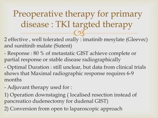
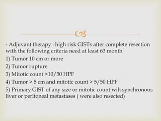
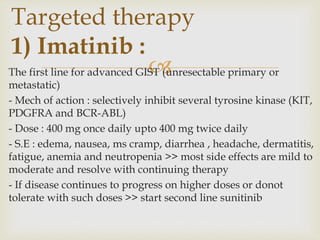
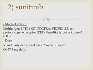
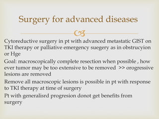
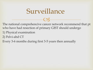
BY
Res / Mahmoud Ali

they are rare tumors (0.1 to 3 % of GIT malignancies )
How ever, they are the most common (80 %)
GIT mesenchymal neoplasm (GIT sarcoma)
In recent years tow key developments changed
understanding and TTT of GIST :
1) Identification of active signals (oncogenic mutations c-KIT
and PDGFRA genes encoding receptor tyrosine kinases )
2) development of therapeutic agent that supress tumor
growth by specifically targeting and inhibiting this signal (
imatinib mesylate , sunitinib malate)
Introduction

Term GIST describes intraabdominal nonepithelial neoplasm ,
that lacked ultra structural features of smooth ms cells and IHC
characters of schwan cells
Histological types:
1) spindle cell : Common in small intestinal, anorectal, colonic
and esophageal GISTs
2) epitheliod : common in Gastric GISTs
3) mixed
Origin : GIST arise from interstitial cells of Cajal ( component of
intestinal autonomic nervous system, intestinal pacemakers)
Pathological features

In 1998 Hirota and colleages reported two critical findings
1) there is near universal expression of transmembrane
receptor tyrosine kinase KIT in GIST
2) presence of function mutation in corresponding c-KIT
protooncogene
With immunohistochemical staining :
- almost all GISTs express CD 117 AG (part of KIT receptor) or
PDGFRA ( gene encoding related receptor tyrosine kinase) as
well as CD34
– almost all msooth ms tumors express actin and desmin
Mutations in receptor tyrosine
kinase

Age : 40 – 80 (median age 60 ) , rare in children may occur as
familial syndrome or as part of caneys triad ( different clinical
presentation)
Sex : equal in both
Incidence :
- American annual incidence 15-20 / million
- European annual incidence 11-15 / million
Hereditary GISTs : majority of cases are sporadic however
some cases were reported with Familial germline KIT or
PDGFRA mutations
Epidemiology

GIST secondary to Familial germline KIT or PDGFRA
mutations:
- Are younger, multifocal, rarely metastatic
- May have :skin hyperpigmentations, diffuse hyperplasia of
intestinal myentric plexus, melanoma, esophageal and breast
cancers
- Gastric GIST maybe component of carneys triad or carney-
stratakis syndrome
Carneys triad :
- GIST, paraganglioma and pulmpnary chondroma (recently
esophageal lieomyoma and adrenal cortical adenomas added as
acomponent of the syndrome)
- 85% in females
- 80% diagnosed before age of 30 years
Carney-Stratakis syndrome :
dyad of gastric GIST and paraganglioma

(A) Site :
1) Stomach (50-70 %) : mostly body but may occur at fundus
and antrum
2)Small intestine (25-35 %)
3) Colon and rectum (5-10 %)
4) Mesentry or omentum (7%)
5) Esophagus (<5% )
6) Ocassionally ( dudenal ampulla, appendix, GB, UB)
Clinical presentation

(B) Haemorrhage :
- Common as it is highly vascular, soft and friable
- Erosin into bowel lumen >> life threatening Hge
- Tumor rupture >> catastrophic IP bleeding or peritoneal
seeding
(C) Intestinal obstruction : may lead to prforatuion
(D) Asymptomatic (31% ) : in small tumors
(E) Metastatic (15-50 %)

A) Radiological:
1) CT : the initial imaging of choice
2) MRI : characterize primary rectal or metastatic liver
lesions
3) PET : characterize ambiguous masses on CT, monitor
response to therapy and detect drug resistant clones
Not specific and not recommended in suspected primary
cases
B) Endoscopy :
1) Upper and lower with biopsy : primary GIST appears as
submucosal lesion with or with out ulceration
2) EUS : FNA biopsy is not consistently diagnostic
Diagnosis

3) Preoperative biopsy
- Primary resectable tumor suspicious for GIST : not
necessary as it may lead to bleeding or dissemination
- Helpful in :
1- if DD include entities like lymphoma
2- if neoadjuvant therapy is considered
3- metastatic cases

1) Tumor size : < 1 cm low risk of recurrence
2) Mitotic index : most important , predictor of recurrence
3) Site of origin : small bowel has higher risk of progression
Acc to size and mitotic count aggressive behavior is :
1) Very low risk : < 2 cm , < 5/50 HPF
2) Low risk : 2-5 cm , <5/50 HPF
3) Intermediate risk : < 5cm , 6-10/50 HPF or 5-10 cm and
>5/50 HPF
4) High risk : >5 cm , > 5/50 HPF or > 10cm with any mitotic
rate or > 10/50HPF with any size
Prognostic factors

1) Surgery
2) Perioperative therapy
Therapy for primary disease

- Curtive for primary, resectable and localised GIST
- Goal of surgery is negative resection margins
- Macroscopically complete resection with negative (R0) or positive
(R1) microscopic margins >> better prognosis than macroscopically
incomplete resection (R2)
- Tumor rupture or capsule violation > increase risk of recurrence
- Extent of resection :
Wedge or segmental resection
More extensive resection may be needed in some cases :
1) Total gastrectomy : large proximal gasric GIST
2) Pancreaticodudenectomy : priampullary GIST
3) APR for low rectal GIST
Rule of surgery for primary
cases

No need for lymphadenectomy in adults (rarely involved)
- Size :
2 cm or more >> resection
< 2 cm debatable management
- Site :
Small bowel and colon > resection regardlrss of size
Stomach < 1 cm >> follow up
- Endoscopic resection : not recommended due to
1) Risk of leaving positive margin
2) May lead to perforation
- Symptomatic GIST or increase in size >> resection regardless
of size

2 effective , well tolerated orally : imatinib mesylate (Gleevec)
and sunitinib malate (Sutent)
- Response : 80 % of metastatic GIST achieve complete or
partial response or stable disease radiographically
- Optimal Duration : still unclear, but data from clinical trials
shows that Maximal radiographic response requires 6-9
months
- Adjuvant therapy used for :
1) Operation downstaging ( localised resection instead of
pancreatico dudenectomy for dudenal GIST)
2) Conversion from open to laparoscopic approach
Preoperative therapy for primary
disease : TKI targted therapy

- Adjuvant therapy : high risk GISTs after complete resection
with the following criteria need at least 63 month
1) Tumor 10 cm or more
2) Tumor rupture
3) Mitotic count >10/50 HPF
4) Tumor > 5 cm and mitotic count > 5/50 HPF
5) Primary GIST of any size or mitotic count wih synchronous
liver or peritoneal metastases ( were also resected)

1) Targeted therapy
2) Surgery
Therapy for advanced disease

The first line for advanced GIST (unresectable primary or
metastatic)
- Mech of action : selectively inhibit several tyrosine kinase (KIT,
PDGFRA and BCR-ABL)
- Dose : 400 mg once daily upto 400 mg twice daily
- S.E : edema, nausea, ms cramp, diarrhea , headache, dermatitis,
fatigue, anemia and neutropenia >> most side effects are mild to
moderate and resolve with continuing therapy
- If disease continues to progress on higher doses or donot
tolerate with such doses >> start second line sunitinib
Targeted therapy
1) Imatinib :

- Mech of action :
Multitargeted TKI : KIT, PDGFRA, VEGFR1,2,3, ret
protooncogene receptor (RET), Fms-like tyrosine kinase-3
(Flt3)
- Dose :
50 md daily in a 4 week on / 2 week off cycle
Or 37.5 mg daily
2) sunitinib

Cytoreductive surgery in pt with advanced metastatic GIST on
TKI therapy or palliative emergency suegery as in obstrucyion
or Hge
Goal: macroscopically complete resection when possible , how
ever tumor may be too extensive to be removed >> orogressive
lesions are removed
Remove all macroscopic lesions is possible in pt with response
to TKI therapy at time of surgery
Pt with generalisrd progresion donot get benefits from
surgery
Surgery for advanced diseases


The national comprehencive cancer network recommend that pt
who have had resection of primary GIST should undergo
1) Physical examination
2) Pelvi-abd CT
Every 3-6 months during first 3-5 years then annually
Surveillance


GIST.pptxmmmmmmmmmmmmmmmmmmmmmmmmmmmmmmm

  • 1.
  • 2.
     they are raretumors (0.1 to 3 % of GIT malignancies ) How ever, they are the most common (80 %) GIT mesenchymal neoplasm (GIT sarcoma) In recent years tow key developments changed understanding and TTT of GIST : 1) Identification of active signals (oncogenic mutations c-KIT and PDGFRA genes encoding receptor tyrosine kinases ) 2) development of therapeutic agent that supress tumor growth by specifically targeting and inhibiting this signal ( imatinib mesylate , sunitinib malate) Introduction
  • 3.
     Term GIST describesintraabdominal nonepithelial neoplasm , that lacked ultra structural features of smooth ms cells and IHC characters of schwan cells Histological types: 1) spindle cell : Common in small intestinal, anorectal, colonic and esophageal GISTs 2) epitheliod : common in Gastric GISTs 3) mixed Origin : GIST arise from interstitial cells of Cajal ( component of intestinal autonomic nervous system, intestinal pacemakers) Pathological features
  • 4.
     In 1998 Hirotaand colleages reported two critical findings 1) there is near universal expression of transmembrane receptor tyrosine kinase KIT in GIST 2) presence of function mutation in corresponding c-KIT protooncogene With immunohistochemical staining : - almost all GISTs express CD 117 AG (part of KIT receptor) or PDGFRA ( gene encoding related receptor tyrosine kinase) as well as CD34 – almost all msooth ms tumors express actin and desmin Mutations in receptor tyrosine kinase
  • 5.
     Age : 40– 80 (median age 60 ) , rare in children may occur as familial syndrome or as part of caneys triad ( different clinical presentation) Sex : equal in both Incidence : - American annual incidence 15-20 / million - European annual incidence 11-15 / million Hereditary GISTs : majority of cases are sporadic however some cases were reported with Familial germline KIT or PDGFRA mutations Epidemiology
  • 6.
     GIST secondary toFamilial germline KIT or PDGFRA mutations: - Are younger, multifocal, rarely metastatic - May have :skin hyperpigmentations, diffuse hyperplasia of intestinal myentric plexus, melanoma, esophageal and breast cancers - Gastric GIST maybe component of carneys triad or carney- stratakis syndrome Carneys triad : - GIST, paraganglioma and pulmpnary chondroma (recently esophageal lieomyoma and adrenal cortical adenomas added as acomponent of the syndrome) - 85% in females - 80% diagnosed before age of 30 years Carney-Stratakis syndrome : dyad of gastric GIST and paraganglioma
  • 7.
     (A) Site : 1)Stomach (50-70 %) : mostly body but may occur at fundus and antrum 2)Small intestine (25-35 %) 3) Colon and rectum (5-10 %) 4) Mesentry or omentum (7%) 5) Esophagus (<5% ) 6) Ocassionally ( dudenal ampulla, appendix, GB, UB) Clinical presentation
  • 8.
     (B) Haemorrhage : -Common as it is highly vascular, soft and friable - Erosin into bowel lumen >> life threatening Hge - Tumor rupture >> catastrophic IP bleeding or peritoneal seeding (C) Intestinal obstruction : may lead to prforatuion (D) Asymptomatic (31% ) : in small tumors (E) Metastatic (15-50 %)
  • 9.
     A) Radiological: 1) CT: the initial imaging of choice 2) MRI : characterize primary rectal or metastatic liver lesions 3) PET : characterize ambiguous masses on CT, monitor response to therapy and detect drug resistant clones Not specific and not recommended in suspected primary cases B) Endoscopy : 1) Upper and lower with biopsy : primary GIST appears as submucosal lesion with or with out ulceration 2) EUS : FNA biopsy is not consistently diagnostic Diagnosis
  • 10.
     3) Preoperative biopsy -Primary resectable tumor suspicious for GIST : not necessary as it may lead to bleeding or dissemination - Helpful in : 1- if DD include entities like lymphoma 2- if neoadjuvant therapy is considered 3- metastatic cases
  • 11.
     1) Tumor size: < 1 cm low risk of recurrence 2) Mitotic index : most important , predictor of recurrence 3) Site of origin : small bowel has higher risk of progression Acc to size and mitotic count aggressive behavior is : 1) Very low risk : < 2 cm , < 5/50 HPF 2) Low risk : 2-5 cm , <5/50 HPF 3) Intermediate risk : < 5cm , 6-10/50 HPF or 5-10 cm and >5/50 HPF 4) High risk : >5 cm , > 5/50 HPF or > 10cm with any mitotic rate or > 10/50HPF with any size Prognostic factors
  • 12.
     1) Surgery 2) Perioperativetherapy Therapy for primary disease
  • 13.
     - Curtive forprimary, resectable and localised GIST - Goal of surgery is negative resection margins - Macroscopically complete resection with negative (R0) or positive (R1) microscopic margins >> better prognosis than macroscopically incomplete resection (R2) - Tumor rupture or capsule violation > increase risk of recurrence - Extent of resection : Wedge or segmental resection More extensive resection may be needed in some cases : 1) Total gastrectomy : large proximal gasric GIST 2) Pancreaticodudenectomy : priampullary GIST 3) APR for low rectal GIST Rule of surgery for primary cases
  • 14.
     No need forlymphadenectomy in adults (rarely involved) - Size : 2 cm or more >> resection < 2 cm debatable management - Site : Small bowel and colon > resection regardlrss of size Stomach < 1 cm >> follow up - Endoscopic resection : not recommended due to 1) Risk of leaving positive margin 2) May lead to perforation - Symptomatic GIST or increase in size >> resection regardless of size
  • 15.
     2 effective ,well tolerated orally : imatinib mesylate (Gleevec) and sunitinib malate (Sutent) - Response : 80 % of metastatic GIST achieve complete or partial response or stable disease radiographically - Optimal Duration : still unclear, but data from clinical trials shows that Maximal radiographic response requires 6-9 months - Adjuvant therapy used for : 1) Operation downstaging ( localised resection instead of pancreatico dudenectomy for dudenal GIST) 2) Conversion from open to laparoscopic approach Preoperative therapy for primary disease : TKI targted therapy
  • 16.
     - Adjuvant therapy: high risk GISTs after complete resection with the following criteria need at least 63 month 1) Tumor 10 cm or more 2) Tumor rupture 3) Mitotic count >10/50 HPF 4) Tumor > 5 cm and mitotic count > 5/50 HPF 5) Primary GIST of any size or mitotic count wih synchronous liver or peritoneal metastases ( were also resected)
  • 17.
     1) Targeted therapy 2)Surgery Therapy for advanced disease
  • 18.
     The first linefor advanced GIST (unresectable primary or metastatic) - Mech of action : selectively inhibit several tyrosine kinase (KIT, PDGFRA and BCR-ABL) - Dose : 400 mg once daily upto 400 mg twice daily - S.E : edema, nausea, ms cramp, diarrhea , headache, dermatitis, fatigue, anemia and neutropenia >> most side effects are mild to moderate and resolve with continuing therapy - If disease continues to progress on higher doses or donot tolerate with such doses >> start second line sunitinib Targeted therapy 1) Imatinib :
  • 19.
     - Mech ofaction : Multitargeted TKI : KIT, PDGFRA, VEGFR1,2,3, ret protooncogene receptor (RET), Fms-like tyrosine kinase-3 (Flt3) - Dose : 50 md daily in a 4 week on / 2 week off cycle Or 37.5 mg daily 2) sunitinib
  • 20.
     Cytoreductive surgery inpt with advanced metastatic GIST on TKI therapy or palliative emergency suegery as in obstrucyion or Hge Goal: macroscopically complete resection when possible , how ever tumor may be too extensive to be removed >> orogressive lesions are removed Remove all macroscopic lesions is possible in pt with response to TKI therapy at time of surgery Pt with generalisrd progresion donot get benefits from surgery Surgery for advanced diseases
  • 21.
  • 22.
     The national comprehencivecancer network recommend that pt who have had resection of primary GIST should undergo 1) Physical examination 2) Pelvi-abd CT Every 3-6 months during first 3-5 years then annually Surveillance
  • 23.