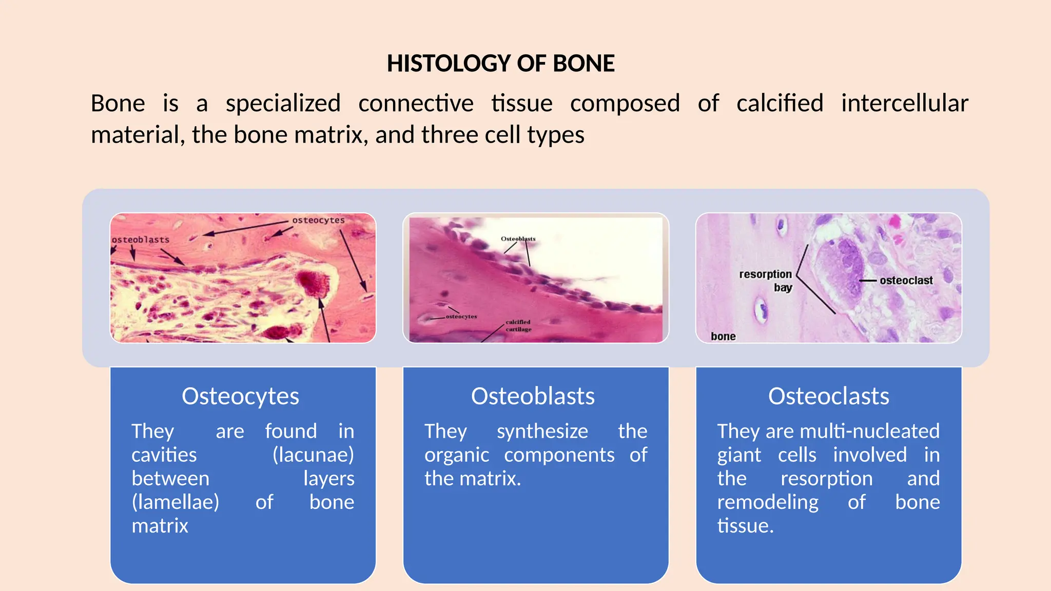
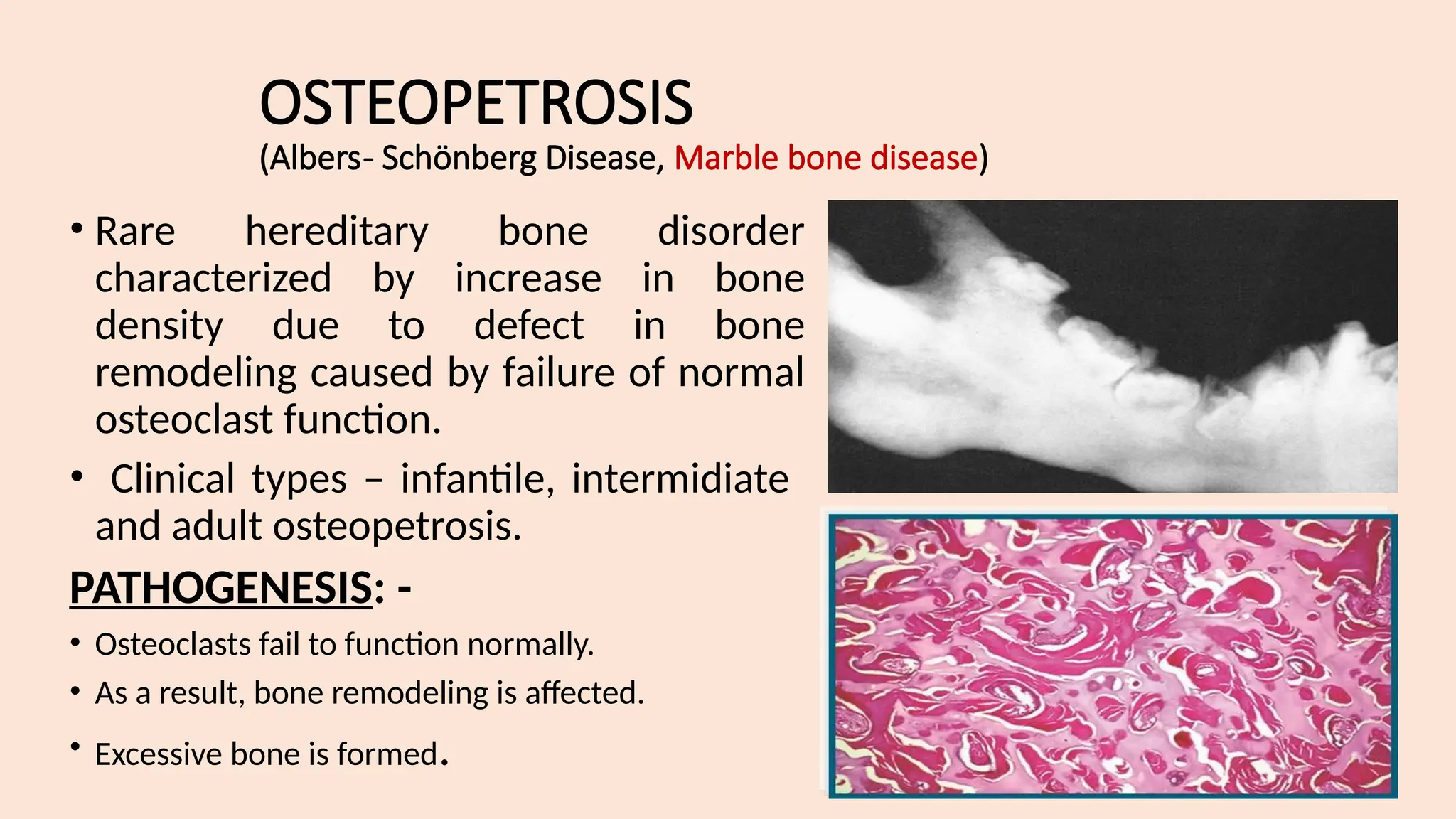
Disease of Bone & Joints
Lecture by : Dr. Diana Prem
Senior Lecturer
Department of Oral Pathology, VMSDC.
Osteocytes
They are found in
cavities (lacunae)
between layers
(lamellae) of bone
matrix
Osteoblasts
They synthesize the
organic components of
the matrix.
Osteoclasts
They are multi-nucleated
giant cells involved in
the resorption and
remodeling of bone
tissue.
Bone is a specialized connective tissue composed of calcified intercellular
material, the bone matrix, and three cell types
HISTOLOGY OF BONE
Bone remodeling
• Bone Remodeling is a continuous process of bone resorption and
bone formation for maintaining normal bone mass.
Reversal line:
A cement line of mineralized matrix delineates the haversian
system
Contains little or no collagen – strongly basophilic  high content
of glycoproteins & proteoglycans
Marks the limit of bone erosion prior to bone formation
Highly irregular  scalloped outline of howship’s lacunae
Resting line
More regular appearance
Denotes the period of rest during formation of bone
Collagen fibres within each lamellae spiral along the length of
lamella but have different orientation to those in adjacent lamellae.
This pattern is to withstand torsion stresses.
Disease Of Bone And Joints
Diseases of skeletal system include infection (osteomyelitis), disordered
growth and development (skeletal dysplasias), metabolic and endocrine
derangements, and tumours and tumour-like
This lecture constitute a group of generalized skeletal diseases which
frequently manifest involvement of the maxilla or mandible conditions.
Should include-
Introduction
Classification
Etiology
Clinical features
Radiographic feature
Oral manifestation
Histological features
Treatment & Prognosis
CLASSIFICATION
A. GENETIC DISEASES OF BONE :
- Cheurbism
- Osteopetrosis
- Osteogenesis imperfecta
- Cleiodocrainal dysplasia
- Achondroplasia
B. ENDOCRINAL DISEASES:
- Hyperparathyroidism, Rickets, Scurvy
C. IDIOPATHIC DISEASES:
- Idiopathic osteosclerosis
- Massive osteolysis
- Langerhan’s cell disease (Histiocytosis-X)
- Paget’s disease
D. REACTIVE DISEASES:
- Giant cell lesion of bone
- Aneurysmal bone cyst
- Simple / Traumatic bone cyst
E. FIBRO-OSSEOUS LESIONS:
(i) Non-neoplastic lesions -
- Fibrous dysplasia
- Cemento-osseous dysplasia
(ii) Neoplasms –
- Ossifying fibroma
F. INFLAMMATORY DISEASES: -
(i) Specific:
- Tuberculosis
- Actinomycosis
(ii) Non specific
- Osteomyelitis
- Dry socket
- Periapical cyst / abscess / granuloma
- Osteoradionecrosis
G. NEOPLASTIC DISEASES: -
(i) Benign:
- Osteoma
- Osteoid osteoma & osteoblastoma
- Chondroma
- Chondromyxoid fibroma
(ii) Malignant:
- Osteosarcoma
- Ewing’s sarcoma
- Chondrosarcoma
FIBRO – OSSEOUS LESIONS
• Fibro-osseous lesions are a diverse group of lesions characterized by
replacement of normal bone by a fibrous tissue containing a newly
formed, mineralized product. It is not a specific diagnosis and
described only as a process.
• These lesions include developmental, reactive and even neoplastic
lesions.
Classification
Fibrous dysplasia
Condition in which normal medullary bone is gradually replaced by an abnormal fibrous
connective tissue proliferation.
1. Hamartomatous
2. Abnormal reaction of bone to a localized traumatic episode.
Types :
Monostotic
• limited to one single
bone, 70%-80%.
• Pain or pathologic
fracture in 10-70yrs.
• Bone deformity less
severe.
• Painless swelling of the
jaw.
Polyostotic
• 20%-30% of fibrous dysplasia.
• Even though skull and jaws
commonly affected, symptoms
are mostly related to
involvement of long bones –
pain, pathological fractures etc.
• Tends to occur in unilateral
distribution.
Craniofacial
• In 10-25% of pt. with
monostotic form.
• In 50% of pt. with polyostotic
form.
• Isolated variety also occurs.
• Hypertelorism, cranial
asymmetry, facial deformity,
visual impairment,
exophthalmos, blindness
Cherubism
• Special variant of of FD,
children - more severe in
boys
• Affects the jaw bilaterally
and symmetrically,
producing so called
cherubic look.
Jaffe’s type
FB involving large
number of bone
Pigmentation of skin
café au lait spots
McCune – Albright syndrome
FB involving large
number of bone.
Pigmentation of skin
café au lait spots.
Endocrine disturbance.
Mazabraud’s syndrome
FB in association with
intramuscular myxoma.
Rare disease.
• Pain in involved limb Spontaneous fracture – structural intergrity is
weak.
• Bowing of weight bearing bones, curvature of femoral neck, proximal
shaft increase - Shepherd’s crook deformity
Polyostotic type
Increased trabeculation –lesion more opaque & mottled
appearance
Opaque with many delicate trabeculae :’ground –glass’
‘or
‘peau d’ orange’ appearance
Cortical bone becomes thinned
Roots of teeth separated or moved out of normal
RADIOGRAPHIC FEATURES
Network of fine bone trabeculae
No significant change iin serum
calcium/phosphorus
Elevated Alkaline phosphatase
LAB FINDING
Moderate increase in Basal
Metabolic Rate
Histopathology features
• Lesion shows typical irregular, shaped trabeculae
of immature woven bone in a cellular, vascular
stroma.
• Theses trabeculae are not connected to each
other.
• They often assume curvilinear shape, which have
been linked to Chinese script writing – C Shaped.
• These trabeculae believed to arise due to
metaplasia and are not bordered by osteoblasts.
• The surrounding stroma is highly cellular and
vascular.
FIBROUS DYSPLASIA
Conservative treatment to prevent deformity.
Management requires a multidisciplinary approach in
polyostotic.
Bisphosphonate therapy may help to improve function,
decrease pain, and lower fracture risk in some patients.
Surgery indicated for confirmatory biopsy, correction of
deformity, prevention of pathologic fracture,
eradication of symptomatic lesions.
TREATMENT
CHERUBISM
 Rare developmental jaw condition, first described by Jones in 1933.
 Jones called it as familial multilocular disease of the jaw.
 Transmitted as an autosomal dominant trait.
 Causes characteristic posterior mandibular swelling due to which the child
appears as a plump cheeked angels called “Cherub” in Renaissance
paintings.
 The jaw lesion remit spontaneously when the child reaches puberty, but
reason for this remission is still unknown.
 The appearance of people with the disorder is caused by a loss of
bone, which the body replaces with excessive amounts of fibrous tissue.
Clinical features
Age incidence: Affected children, are normal at the birth and
are without any clinically or radiographically evident
disease until 14 months to 3 yrs of age.
Sex incidence: males = females
Site predilection:
• Mostly bilateral involvement
• Mandible affected more commonly than maxilla.
• In maxilla, tuberosity region is affected frequently,
resulting in respiratory obstruction and impairment of
vision & hearing.
• The lesion are painless and symmetrical.
• Cervical lymphadenopathy contributes to the patients full
faced appearance, it is said to be caused due to lymphoid
hyperplasia with fibrosis.
Oral manifestations
• Progressive, extensive bone involvement
causes widening and distortion of alveoli.
• As a result, developing teeth displaced, fail to
erupt.
• Numerous dental abnormalities have been
reported, such as agenesis of the 2nd
& 3rd
mandibular molars, displacement of the teeth,
premature exfoliation of the primary teeth,
delayed eruption of the permanent teeth, and
transposition and rotation of the teeth.
• The permanent dentition is often defective.
• In sever cases root resorption occurs.
• It is been connected to NOONAN’S SYNDROME.
Radiological features
• The presence of numerous unerupted teeth and the
destruction of the alveolar bone may displace the teeth,
producing a radiographic appearance referred as FLOATING
TOOTH SYNDROME.
• With adulthood, the cystic areas in the jaws become re-
ossified, which results in irregular patchy sclerosis.
• There is classic but non specific ground glass appearance
because of the small, tightly compressed trabecular pattern.
Histopathology
• Features are similar to giant cell tumors.
• In cherubism, normal bone is partly replaced
by pathologic tissue.
• Under the microscope, it contains numerous
randomly distributed multinucleated giant
cells and vascular spaces within a fibrous
connective tissue stroma.
• An increase in osteoid and newly formed
bone matrix is found in the peripheral region
of the fibrotic stroma in patients above the
age of 20 years.
• Commonest type of fibro-osseous lesions in head and neck
region, occurring in the tooth bearing area.
• Believed to represent some form of reactive process.
• Cemento-osseous dysplasia arises in close approximation to
the periodontal ligament and exhibits histopathological
similarities with the structure. Hence some investigators
have suggested these lesion are of periodontal ligament
origin.
CEMENTO-OSSEOUS DYSPLASIAS
• 30 – 50 years, marked female predilection,
predominantly mandibular anteriors.
• solitary lesion, but mostly as multiple lesions.
• Almost always asymptomatic.
• Teeth associated with the lesions are always vital.
Periapical
cemento-
osseous
dysplasia
• Commonest form, Features intermediate between
those of periapical and florid cemento-osseous
dysplasia.
• Mostly posterior mandible, asymptomatic,
detected during routine radiographic examination.
Focal cemento-
osseous
dysplasia
• Widespread disease affecting greater area of jaw bones.
• Secondary infections commonly occur (low grade
osteomyelitis)
 Affected jaw bone may show expansion.
Florid cemento-
osseous
dysplasia
Radiographic
Lesions are well defined and radiopaque, often
mixed with areas of less well defined mixed
radiopaque-radiolucent regions.
In early stages, lesion shows fibroblastic proliferation
In later stages, the lesion shows increasing deposition of
bone or cementum like material.
In the final stages, the entire lesion may be composed
of dense mineralized tissue.
Paget’s disease
Paget’s disease is a chronic condition of bone
characterized by disorder of the normal bone
remodeling process. Named after the England
Surgeons Sir James Paget.
Characterized by excessive breakdown of bone
tissue followed by abnormal bone formation.
Also known as Osteitis Deformans is a bone
disease unknown cause.
Etiology :
• The cause of Paget’s disease is unknown. Disease may be caused by a
virus.
• The other predisposing factors could be – inflammatory, genetic,
endocrine factors or a slow virus infection, autoimmune, connective
tissue or vascular disorder.
Pathogenesis
Hyper vascular/
Osteolytic phase
Initial phase of
Disorder involves
Bone resorption by
osteoclasts
Intermediate
phase
Osteoclytic activity
+
Osteoblastic activity
Exhaustive
(burn out)
stage
Abnormal matrix
Persists but cellular
Activity is nearly
Absent.
Paget’s disease
Clinical features
Age incidence: Middle aged individuals Sex incidence: Male to female
ratio is 2:1
• Severe bone pain and limitation of movement, especially of joints.
• Affected bones – thickened, enlarged and weak.
Oral manifestations :
• Maxilla affected more than mandible.
• Maxilla – enlargement of middle third of face (leontiasis ossea)
• Nasal obstruction, obliterated sinuses and deviated septum also
occur.
• In dentulous patients spacing of teeth is seen, while edentulous
patients complains of tightness of the dentures.
• Mandible involved rarely – may cause prognathism.
Radiological feature
• Early stage (lytic) -radiolucency
and alteration of trabecular
pattern.
• Late stage (osteoblastic) – patchy
areas of sclerotic bone is formed,
called “cotton wool” appearance.
• Dental radiographs also show the
classical cotton wool appearance.
• Extensive hypercementosis can
be noted.
Histopathology
Lytic phase
• Marked by disordered areas of resorption
• Presence of Increased number of large
Osteoclasts
Mixed Lytic &
Blastic phase
• Harphazard laying of new bone matrix - Formation of Woven
bone
• Small irregular shaped bone fragments – Jigsaw or mosaic
pattern with deeply staining hematoxyphilic reversal lines.
Sclerotic or
burned out phase
• No tendency to form haversian system – bones are hard and
dense.
• Osteoblastic activity diminishes and osteoporotic phase
predominates.
Laboratory findings :
• Abnormally elevated serum alkaline phosphatase level upto 250
Bodansky units (normal – 30 to 40).
• But normal calcium and phosphorous levels).
Treatment :
• It is a chronic and slow growing diease, it is seldom the cause of death.
• In patient with no symptom and less involvement treatment is not
required.
• Bone pain is mostly controlled by anti-inflammatory drugs.
OSTEOGENESIS IMPERFECTA
• It is a condition resulting from abnormality in the type I collagen,
which most commonly manifests as fragility of bones.
Marfan syndrome
• It is a spectrum of disorders caused
by a heritable genetic defect of
connective tissue that has an
autosomal dominant mode of
transmission.
• FBN1 gene on chromosome 15,
bands q15–q23 - fibrillin.
• Abnormalities in this protein cause
a myriad of distinct clinical
problems, of which the
musculoskeletal, cardiac, and
ocular problems predominate.
OSTEOPETROSIS
(Albers- Schönberg Disease, Marble bone disease)
• Rare hereditary bone disorder
characterized by increase in bone
density due to defect in bone
remodeling caused by failure of normal
osteoclast function.
• Clinical types – infantile, intermidiate
and adult osteopetrosis.
PATHOGENESIS: -
• Osteoclasts fail to function normally.
• As a result, bone remodeling is affected.
• Excessive bone is formed.
To know-
• Achondrogenesis
• Hypophosphatasia
• Mucopolysaccharidoses Type I – VII
• Rickets
• Hyperparathyroidism
• Hypoparathyroidism
Mandibulofacial Dysostosis (Treacher Collins-
Franceschetti syndrome)
• The mandibulofacial dysostosis syndrome encom passes a
group of closely related defects of the head and face, often
hereditary or familial in pattern, following an irregular
form of dominant transmission.
• The characteristic faces of the patients have often been
described as being bird like or fish like in nature.
The important clinical manifestations of the disease are:
Antimongoloid palpebral fissures with a coloboma of the outer portion of the lower lids, and
deficiency of the eyelashes (and sometimes the upper lids).
Hypoplasia of the facial bones, especially of the malar bones and mandible.
Malformation of the external ear, and occasionally of the middle and internal ears.
Macrostomia, high palate (sometimes cleft) and abnormal position and malocclusion of the
teeth.
Blind fistulas between the angles of the ears and the angles of the mouth.
Pierre Robin Malformation / Syndrome
• Three essential components which include:
Micrognathia or retrognathia
Cleft palate
Glossoptosis, often accompanied by airway obstruction.
Apert Syndrome (Acrocephalosyndactyly)
• It is a rare autosomal dominant disorder characterized by
craniosynostosis, craniofacial anomalies, and severe
symmetrical syndactyly (cutaneous and bony fusion) of the
hands and feet.
Oral manifestation :
• The jaw shows a prominent mandible and maxillary
hypoplasia
• drooping angles of the mouth
• high arched palate, bifid uvula, cleft palate
• crowded upper teeth, malocclusion, delayed and ectopic
eruption
• shovel-shaped incisors, supernumerary teeth
• V shaped maxillary dental arch, and bulging alveolar ridges.
Cleidocranial Dysplasia
• A congenital disorder of bone formation manifested with clavicular
hypoplasia or agenesis with a narrow thorax, which allows approximation
of the shoulders in front of the chest.
Oral manifestation
High, narrow, arched palate, and actual
cleft palate
Underdeveloped maxilla, enlarged
mandibles
Lacrimal and zygomatic bones are also
reported to be underdeveloped.
Prolonged retention of the deciduous
teeth and subsequent delay in eruption
of the succedaneous teeth.
Down Syndrome (Down’s syndrome, trisomy 21
syndrome, mongolism, congenital acromicria syndrome)
• Down syndrome is a frequent form of mental retardation associated with characteristic
morphologic features (mongolism
• Three cytogenetic variants cause Down syndrome: trisomy 21, chromosomal
translocation, and mosaicism. Trisomy 21 accounts for nearly 95% of all patients with
Down syndrome.
Oral manifestations
Small mouth, Macroglossia, scrotal tongue
(80%)
Hypoplasia of the maxilla, delayed tooth
eruption, partial anodontia, enamel hypoplasia
Juvenile periodontitis and
Cleft lip or palate (rare) are noticed commonly.
Fissuring and thickening of the lips and angular
cheilitis
LANGERHANS CELL DISEASE
• The term HISTOCYTOSIS was introduce as a collective designation for
a spectrum of clinicopathologic disorders characterized by
proliferation of histiocytes-like cells.
• It is an idiopathic disease characterized by proliferation of histiocyte
like cells (Langerhan's cells), that are accompanied by varying
numbers of eosinophils, lymphocytes, plasma cells & multinucleated
giant cell.
• Believed to be a non neoplastic process.
Types
acute disseminated
disease with bone,
visceral and skin
involvement, occurring
mainly in infants.
Letterer-Siwe disease:
Chronic disseminated
disease involving
bones, viscera and
skin. It shows triad of
lytic skull lesion,
exophthalmous, and
diabetes insipidus.
Hand-Schüller-
Christian disease:
solitary / multiple bone
involvement without
systemic organ
involvement. It causes
localized bone destruction
with swelling and often
pain.
Eosinophilic
granuloma of bone:
To know-
• Craniofacial dysostosis
• Acgondroplasia
• Chondroectodermanl dysplasia
• Infantile cortical hyperostosis
• Massive osteolysis
• Cementoblastoma
Disease Temporomandibular joint
• TMJs can occur either before or after birth.
Developmental disturbances as follows:
1. Hypoplasia or aplasia of the condyle
a. Congenital or primary hypoplasia or aplasia
b. Acquired or secondary hypoplasia or aplasia
2. Condylar hyperplasia
3. Bifid condyle.
Traumatic Disturbances Of Temporomandibular Joint
• Dislocation of the Condyles
• Ankylosis (Hypomobility)
• Injuries of Articular Disk: Internal Derangement
Inflammatory Disturbances Of Temporomandibular Joint
• Arthritis or inflammation of the joints is one of the most
prevalent chronic diseases. Temporomandibular joint may
suffer from any form of arthritis.
• Following three are the most common types of arthritis of
TMJ:
• Osteoarthritis, or degenerative joint disease
• Rheumatoid arthritis
• Septic arthritis.
BONE PATHOLOGY / DISEASE OD BONE & JOINTS.pptx

BONE PATHOLOGY / DISEASE OD BONE & JOINTS.pptx

  • 1.
    Disease of Bone& Joints Lecture by : Dr. Diana Prem Senior Lecturer Department of Oral Pathology, VMSDC.
  • 2.
    Osteocytes They are foundin cavities (lacunae) between layers (lamellae) of bone matrix Osteoblasts They synthesize the organic components of the matrix. Osteoclasts They are multi-nucleated giant cells involved in the resorption and remodeling of bone tissue. Bone is a specialized connective tissue composed of calcified intercellular material, the bone matrix, and three cell types HISTOLOGY OF BONE
  • 3.
    Bone remodeling • BoneRemodeling is a continuous process of bone resorption and bone formation for maintaining normal bone mass. Reversal line: A cement line of mineralized matrix delineates the haversian system Contains little or no collagen – strongly basophilic  high content of glycoproteins & proteoglycans Marks the limit of bone erosion prior to bone formation Highly irregular  scalloped outline of howship’s lacunae Resting line More regular appearance Denotes the period of rest during formation of bone Collagen fibres within each lamellae spiral along the length of lamella but have different orientation to those in adjacent lamellae. This pattern is to withstand torsion stresses.
  • 4.
    Disease Of BoneAnd Joints Diseases of skeletal system include infection (osteomyelitis), disordered growth and development (skeletal dysplasias), metabolic and endocrine derangements, and tumours and tumour-like This lecture constitute a group of generalized skeletal diseases which frequently manifest involvement of the maxilla or mandible conditions.
  • 5.
    Should include- Introduction Classification Etiology Clinical features Radiographicfeature Oral manifestation Histological features Treatment & Prognosis
  • 6.
  • 7.
    A. GENETIC DISEASESOF BONE : - Cheurbism - Osteopetrosis - Osteogenesis imperfecta - Cleiodocrainal dysplasia - Achondroplasia B. ENDOCRINAL DISEASES: - Hyperparathyroidism, Rickets, Scurvy C. IDIOPATHIC DISEASES: - Idiopathic osteosclerosis - Massive osteolysis - Langerhan’s cell disease (Histiocytosis-X) - Paget’s disease
  • 8.
    D. REACTIVE DISEASES: -Giant cell lesion of bone - Aneurysmal bone cyst - Simple / Traumatic bone cyst E. FIBRO-OSSEOUS LESIONS: (i) Non-neoplastic lesions - - Fibrous dysplasia - Cemento-osseous dysplasia (ii) Neoplasms – - Ossifying fibroma
  • 9.
    F. INFLAMMATORY DISEASES:- (i) Specific: - Tuberculosis - Actinomycosis (ii) Non specific - Osteomyelitis - Dry socket - Periapical cyst / abscess / granuloma - Osteoradionecrosis
  • 10.
    G. NEOPLASTIC DISEASES:- (i) Benign: - Osteoma - Osteoid osteoma & osteoblastoma - Chondroma - Chondromyxoid fibroma (ii) Malignant: - Osteosarcoma - Ewing’s sarcoma - Chondrosarcoma
  • 11.
    FIBRO – OSSEOUSLESIONS • Fibro-osseous lesions are a diverse group of lesions characterized by replacement of normal bone by a fibrous tissue containing a newly formed, mineralized product. It is not a specific diagnosis and described only as a process. • These lesions include developmental, reactive and even neoplastic lesions.
  • 12.
  • 14.
    Fibrous dysplasia Condition inwhich normal medullary bone is gradually replaced by an abnormal fibrous connective tissue proliferation. 1. Hamartomatous 2. Abnormal reaction of bone to a localized traumatic episode. Types : Monostotic • limited to one single bone, 70%-80%. • Pain or pathologic fracture in 10-70yrs. • Bone deformity less severe. • Painless swelling of the jaw. Polyostotic • 20%-30% of fibrous dysplasia. • Even though skull and jaws commonly affected, symptoms are mostly related to involvement of long bones – pain, pathological fractures etc. • Tends to occur in unilateral distribution. Craniofacial • In 10-25% of pt. with monostotic form. • In 50% of pt. with polyostotic form. • Isolated variety also occurs. • Hypertelorism, cranial asymmetry, facial deformity, visual impairment, exophthalmos, blindness Cherubism • Special variant of of FD, children - more severe in boys • Affects the jaw bilaterally and symmetrically, producing so called cherubic look.
  • 15.
    Jaffe’s type FB involvinglarge number of bone Pigmentation of skin café au lait spots McCune – Albright syndrome FB involving large number of bone. Pigmentation of skin café au lait spots. Endocrine disturbance. Mazabraud’s syndrome FB in association with intramuscular myxoma. Rare disease. • Pain in involved limb Spontaneous fracture – structural intergrity is weak. • Bowing of weight bearing bones, curvature of femoral neck, proximal shaft increase - Shepherd’s crook deformity Polyostotic type
  • 16.
    Increased trabeculation –lesionmore opaque & mottled appearance Opaque with many delicate trabeculae :’ground –glass’ ‘or ‘peau d’ orange’ appearance Cortical bone becomes thinned Roots of teeth separated or moved out of normal RADIOGRAPHIC FEATURES Network of fine bone trabeculae
  • 17.
    No significant changeiin serum calcium/phosphorus Elevated Alkaline phosphatase LAB FINDING Moderate increase in Basal Metabolic Rate
  • 18.
    Histopathology features • Lesionshows typical irregular, shaped trabeculae of immature woven bone in a cellular, vascular stroma. • Theses trabeculae are not connected to each other. • They often assume curvilinear shape, which have been linked to Chinese script writing – C Shaped. • These trabeculae believed to arise due to metaplasia and are not bordered by osteoblasts. • The surrounding stroma is highly cellular and vascular.
  • 19.
  • 20.
    Conservative treatment toprevent deformity. Management requires a multidisciplinary approach in polyostotic. Bisphosphonate therapy may help to improve function, decrease pain, and lower fracture risk in some patients. Surgery indicated for confirmatory biopsy, correction of deformity, prevention of pathologic fracture, eradication of symptomatic lesions. TREATMENT
  • 21.
    CHERUBISM  Rare developmentaljaw condition, first described by Jones in 1933.  Jones called it as familial multilocular disease of the jaw.  Transmitted as an autosomal dominant trait.  Causes characteristic posterior mandibular swelling due to which the child appears as a plump cheeked angels called “Cherub” in Renaissance paintings.  The jaw lesion remit spontaneously when the child reaches puberty, but reason for this remission is still unknown.  The appearance of people with the disorder is caused by a loss of bone, which the body replaces with excessive amounts of fibrous tissue.
  • 22.
    Clinical features Age incidence:Affected children, are normal at the birth and are without any clinically or radiographically evident disease until 14 months to 3 yrs of age. Sex incidence: males = females Site predilection: • Mostly bilateral involvement • Mandible affected more commonly than maxilla. • In maxilla, tuberosity region is affected frequently, resulting in respiratory obstruction and impairment of vision & hearing. • The lesion are painless and symmetrical. • Cervical lymphadenopathy contributes to the patients full faced appearance, it is said to be caused due to lymphoid hyperplasia with fibrosis.
  • 23.
    Oral manifestations • Progressive,extensive bone involvement causes widening and distortion of alveoli. • As a result, developing teeth displaced, fail to erupt. • Numerous dental abnormalities have been reported, such as agenesis of the 2nd & 3rd mandibular molars, displacement of the teeth, premature exfoliation of the primary teeth, delayed eruption of the permanent teeth, and transposition and rotation of the teeth. • The permanent dentition is often defective. • In sever cases root resorption occurs. • It is been connected to NOONAN’S SYNDROME. Radiological features • The presence of numerous unerupted teeth and the destruction of the alveolar bone may displace the teeth, producing a radiographic appearance referred as FLOATING TOOTH SYNDROME. • With adulthood, the cystic areas in the jaws become re- ossified, which results in irregular patchy sclerosis. • There is classic but non specific ground glass appearance because of the small, tightly compressed trabecular pattern.
  • 24.
    Histopathology • Features aresimilar to giant cell tumors. • In cherubism, normal bone is partly replaced by pathologic tissue. • Under the microscope, it contains numerous randomly distributed multinucleated giant cells and vascular spaces within a fibrous connective tissue stroma. • An increase in osteoid and newly formed bone matrix is found in the peripheral region of the fibrotic stroma in patients above the age of 20 years.
  • 25.
    • Commonest typeof fibro-osseous lesions in head and neck region, occurring in the tooth bearing area. • Believed to represent some form of reactive process. • Cemento-osseous dysplasia arises in close approximation to the periodontal ligament and exhibits histopathological similarities with the structure. Hence some investigators have suggested these lesion are of periodontal ligament origin. CEMENTO-OSSEOUS DYSPLASIAS
  • 26.
    • 30 –50 years, marked female predilection, predominantly mandibular anteriors. • solitary lesion, but mostly as multiple lesions. • Almost always asymptomatic. • Teeth associated with the lesions are always vital. Periapical cemento- osseous dysplasia • Commonest form, Features intermediate between those of periapical and florid cemento-osseous dysplasia. • Mostly posterior mandible, asymptomatic, detected during routine radiographic examination. Focal cemento- osseous dysplasia • Widespread disease affecting greater area of jaw bones. • Secondary infections commonly occur (low grade osteomyelitis)  Affected jaw bone may show expansion. Florid cemento- osseous dysplasia
  • 27.
    Radiographic Lesions are welldefined and radiopaque, often mixed with areas of less well defined mixed radiopaque-radiolucent regions. In early stages, lesion shows fibroblastic proliferation In later stages, the lesion shows increasing deposition of bone or cementum like material. In the final stages, the entire lesion may be composed of dense mineralized tissue.
  • 28.
    Paget’s disease Paget’s diseaseis a chronic condition of bone characterized by disorder of the normal bone remodeling process. Named after the England Surgeons Sir James Paget. Characterized by excessive breakdown of bone tissue followed by abnormal bone formation. Also known as Osteitis Deformans is a bone disease unknown cause.
  • 29.
    Etiology : • Thecause of Paget’s disease is unknown. Disease may be caused by a virus. • The other predisposing factors could be – inflammatory, genetic, endocrine factors or a slow virus infection, autoimmune, connective tissue or vascular disorder.
  • 30.
    Pathogenesis Hyper vascular/ Osteolytic phase Initialphase of Disorder involves Bone resorption by osteoclasts Intermediate phase Osteoclytic activity + Osteoblastic activity Exhaustive (burn out) stage Abnormal matrix Persists but cellular Activity is nearly Absent. Paget’s disease
  • 31.
    Clinical features Age incidence:Middle aged individuals Sex incidence: Male to female ratio is 2:1 • Severe bone pain and limitation of movement, especially of joints. • Affected bones – thickened, enlarged and weak. Oral manifestations : • Maxilla affected more than mandible. • Maxilla – enlargement of middle third of face (leontiasis ossea) • Nasal obstruction, obliterated sinuses and deviated septum also occur. • In dentulous patients spacing of teeth is seen, while edentulous patients complains of tightness of the dentures. • Mandible involved rarely – may cause prognathism.
  • 32.
    Radiological feature • Earlystage (lytic) -radiolucency and alteration of trabecular pattern. • Late stage (osteoblastic) – patchy areas of sclerotic bone is formed, called “cotton wool” appearance. • Dental radiographs also show the classical cotton wool appearance. • Extensive hypercementosis can be noted.
  • 33.
    Histopathology Lytic phase • Markedby disordered areas of resorption • Presence of Increased number of large Osteoclasts Mixed Lytic & Blastic phase • Harphazard laying of new bone matrix - Formation of Woven bone • Small irregular shaped bone fragments – Jigsaw or mosaic pattern with deeply staining hematoxyphilic reversal lines. Sclerotic or burned out phase • No tendency to form haversian system – bones are hard and dense. • Osteoblastic activity diminishes and osteoporotic phase predominates.
  • 34.
    Laboratory findings : •Abnormally elevated serum alkaline phosphatase level upto 250 Bodansky units (normal – 30 to 40). • But normal calcium and phosphorous levels). Treatment : • It is a chronic and slow growing diease, it is seldom the cause of death. • In patient with no symptom and less involvement treatment is not required. • Bone pain is mostly controlled by anti-inflammatory drugs.
  • 35.
    OSTEOGENESIS IMPERFECTA • Itis a condition resulting from abnormality in the type I collagen, which most commonly manifests as fragility of bones.
  • 36.
    Marfan syndrome • Itis a spectrum of disorders caused by a heritable genetic defect of connective tissue that has an autosomal dominant mode of transmission. • FBN1 gene on chromosome 15, bands q15–q23 - fibrillin. • Abnormalities in this protein cause a myriad of distinct clinical problems, of which the musculoskeletal, cardiac, and ocular problems predominate.
  • 37.
    OSTEOPETROSIS (Albers- Schönberg Disease,Marble bone disease) • Rare hereditary bone disorder characterized by increase in bone density due to defect in bone remodeling caused by failure of normal osteoclast function. • Clinical types – infantile, intermidiate and adult osteopetrosis. PATHOGENESIS: - • Osteoclasts fail to function normally. • As a result, bone remodeling is affected. • Excessive bone is formed.
  • 38.
    To know- • Achondrogenesis •Hypophosphatasia • Mucopolysaccharidoses Type I – VII • Rickets • Hyperparathyroidism • Hypoparathyroidism
  • 39.
    Mandibulofacial Dysostosis (TreacherCollins- Franceschetti syndrome) • The mandibulofacial dysostosis syndrome encom passes a group of closely related defects of the head and face, often hereditary or familial in pattern, following an irregular form of dominant transmission. • The characteristic faces of the patients have often been described as being bird like or fish like in nature. The important clinical manifestations of the disease are: Antimongoloid palpebral fissures with a coloboma of the outer portion of the lower lids, and deficiency of the eyelashes (and sometimes the upper lids). Hypoplasia of the facial bones, especially of the malar bones and mandible. Malformation of the external ear, and occasionally of the middle and internal ears. Macrostomia, high palate (sometimes cleft) and abnormal position and malocclusion of the teeth. Blind fistulas between the angles of the ears and the angles of the mouth.
  • 40.
    Pierre Robin Malformation/ Syndrome • Three essential components which include: Micrognathia or retrognathia Cleft palate Glossoptosis, often accompanied by airway obstruction.
  • 41.
    Apert Syndrome (Acrocephalosyndactyly) •It is a rare autosomal dominant disorder characterized by craniosynostosis, craniofacial anomalies, and severe symmetrical syndactyly (cutaneous and bony fusion) of the hands and feet. Oral manifestation : • The jaw shows a prominent mandible and maxillary hypoplasia • drooping angles of the mouth • high arched palate, bifid uvula, cleft palate • crowded upper teeth, malocclusion, delayed and ectopic eruption • shovel-shaped incisors, supernumerary teeth • V shaped maxillary dental arch, and bulging alveolar ridges.
  • 42.
    Cleidocranial Dysplasia • Acongenital disorder of bone formation manifested with clavicular hypoplasia or agenesis with a narrow thorax, which allows approximation of the shoulders in front of the chest. Oral manifestation High, narrow, arched palate, and actual cleft palate Underdeveloped maxilla, enlarged mandibles Lacrimal and zygomatic bones are also reported to be underdeveloped. Prolonged retention of the deciduous teeth and subsequent delay in eruption of the succedaneous teeth.
  • 43.
    Down Syndrome (Down’ssyndrome, trisomy 21 syndrome, mongolism, congenital acromicria syndrome) • Down syndrome is a frequent form of mental retardation associated with characteristic morphologic features (mongolism • Three cytogenetic variants cause Down syndrome: trisomy 21, chromosomal translocation, and mosaicism. Trisomy 21 accounts for nearly 95% of all patients with Down syndrome. Oral manifestations Small mouth, Macroglossia, scrotal tongue (80%) Hypoplasia of the maxilla, delayed tooth eruption, partial anodontia, enamel hypoplasia Juvenile periodontitis and Cleft lip or palate (rare) are noticed commonly. Fissuring and thickening of the lips and angular cheilitis
  • 44.
    LANGERHANS CELL DISEASE •The term HISTOCYTOSIS was introduce as a collective designation for a spectrum of clinicopathologic disorders characterized by proliferation of histiocytes-like cells. • It is an idiopathic disease characterized by proliferation of histiocyte like cells (Langerhan's cells), that are accompanied by varying numbers of eosinophils, lymphocytes, plasma cells & multinucleated giant cell. • Believed to be a non neoplastic process.
  • 45.
    Types acute disseminated disease withbone, visceral and skin involvement, occurring mainly in infants. Letterer-Siwe disease: Chronic disseminated disease involving bones, viscera and skin. It shows triad of lytic skull lesion, exophthalmous, and diabetes insipidus. Hand-Schüller- Christian disease: solitary / multiple bone involvement without systemic organ involvement. It causes localized bone destruction with swelling and often pain. Eosinophilic granuloma of bone:
  • 46.
    To know- • Craniofacialdysostosis • Acgondroplasia • Chondroectodermanl dysplasia • Infantile cortical hyperostosis • Massive osteolysis • Cementoblastoma
  • 47.
    Disease Temporomandibular joint •TMJs can occur either before or after birth. Developmental disturbances as follows: 1. Hypoplasia or aplasia of the condyle a. Congenital or primary hypoplasia or aplasia b. Acquired or secondary hypoplasia or aplasia 2. Condylar hyperplasia 3. Bifid condyle. Traumatic Disturbances Of Temporomandibular Joint • Dislocation of the Condyles • Ankylosis (Hypomobility) • Injuries of Articular Disk: Internal Derangement
  • 48.
    Inflammatory Disturbances OfTemporomandibular Joint • Arthritis or inflammation of the joints is one of the most prevalent chronic diseases. Temporomandibular joint may suffer from any form of arthritis. • Following three are the most common types of arthritis of TMJ: • Osteoarthritis, or degenerative joint disease • Rheumatoid arthritis • Septic arthritis.