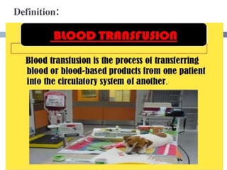
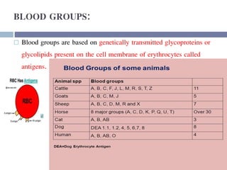
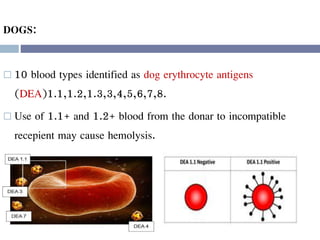
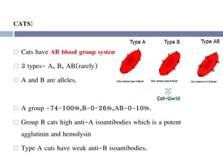
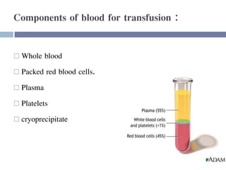
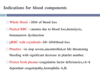
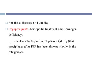
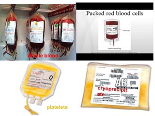
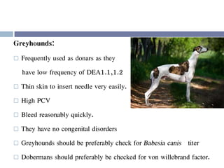
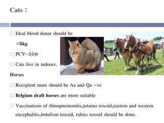
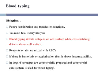
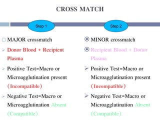
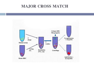
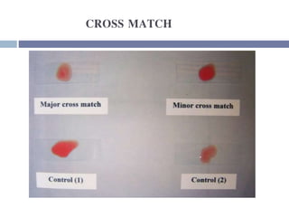
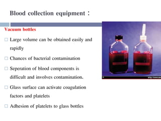
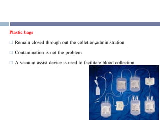
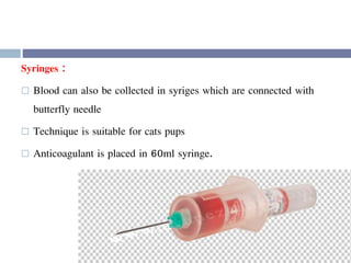
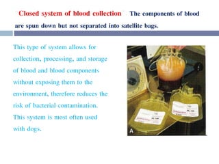
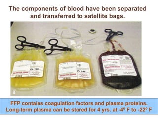
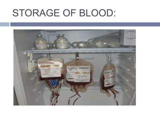
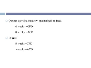
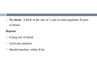
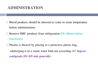
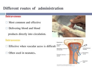
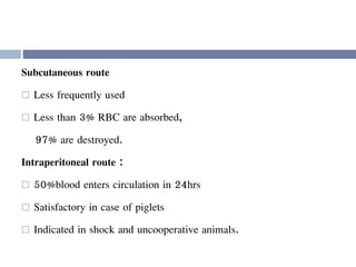
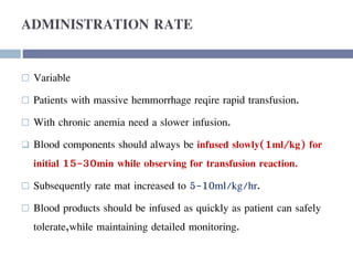
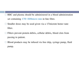
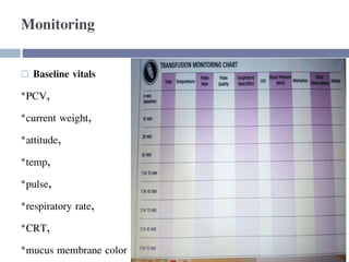
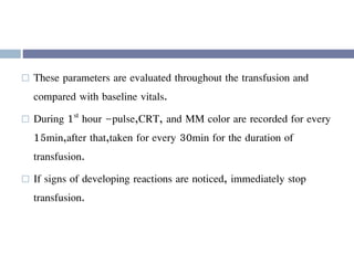
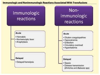
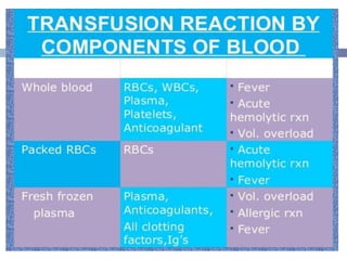
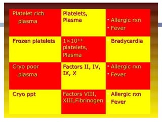
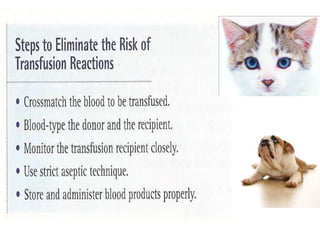
HYPOVOLEMIA
&
BLOOD TRANSFUSION
Y.D.HUSSAINI,
GVM/2018-22,
DEPT.OF VEYTERINARY
GYNAECOLOGY &OBSTETRICS.
 Hypovolemia is a state of decreased blood volume
 more specifically, decrease in volume of blood plasma.
 It is thus the intravascular component of volume contraction
(Or loss of blood volume due to things such as bleeding or
dehydration)
 hypovolemia and volume contraction are sometimes used
synonymously.
 Causes
 Common causes of hypovolemia are:
 Loss of blood (external or internal bleeding or blood donation)
 Loss of plasma (severe burns and lesions discharging fluid)
 Loss of body sodium and consequent intravascular water;
e.g. diarrhea or vomiting
 Signs and symptoms
 Symptoms may not be present until 10–20% of total whole-
blood volume is lost.
 fatigue, nausea, profuse sweating, dizziness may occur as the
condition develops.
 Diagnosis
 Hypovolemia can be recognized by tachycardia,
 diminished blood pressure,
 absence of perfusion as assessed by skin signs (skin turning pale)
and/or capillary refill on forehead, lips and nail beds.
 The patient may feel dizzy, faint, nauseated, or very thirsty.
These signs are also characteristic of most types of shock.
Usually referred to as a "class" of shock.
there are 4 stages of hypovolemic shock
Stage 1 Stage 2 Stage 3 Stage 4
Blood loss Up to 15% (750 mL) 15–30% (750–1500 mL) 30–40% (1500–2000 mL)
Over 40% (over 2000
mL)
Blood pressure
Normal (Maintained
by vasoconstriction)
Increased diastolic BP Systolic BP < 100 Systolic BP < 70
Heart rate Normal
Slight tachycardia (> 100
bpm)
Tachycardia (> 120 bpm)
Extreme tachycardia (>
140 bpm) with weak
pulse
Respiratory rate Normal Increased (> 20) Tachypneic (> 30) Extreme tachypnea
Mental status Normal Slight anxiety, restless Altered, confused
Decreased LOC, lethargy,
coma
Skin Pallor Pale, cool, clammy Increased diaphoresis
Extreme diaphoresis;
mottling possible
Capillary refill Normal Delayed Delayed Absent
Urine output Normal 20–30 mL/h 20 mL/h Negligible
 Treatment
 Fluid replacement is beneficial in hypovolemia of stage 2,
and is necessary in stage 3 and 4.
 The following interventions are carried out:
 IV access
 Oxygen as required
 Fresh frozen plasma or blood transfusion
 Surgical repair at sites of bleeding
 Vasopressors (like (dopamine and noradrenaline) should generally
be avoided, as they may result in further tissue ischemia and don't
correct the primary problem.
 Fluids are the preferred choice of therapy
BLOOD TRANSFUSION IN
ANIMALS
Definition:
INTRODUCTION:
 Indications in animals
 Animal blood groups
 different components of blood for transfusion
 selection of donar
 Blood typing
 Cross match
 Canine and feline collection
 Administration
 Reactions of transfusion
Indications :
 Acute blood loss
 Deficiency of blood constituents.
 Decreased carrying capacity of blood.
 Precautionary measure for surgery.
 Auto immune hemolytic anemia.
 Disseminated intravascular coagulopathy.
Cattle :
 Pulmonary hemorrhage
 Abomasal ulcer
 Enzootic hematuria with bladder bleeding
 Rupture of mid uterine artery in prolapse.
 Very heavy infestation of hookworms
 Thieleriasis. anaplasmosis ,babesiosis.
Horses :
 PCV with 12%
 Hb concentrations >8g/dl
 Traumatic injury
 Hemophilia
 Heavy infestation with strongyles
BLOOD GROUPS:
 Blood groups are based on genetically transmitted glycoproteins or
glycolipids present on the cell membrane of erythrocytes called
antigens.
DOGS:
 10 blood types identified as dog erythrocyte antigens
(DEA)1.1,1.2,1.3,3,4,5,6,7,8.
 Use of 1.1+ and 1.2+ blood from the donar to incompatible
recepient may cause hemolysis.
CATS:
 Cats have AB blood group system
 3 types- A, B, AB(rarely)
 A and B are alleles.
 A group -74-100%,B-0-26%,AB-0-10%.
 Group B cats high anti-A isoantibodies which is a potent
agglutinin and hemolysin
 Type A cats have weak anti-B isoantibodies.
HORSES:
 There are 8 recognised blood groups in horses
A,C,D,K,P,Q,T, and U.
BOVINES:
 12 major blood groups
 Variation in each group lead to 60 blood groups.
Components of blood for transfusion :
 Whole blood
 Packed red blood cells.
 Plasma
 Platelets
 cryoprecipitate
Indications for blood components
 Whole blood –30% of blood loss
 Packed RBC —anemia due to blood loss,hemolysis,
bonemarrow dysfunction
 pRBC with crytalloids-30-50%blood loss
 Platelets –to stop severe,uncontrolled,or life threatening
bleeding with significant decrease in platelet number.
 Frozen fresh plasma-coagulation factor deficiencies,vit-k
dependant coagulopathy,hemophilia A,B.
 For these diseases 6-10ml/kg
 Cryoprecipitate-hemophilia treatment and fibrinogen
deficiency.
It is cold insoluble portion of plasma (slushy)that
precipitates after FFP has been thawed slowly in the
refrigerator.
platelets
Whole blood
Frozen fresh plasma
DONAR SELECTION:
 Minimize risk of transfusion reaction.
 To avoid blood transmissible diseases.
Characteristics :
 Adult animal with good body condition and body size.
 No history of blood transfusion
 No history of blood based vaccines.
 Preferably a male,if female,nulliparous animal.
 Female should preferably be spayed.
 Genetically related or of same breed.
 Free from blood transmitted diseases.
DOGS:
 Co operative temperment,short hair coat, not be obese.
 Ideal blood donar should be above 25kg and able to
donate450ml of blood at one time after 3 weeks inteval.
 Age-1-8years
 Dog PCV-40%
 Must free from brucellosis, ehrlichiosis, microfilaria,
trypanosomiasis.
 Donar should be preferably –ve for DEA1.1,1.2 and 7
 (40%dogs are –ve for DEA1.1,1.2.)(60% +ve)
 Universal recipient in dogs: DEA 1.1 +ve
 Universal donor: DEA 1.1,DEA 1.2 -ve
 Dogs +ve for DEA 4 and –ve for
all others are Best as universal donars.
Greyhounds:
 Frequently used as donars as they
have low frequency of DEA1.1,1.2
 Thin skin to insert needle very easily.
 High PCV
 Bleed reasonably quickly.
 They have no congenital disorders
 Greyhounds should be preferably check for Babesia canis titer
 Dobermans should preferably be checked for von willebrand factor.
Hemolytic reactions are unlikely to occur after first transfusion
even after –ve DEA1.1,1.2 recepient is given blood from
+ve 1.1,1.2.
But the animals would be sensitized and there would be
immediate hemolysis following second transfusion of
1.1,1.2 +ve donar.
Cats :
 Ideal blood donar should be
>5kg
 PCV-35%
 Cats live in indoors.
Horses
 Recepient mare should be Aa and Qa –ve
 Belgium draft horses are more suitable
 Vaccinations of rhinopnemonitis,tetanus toxoid,eastern and western
encephalitis,botulism toxoid, rabies toxoid should be done.
 Alloantigens Aa of the A system and Qa of the Q system
are the most immunogenic of the equine alloantigens and
are highly prevalent among light breeds.
 Transfusion of blood from a donar +ve with these
alloantigens will result in the development of high
alloantibody titer in the recepient that can cause severe
hemolysis upon subsequent exposure
 Donars lacking these alloantigens are considered as safe
blood donars.
Blood typing
Objectives :
 Future sensitization and transfusion reactions.
 To avoid fetal isoerythrolysis
 Blood typing detects antigens on cell surface while crossmatching
detects abs on cell surface.
 Reagents or abs are mixed with RBCs
 If there is hemolysis or agglutination then it shows incompatability.
 In dogs 6 serotypes are commercially prepared and commercial
card system is used for blood typing.
CROSS MATCH
 MAJOR crossmatch
 Donor Blood + Recipient
Plasma
 Positive Test=Macro or
Microagglutination present
(Incompatible)
 Negative Test=Macro or
Microagglutination Absent
(Compatible)
 MINOR crossmatch
 Recipient Blood + Donor
Plasma
 Positive Test=Macro or
Microagglutination present
(Incompatible)
 Negative Test=Macro or
Microagglutination Absent
(Compatible)
Step 1 Step 2
MAJOR CROSS MATCH
CROSS MATCH
Blood collection equipment :
Vacuum bottles
 Large volume can be obtained easily and
rapidly
 Chances of bacterial contamination
 Seperation of blood components is
difficult and involves contamination.
 Glass surface can activate coagulation
factors and platelets
 Adhesion of platelets to glass bottles
Plastic bags
 Remain closed through out the colletion,administration
 Contamination is not the problem
 A vacuum assist device is used to facilitate blood collection
Syringes :
 Blood can also be collected in syriges which are connected with
butterfly needle
 Technique is suitable for cats pups
 Anticoagulant is placed in 60ml syringe.
CANINE BLOOD COLLECTION
 Physical examination and PCV prior to each donation.
 Donar in lateral recumbency.
 Fur over the jugular vein is clipped and site is prepared
aseptically.
 16g needle is inserted into jugular vein
 Blood is allowed to flow by gravity in collection bag
 The bag is gently rocked back and forth as it fills, distributing the
anticoagulant evenly.
 Sample is periodically weighed until the proper volume is
achieved.
After the donor dog has been clipped and surgically prepared, the
collection needle is inserted into the jugular vein.
site for withdrawing blood in blood transfusion
As blood is collected, it should be mixed gently in a back and forth
motion as seen above.
 Canine units are usually 450ml or 450g
 Hemostat is clamped below tubing and the needle is
removed from the vein.
 Pressure is applied to jugular vein to prevent hematoma
formation, then light pressure wrap is applied for 30min -
1hr.
 Remaining blood in the line is stripped back into collection
tube and mixed with anti coagulant.
FELINE BLOOD COLLECTION
 Physical examination and PCV performed
before collection.
 Donar in lateral recumbency.
 19g butterfly needle is inserted into jugular
vein and blood aspirated with gentle pressure,
avoiding venous collapse.
 Total volume of 53ml is collected.
 Blood can be immediately transfused or
placed into small collection bag for storage.
Blood collection systems
“Closed system of blood collection”. The components of blood
are spun down but not separated into satellite bags.
This type of system allows for
collection, processing, and storage
of blood and blood components
without exposing them to the
environment, therefore reduces the
risk of bacterial contamination.
This system is most often used
with dogs.
The components of blood have been separated
and transferred to satellite bags.
FFP contains coagulation factors and plasma proteins.
Long-term plasma can be stored for 4 yrs. at -4º F to -22º F
STORAGE OF BLOOD:
Anticoagulants :
 Acid ,citrate,dextrose (ACD) or citrate,phosphate,dextrose (CPD)
are recommended esp.blood products are stored.
 These also provide nutrients for stabilization and metabolism of
RBCs
 14ml of ACD or CPD are used per 100ml of blood.
 Erythrocyte preservation is better in CPD.
 All preservative solutions contain citrate as anticoagulant.
 Oxygen carrying capacity maintained in dogs:
4 weeks –CPD
3 weeks –ACD
 In cats:
5 weeks—CPD
4weeks—ACD
 Na citrate 3.85% at the rate of 1 part of anticoagulantto 9 parts
of blood.
Heparin
 0.2mg/ml of blood
 Acrtivates platelets
 Should transfuse within 8 hrs
ADMINISTRATION
 Blood products should be allowed to come to room temperature
before administration
 Remove RBC products from refrigeration 30-60min before
transfusion.
 Plasma is thawed by placing in a protective plastic bag,
submerging it in a warm water bath not exceeding 37 degrees
centigrade,30-60 min generally.
Different routes of administration
Intravenous
 Most common and effective
 Delivering blood and blood
products directly into circulation.
Intraosseus
 Effective when vascular acess is difficult
 Often used in neanates.
Subcutaneous route
 Less frequently used
 Less than 3% RBC are absorbed,
97% are destroyed.
Intraperitoneal route :
 50%blood enters circulation in 24hrs
 Satisfactory in case of piglets
 Indicated in shock and uncooperative animals.
ADMINISTRATION RATE
 Variable
 Patients with massive hemmorrhage reqire rapid transfusion.
 With chronic anemia need a slower infusion.
 Blood components should always be infused slowly(1ml/kg) for
initial 15-30min while observing for transfusion reaction.
 Subsequently rate mat increased to 5-10ml/kg/hr.
 Blood products should be infused as quickly as patient can safely
tolerate,while maintaining detailed monitoring.
 RBC and plasma should be administered in a blood administration
set containing 170-220micro mm in line filter.
 Smaller doses may be used given via a 17microm hemo-nate
filter.
 Filters prevent protein debris, cellular debris, blood clots from
passing to patient.
 Blood products may be infused via free drip, syringe pump, fluid
pump.
Monitoring
 Baseline vitals
*PCV,
*current weight,
*attitude,
*temp,
*pulse,
*respiratory rate,
*CRT,
*mucus membrane color
 These parameters are evaluated throughout the transfusion and
compared with baseline vitals.
 During 1st hour –pulse,CRT, and MM color are recorded for every
15min,after that,taken for every 30min for the duration of
transfusion.
 If signs of developing reactions are noticed, immediately stop
transfusion.
Blood transfusion
Blood transfusion
Blood transfusion
Blood transfusion
Blood transfusion

Blood transfusion

  • 1.
  • 2.
     Hypovolemia isa state of decreased blood volume  more specifically, decrease in volume of blood plasma.  It is thus the intravascular component of volume contraction (Or loss of blood volume due to things such as bleeding or dehydration)  hypovolemia and volume contraction are sometimes used synonymously.
  • 3.
     Causes  Commoncauses of hypovolemia are:  Loss of blood (external or internal bleeding or blood donation)  Loss of plasma (severe burns and lesions discharging fluid)  Loss of body sodium and consequent intravascular water; e.g. diarrhea or vomiting
  • 4.
     Signs andsymptoms  Symptoms may not be present until 10–20% of total whole- blood volume is lost.  fatigue, nausea, profuse sweating, dizziness may occur as the condition develops.
  • 5.
     Diagnosis  Hypovolemiacan be recognized by tachycardia,  diminished blood pressure,  absence of perfusion as assessed by skin signs (skin turning pale) and/or capillary refill on forehead, lips and nail beds.  The patient may feel dizzy, faint, nauseated, or very thirsty. These signs are also characteristic of most types of shock.
  • 6.
    Usually referred toas a "class" of shock. there are 4 stages of hypovolemic shock Stage 1 Stage 2 Stage 3 Stage 4 Blood loss Up to 15% (750 mL) 15–30% (750–1500 mL) 30–40% (1500–2000 mL) Over 40% (over 2000 mL) Blood pressure Normal (Maintained by vasoconstriction) Increased diastolic BP Systolic BP < 100 Systolic BP < 70 Heart rate Normal Slight tachycardia (> 100 bpm) Tachycardia (> 120 bpm) Extreme tachycardia (> 140 bpm) with weak pulse Respiratory rate Normal Increased (> 20) Tachypneic (> 30) Extreme tachypnea Mental status Normal Slight anxiety, restless Altered, confused Decreased LOC, lethargy, coma Skin Pallor Pale, cool, clammy Increased diaphoresis Extreme diaphoresis; mottling possible Capillary refill Normal Delayed Delayed Absent Urine output Normal 20–30 mL/h 20 mL/h Negligible
  • 7.
     Treatment  Fluidreplacement is beneficial in hypovolemia of stage 2, and is necessary in stage 3 and 4.  The following interventions are carried out:  IV access  Oxygen as required  Fresh frozen plasma or blood transfusion  Surgical repair at sites of bleeding  Vasopressors (like (dopamine and noradrenaline) should generally be avoided, as they may result in further tissue ischemia and don't correct the primary problem.  Fluids are the preferred choice of therapy
  • 8.
  • 9.
  • 10.
    INTRODUCTION:  Indications inanimals  Animal blood groups  different components of blood for transfusion  selection of donar  Blood typing  Cross match  Canine and feline collection  Administration  Reactions of transfusion
  • 11.
    Indications :  Acuteblood loss  Deficiency of blood constituents.  Decreased carrying capacity of blood.  Precautionary measure for surgery.  Auto immune hemolytic anemia.  Disseminated intravascular coagulopathy.
  • 12.
    Cattle :  Pulmonaryhemorrhage  Abomasal ulcer  Enzootic hematuria with bladder bleeding  Rupture of mid uterine artery in prolapse.  Very heavy infestation of hookworms  Thieleriasis. anaplasmosis ,babesiosis.
  • 13.
    Horses :  PCVwith 12%  Hb concentrations >8g/dl  Traumatic injury  Hemophilia  Heavy infestation with strongyles
  • 14.
    BLOOD GROUPS:  Bloodgroups are based on genetically transmitted glycoproteins or glycolipids present on the cell membrane of erythrocytes called antigens.
  • 15.
    DOGS:  10 bloodtypes identified as dog erythrocyte antigens (DEA)1.1,1.2,1.3,3,4,5,6,7,8.  Use of 1.1+ and 1.2+ blood from the donar to incompatible recepient may cause hemolysis.
  • 16.
    CATS:  Cats haveAB blood group system  3 types- A, B, AB(rarely)  A and B are alleles.  A group -74-100%,B-0-26%,AB-0-10%.  Group B cats high anti-A isoantibodies which is a potent agglutinin and hemolysin  Type A cats have weak anti-B isoantibodies.
  • 17.
    HORSES:  There are8 recognised blood groups in horses A,C,D,K,P,Q,T, and U. BOVINES:  12 major blood groups  Variation in each group lead to 60 blood groups.
  • 18.
    Components of bloodfor transfusion :  Whole blood  Packed red blood cells.  Plasma  Platelets  cryoprecipitate
  • 19.
    Indications for bloodcomponents  Whole blood –30% of blood loss  Packed RBC —anemia due to blood loss,hemolysis, bonemarrow dysfunction  pRBC with crytalloids-30-50%blood loss  Platelets –to stop severe,uncontrolled,or life threatening bleeding with significant decrease in platelet number.  Frozen fresh plasma-coagulation factor deficiencies,vit-k dependant coagulopathy,hemophilia A,B.
  • 20.
     For thesediseases 6-10ml/kg  Cryoprecipitate-hemophilia treatment and fibrinogen deficiency. It is cold insoluble portion of plasma (slushy)that precipitates after FFP has been thawed slowly in the refrigerator.
  • 21.
  • 22.
  • 23.
    DONAR SELECTION:  Minimizerisk of transfusion reaction.  To avoid blood transmissible diseases. Characteristics :  Adult animal with good body condition and body size.  No history of blood transfusion  No history of blood based vaccines.  Preferably a male,if female,nulliparous animal.  Female should preferably be spayed.  Genetically related or of same breed.  Free from blood transmitted diseases.
  • 24.
    DOGS:  Co operativetemperment,short hair coat, not be obese.  Ideal blood donar should be above 25kg and able to donate450ml of blood at one time after 3 weeks inteval.  Age-1-8years  Dog PCV-40%
  • 25.
     Must freefrom brucellosis, ehrlichiosis, microfilaria, trypanosomiasis.  Donar should be preferably –ve for DEA1.1,1.2 and 7  (40%dogs are –ve for DEA1.1,1.2.)(60% +ve)  Universal recipient in dogs: DEA 1.1 +ve  Universal donor: DEA 1.1,DEA 1.2 -ve  Dogs +ve for DEA 4 and –ve for all others are Best as universal donars.
  • 26.
    Greyhounds:  Frequently usedas donars as they have low frequency of DEA1.1,1.2  Thin skin to insert needle very easily.  High PCV  Bleed reasonably quickly.  They have no congenital disorders  Greyhounds should be preferably check for Babesia canis titer  Dobermans should preferably be checked for von willebrand factor.
  • 27.
    Hemolytic reactions areunlikely to occur after first transfusion even after –ve DEA1.1,1.2 recepient is given blood from +ve 1.1,1.2. But the animals would be sensitized and there would be immediate hemolysis following second transfusion of 1.1,1.2 +ve donar.
  • 28.
    Cats :  Idealblood donar should be >5kg  PCV-35%  Cats live in indoors. Horses  Recepient mare should be Aa and Qa –ve  Belgium draft horses are more suitable  Vaccinations of rhinopnemonitis,tetanus toxoid,eastern and western encephalitis,botulism toxoid, rabies toxoid should be done.
  • 29.
     Alloantigens Aaof the A system and Qa of the Q system are the most immunogenic of the equine alloantigens and are highly prevalent among light breeds.  Transfusion of blood from a donar +ve with these alloantigens will result in the development of high alloantibody titer in the recepient that can cause severe hemolysis upon subsequent exposure  Donars lacking these alloantigens are considered as safe blood donars.
  • 31.
    Blood typing Objectives : Future sensitization and transfusion reactions.  To avoid fetal isoerythrolysis  Blood typing detects antigens on cell surface while crossmatching detects abs on cell surface.  Reagents or abs are mixed with RBCs  If there is hemolysis or agglutination then it shows incompatability.  In dogs 6 serotypes are commercially prepared and commercial card system is used for blood typing.
  • 32.
    CROSS MATCH  MAJORcrossmatch  Donor Blood + Recipient Plasma  Positive Test=Macro or Microagglutination present (Incompatible)  Negative Test=Macro or Microagglutination Absent (Compatible)  MINOR crossmatch  Recipient Blood + Donor Plasma  Positive Test=Macro or Microagglutination present (Incompatible)  Negative Test=Macro or Microagglutination Absent (Compatible) Step 1 Step 2
  • 33.
  • 34.
  • 35.
    Blood collection equipment: Vacuum bottles  Large volume can be obtained easily and rapidly  Chances of bacterial contamination  Seperation of blood components is difficult and involves contamination.  Glass surface can activate coagulation factors and platelets  Adhesion of platelets to glass bottles
  • 36.
    Plastic bags  Remainclosed through out the colletion,administration  Contamination is not the problem  A vacuum assist device is used to facilitate blood collection
  • 37.
    Syringes :  Bloodcan also be collected in syriges which are connected with butterfly needle  Technique is suitable for cats pups  Anticoagulant is placed in 60ml syringe.
  • 38.
    CANINE BLOOD COLLECTION Physical examination and PCV prior to each donation.  Donar in lateral recumbency.  Fur over the jugular vein is clipped and site is prepared aseptically.  16g needle is inserted into jugular vein  Blood is allowed to flow by gravity in collection bag  The bag is gently rocked back and forth as it fills, distributing the anticoagulant evenly.  Sample is periodically weighed until the proper volume is achieved.
  • 39.
    After the donordog has been clipped and surgically prepared, the collection needle is inserted into the jugular vein. site for withdrawing blood in blood transfusion
  • 40.
    As blood iscollected, it should be mixed gently in a back and forth motion as seen above.
  • 41.
     Canine unitsare usually 450ml or 450g  Hemostat is clamped below tubing and the needle is removed from the vein.  Pressure is applied to jugular vein to prevent hematoma formation, then light pressure wrap is applied for 30min - 1hr.  Remaining blood in the line is stripped back into collection tube and mixed with anti coagulant.
  • 42.
    FELINE BLOOD COLLECTION Physical examination and PCV performed before collection.  Donar in lateral recumbency.  19g butterfly needle is inserted into jugular vein and blood aspirated with gentle pressure, avoiding venous collapse.  Total volume of 53ml is collected.  Blood can be immediately transfused or placed into small collection bag for storage.
  • 43.
  • 44.
    “Closed system ofblood collection”. The components of blood are spun down but not separated into satellite bags. This type of system allows for collection, processing, and storage of blood and blood components without exposing them to the environment, therefore reduces the risk of bacterial contamination. This system is most often used with dogs.
  • 45.
    The components ofblood have been separated and transferred to satellite bags. FFP contains coagulation factors and plasma proteins. Long-term plasma can be stored for 4 yrs. at -4º F to -22º F
  • 47.
  • 48.
    Anticoagulants :  Acid,citrate,dextrose (ACD) or citrate,phosphate,dextrose (CPD) are recommended esp.blood products are stored.  These also provide nutrients for stabilization and metabolism of RBCs  14ml of ACD or CPD are used per 100ml of blood.  Erythrocyte preservation is better in CPD.  All preservative solutions contain citrate as anticoagulant.
  • 49.
     Oxygen carryingcapacity maintained in dogs: 4 weeks –CPD 3 weeks –ACD  In cats: 5 weeks—CPD 4weeks—ACD
  • 50.
     Na citrate3.85% at the rate of 1 part of anticoagulantto 9 parts of blood. Heparin  0.2mg/ml of blood  Acrtivates platelets  Should transfuse within 8 hrs
  • 51.
    ADMINISTRATION  Blood productsshould be allowed to come to room temperature before administration  Remove RBC products from refrigeration 30-60min before transfusion.  Plasma is thawed by placing in a protective plastic bag, submerging it in a warm water bath not exceeding 37 degrees centigrade,30-60 min generally.
  • 53.
    Different routes ofadministration Intravenous  Most common and effective  Delivering blood and blood products directly into circulation. Intraosseus  Effective when vascular acess is difficult  Often used in neanates.
  • 54.
    Subcutaneous route  Lessfrequently used  Less than 3% RBC are absorbed, 97% are destroyed. Intraperitoneal route :  50%blood enters circulation in 24hrs  Satisfactory in case of piglets  Indicated in shock and uncooperative animals.
  • 55.
    ADMINISTRATION RATE  Variable Patients with massive hemmorrhage reqire rapid transfusion.  With chronic anemia need a slower infusion.  Blood components should always be infused slowly(1ml/kg) for initial 15-30min while observing for transfusion reaction.  Subsequently rate mat increased to 5-10ml/kg/hr.  Blood products should be infused as quickly as patient can safely tolerate,while maintaining detailed monitoring.
  • 56.
     RBC andplasma should be administered in a blood administration set containing 170-220micro mm in line filter.  Smaller doses may be used given via a 17microm hemo-nate filter.  Filters prevent protein debris, cellular debris, blood clots from passing to patient.  Blood products may be infused via free drip, syringe pump, fluid pump.
  • 57.
    Monitoring  Baseline vitals *PCV, *currentweight, *attitude, *temp, *pulse, *respiratory rate, *CRT, *mucus membrane color
  • 59.
     These parametersare evaluated throughout the transfusion and compared with baseline vitals.  During 1st hour –pulse,CRT, and MM color are recorded for every 15min,after that,taken for every 30min for the duration of transfusion.  If signs of developing reactions are noticed, immediately stop transfusion.