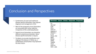
New biomarkers in SLE:
from bench to bedside
By Pankaj Ombase
M.S. ( Pharm)
NIPER, Reabareli.
( lucknow)
Department of Biotechnology 1
Systemic Lupus Erythematosus
• Systemic lupus erythematosus (SLE) is an autoimmune disease. In this disease, the immune
system of the body mistakenly attacks healthy tissue. It can affect the skin, joints, kidneys,
brain, and other organs.
Causes :
• The cause of SLE is not clearly known. It may be linked to the following factors:
a)Genetic
b)Environmental
c)Hormonal
d)Certain medicines
Department of Biotechnology 2
Pathophysiology
Department of Biotechnology 3
Introduction
• Biomarker is termed as ‘a defined characteristic that is measured as an
indicator of normal biological processes, pathogenic processes or responses
to an exposure or intervention
• Novel biomarkers are helpful in early diagnosis and in differential diagnosis
from clinically overlapped disorders, in correct evaluation and staging of
organ involvement, and in predicting and monitoring response to therapies
Department of Biotechnology 4
Complement :
 Complement is a double-edged sword in SLE, protecting from the disease on one hand but
mediating organ damage on the other. Complement activation is increased in active
disease.
Decreased concentration of complement components in blood is strongly associated with
active disease. Detection of C3 and C4 levels in blood is act as biomarker.
Thus, measurement of C3 and C4 levels is not a sensitive tool to study.
The measurement of complement split products represents a powerful tool to get a full
picture of complement status in SLE. The rapid decay of these products has been the main
obstacle to their detection, but novel approaches, including evaluation of cell-bound
complement products, have been proposed to overcome it.
Department of Biotechnology 5
Conti….
• Detection by cytometry of C4d levels on erythrocytes and B cells (cell-
bound complement activation products, CB-CAP) is a sensitive assay for the
diagnosis and follow up of SLE patients.
• iC3b is the breakdown product of C3b. Its half-life in serum (90 min) is long
enough to allow detection.
• iC3b detection by lateral flow assay minimizes serum handling and in vitro
activation of complement . Its evaluation, expressed as ratio of blood iC3b
to serum C3 levels, has been proposed as a useful tool to analyse
complement activation in sera. In fact, this ratio identifies active disease.
Department of Biotechnology 6
Serum biomarkers :
• The genome-wide expression studies have indicated that the majority of SLE patients are
characterized by an increased expression of type I IFN-regulated genes, indicated as an IFN
gene signature.
• Direct measurement of type I IFN has always been difficult, because it is present in minute
amounts either as protein in serum .
• Recently, a novel assay has been proposed based on a new digital ELISA that allows a 5000-
fold increase in sensitivity . By this assay, high circulating levels of IFN alfa were detected in
SLE sera
• Most data on SLE have been obtained by transcriptomic analysis, the tool to analyse gene
expression, that has been performed in whole blood and tissues from SLE patients. These
studies led to the identification of gene modules hyperexpressed in SLE patients
Department of Biotechnology 7
CXCL10, Galectin-9 and SIGLEC-1 :
• Gene expression analysis is not yet applicable to routine patient evaluation,
suggesting the utility of searching for surrogate markers of IFN signature like
CXCL10, Galectin-9 and SIGLEC-1 .
• C-X-C motif chemokine ligand 10 (CXCL10) is present at increased levels in
SLE sera and in affected tissue. CXCL10 has the highest correlation with
disease activity and the best predictive ability for disease flare.
• beta-galactosidebinding lectin (Galectin-9) reflect organ damage as well as
disease activity .
Department of Biotechnology 8
IL-1 family and BAFF Family
• The major inducer of type II IFN is IL-18, an IL-1 family cytokine that has been
extensively investigated in SLE and proposed as a biomarker of disease activity.
• Overproduction of the soluble inhibitor IL-18 binding protein (IL-18BP), IL-18 and
free IL18 levels are increased and correlate with disease activity indexes
• Among IL-1 family cytokines and receptors, the soluble form of ST2/IL-1 receptor
4 (IL-1R4) has been recently proposed as new biomarker in SLE. Its level is
increasing during disease state.
• BAFF levels have been extensively investigated in SLE, increased serum levels have
been reported.
Department of Biotechnology 9
Urine biomarkers :
• Cytokines and chemokines are locally produced by infiltrating inflammatory cells
and can be easily detected in urine.
• Among inflammatory chemokines that mediate leucocyte infiltration and play a
significant role in the progression of nephritis, monocyte chemoattractant protein-
1 (MCP-1), CXCL10, CXCL4 and CXCL16 are very interesting candidates.
• Urinary levels of CXCL10 and CXCL16 are highly increased in lupus nephritis.
• CXCR3 and CXCR6 are CXCL10 and CXCL16 receptors overexpressed on urine CD4 T
cells. Urinary CD4 T cells are a very sensitive and specific marker of active lupus
nephritis.
Department of Biotechnology 10
Conti…
• In lupus nephritis, vascular cell adhesion molecule 1 (VCAM-1) expression is
upregulated in glomerular epithelial cells.
• Activated leucocyte CAM (ALCAM) is also hyperexpressed on macrophages
and glomerular endothelia.
• Urinary neutrophil gelatinase associated lipocalin (NGAL) has been
validated as biomarker of active nephritis in adult disease.NGAL, which is
upregulated in intrinsic renal cells in response to acute injury, enhances
local inflammation and induces apoptosis of tubular cells
Department of Biotechnology 11
Conclusion and Perspectives
• Combination of novel and traditional
disease-specific biomarkers may improve
diagnosis and management of SLE
• Most of the proposed urinary biomarkers
are associated with active nephritis,
irrespective of the underlying disease
• Several serum biomarkers are shared by
different autoimmune disorders. More
disease specific biomarker is require.
• To obtain an accurate classification and
staging of the disease in follow-up, a
combination of biomarkers may represent
the best choice.
Department of Biotechnology 12
References
• Capecchi R, Puxeddu I,
Pratesi F. and Migliorini P,
2020. New biomarkers in SLE:
from bench to
bedside. Rheumatology,
59(Supplement 5), pp.v12-v18.
Department of Biotechnology 13
Department of Biotechnology 14

biomarkers in SLE ( Systemic Lupus Erythematosus )

  • 1.
    New biomarkers inSLE: from bench to bedside By Pankaj Ombase M.S. ( Pharm) NIPER, Reabareli. ( lucknow) Department of Biotechnology 1
  • 2.
    Systemic Lupus Erythematosus •Systemic lupus erythematosus (SLE) is an autoimmune disease. In this disease, the immune system of the body mistakenly attacks healthy tissue. It can affect the skin, joints, kidneys, brain, and other organs. Causes : • The cause of SLE is not clearly known. It may be linked to the following factors: a)Genetic b)Environmental c)Hormonal d)Certain medicines Department of Biotechnology 2
  • 3.
  • 4.
    Introduction • Biomarker istermed as ‘a defined characteristic that is measured as an indicator of normal biological processes, pathogenic processes or responses to an exposure or intervention • Novel biomarkers are helpful in early diagnosis and in differential diagnosis from clinically overlapped disorders, in correct evaluation and staging of organ involvement, and in predicting and monitoring response to therapies Department of Biotechnology 4
  • 5.
    Complement :  Complementis a double-edged sword in SLE, protecting from the disease on one hand but mediating organ damage on the other. Complement activation is increased in active disease. Decreased concentration of complement components in blood is strongly associated with active disease. Detection of C3 and C4 levels in blood is act as biomarker. Thus, measurement of C3 and C4 levels is not a sensitive tool to study. The measurement of complement split products represents a powerful tool to get a full picture of complement status in SLE. The rapid decay of these products has been the main obstacle to their detection, but novel approaches, including evaluation of cell-bound complement products, have been proposed to overcome it. Department of Biotechnology 5
  • 6.
    Conti…. • Detection bycytometry of C4d levels on erythrocytes and B cells (cell- bound complement activation products, CB-CAP) is a sensitive assay for the diagnosis and follow up of SLE patients. • iC3b is the breakdown product of C3b. Its half-life in serum (90 min) is long enough to allow detection. • iC3b detection by lateral flow assay minimizes serum handling and in vitro activation of complement . Its evaluation, expressed as ratio of blood iC3b to serum C3 levels, has been proposed as a useful tool to analyse complement activation in sera. In fact, this ratio identifies active disease. Department of Biotechnology 6
  • 7.
    Serum biomarkers : •The genome-wide expression studies have indicated that the majority of SLE patients are characterized by an increased expression of type I IFN-regulated genes, indicated as an IFN gene signature. • Direct measurement of type I IFN has always been difficult, because it is present in minute amounts either as protein in serum . • Recently, a novel assay has been proposed based on a new digital ELISA that allows a 5000- fold increase in sensitivity . By this assay, high circulating levels of IFN alfa were detected in SLE sera • Most data on SLE have been obtained by transcriptomic analysis, the tool to analyse gene expression, that has been performed in whole blood and tissues from SLE patients. These studies led to the identification of gene modules hyperexpressed in SLE patients Department of Biotechnology 7
  • 8.
    CXCL10, Galectin-9 andSIGLEC-1 : • Gene expression analysis is not yet applicable to routine patient evaluation, suggesting the utility of searching for surrogate markers of IFN signature like CXCL10, Galectin-9 and SIGLEC-1 . • C-X-C motif chemokine ligand 10 (CXCL10) is present at increased levels in SLE sera and in affected tissue. CXCL10 has the highest correlation with disease activity and the best predictive ability for disease flare. • beta-galactosidebinding lectin (Galectin-9) reflect organ damage as well as disease activity . Department of Biotechnology 8
  • 9.
    IL-1 family andBAFF Family • The major inducer of type II IFN is IL-18, an IL-1 family cytokine that has been extensively investigated in SLE and proposed as a biomarker of disease activity. • Overproduction of the soluble inhibitor IL-18 binding protein (IL-18BP), IL-18 and free IL18 levels are increased and correlate with disease activity indexes • Among IL-1 family cytokines and receptors, the soluble form of ST2/IL-1 receptor 4 (IL-1R4) has been recently proposed as new biomarker in SLE. Its level is increasing during disease state. • BAFF levels have been extensively investigated in SLE, increased serum levels have been reported. Department of Biotechnology 9
  • 10.
    Urine biomarkers : •Cytokines and chemokines are locally produced by infiltrating inflammatory cells and can be easily detected in urine. • Among inflammatory chemokines that mediate leucocyte infiltration and play a significant role in the progression of nephritis, monocyte chemoattractant protein- 1 (MCP-1), CXCL10, CXCL4 and CXCL16 are very interesting candidates. • Urinary levels of CXCL10 and CXCL16 are highly increased in lupus nephritis. • CXCR3 and CXCR6 are CXCL10 and CXCL16 receptors overexpressed on urine CD4 T cells. Urinary CD4 T cells are a very sensitive and specific marker of active lupus nephritis. Department of Biotechnology 10
  • 11.
    Conti… • In lupusnephritis, vascular cell adhesion molecule 1 (VCAM-1) expression is upregulated in glomerular epithelial cells. • Activated leucocyte CAM (ALCAM) is also hyperexpressed on macrophages and glomerular endothelia. • Urinary neutrophil gelatinase associated lipocalin (NGAL) has been validated as biomarker of active nephritis in adult disease.NGAL, which is upregulated in intrinsic renal cells in response to acute injury, enhances local inflammation and induces apoptosis of tubular cells Department of Biotechnology 11
  • 12.
    Conclusion and Perspectives •Combination of novel and traditional disease-specific biomarkers may improve diagnosis and management of SLE • Most of the proposed urinary biomarkers are associated with active nephritis, irrespective of the underlying disease • Several serum biomarkers are shared by different autoimmune disorders. More disease specific biomarker is require. • To obtain an accurate classification and staging of the disease in follow-up, a combination of biomarkers may represent the best choice. Department of Biotechnology 12
  • 13.
    References • Capecchi R,Puxeddu I, Pratesi F. and Migliorini P, 2020. New biomarkers in SLE: from bench to bedside. Rheumatology, 59(Supplement 5), pp.v12-v18. Department of Biotechnology 13
  • 14.