🧬 Biological Classification – Class 11 Biology | NCERT | CBSE | NEET
This PowerPoint presentation is a complete and concept-rich resource based on Chapter 2 – Biological Classification from the NCERT Class 11 Biology textbook. Designed with the dual aim of supporting both CBSE Board Exam preparation and NEET UG entrance studies, it offers structured explanations, well-labeled diagrams, and memory aids to help students master this foundational topic in biology.
📚 What This PPT Covers:
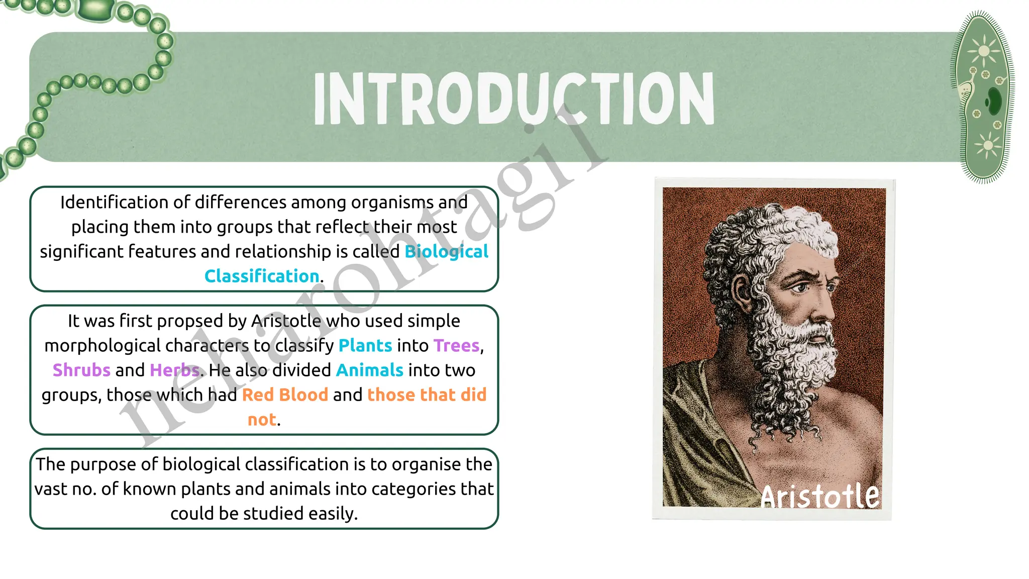
Introduction to Biological Classification
Need for classification
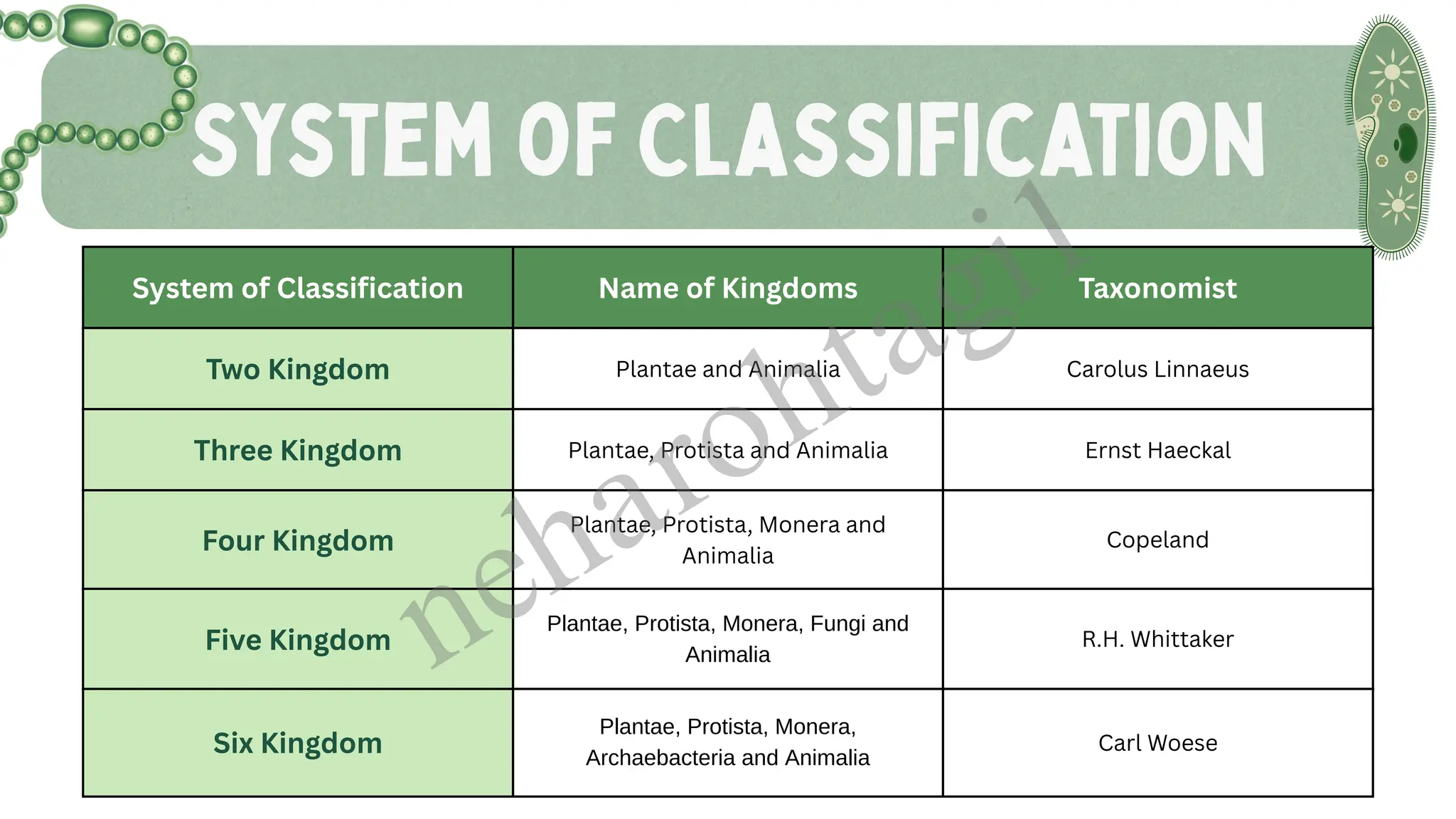
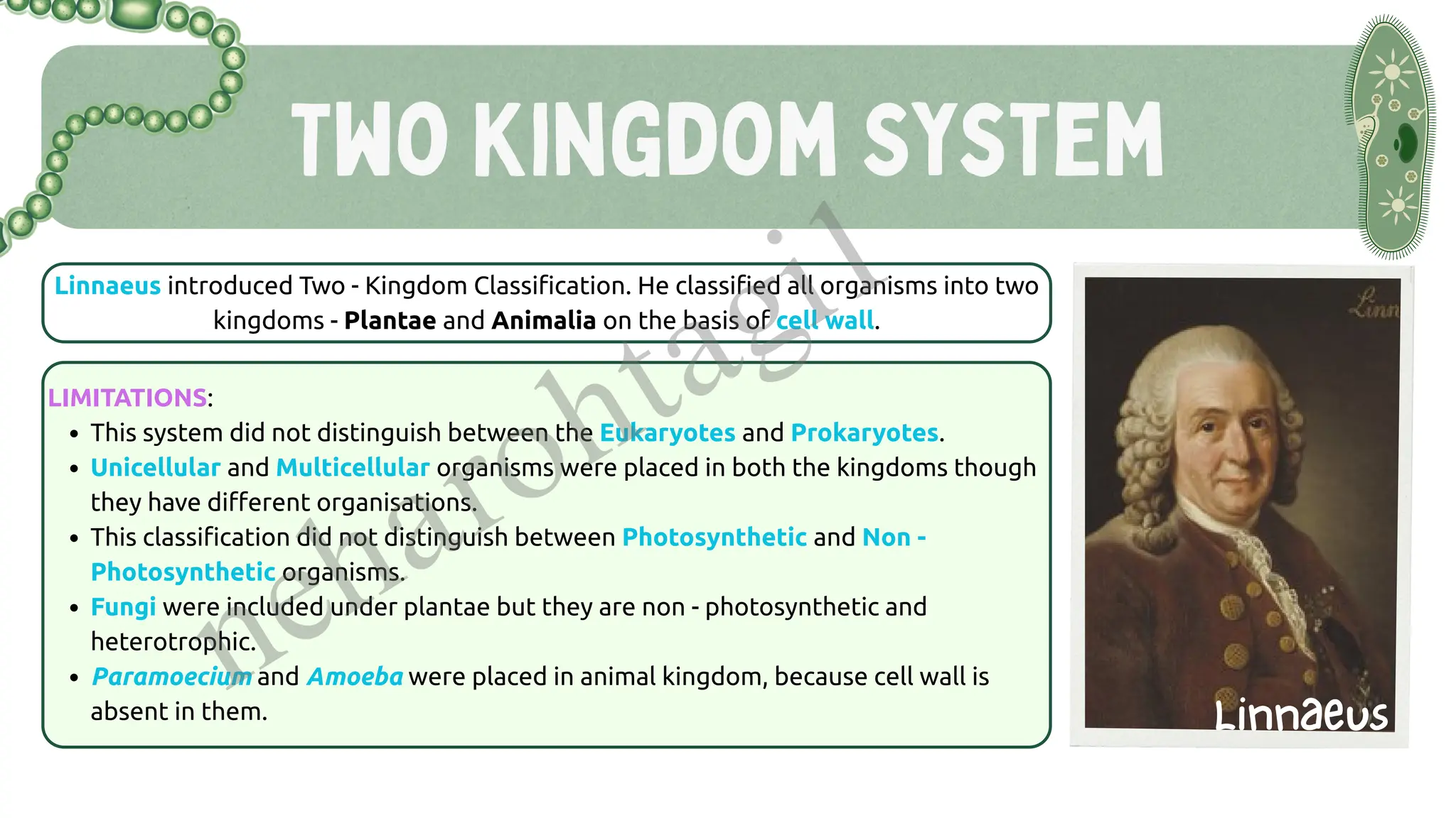
Historical background (Aristotle, Linnaeus)
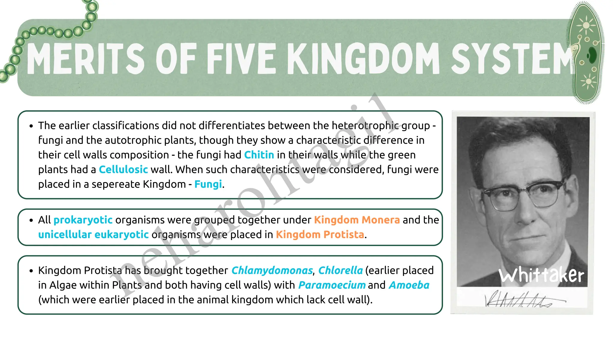
Five Kingdom Classification (R.H. Whittaker)
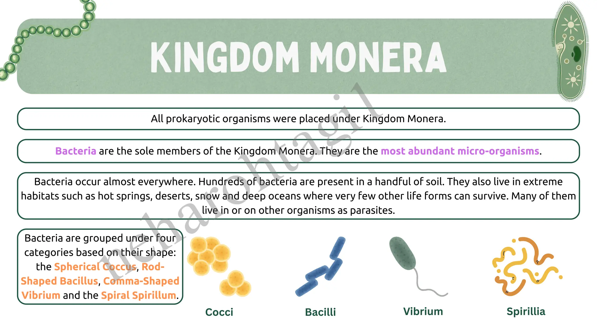
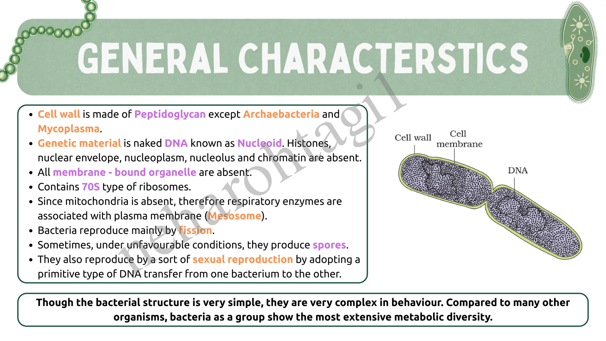
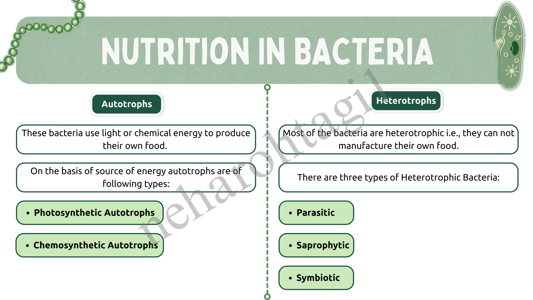
Kingdom Monera
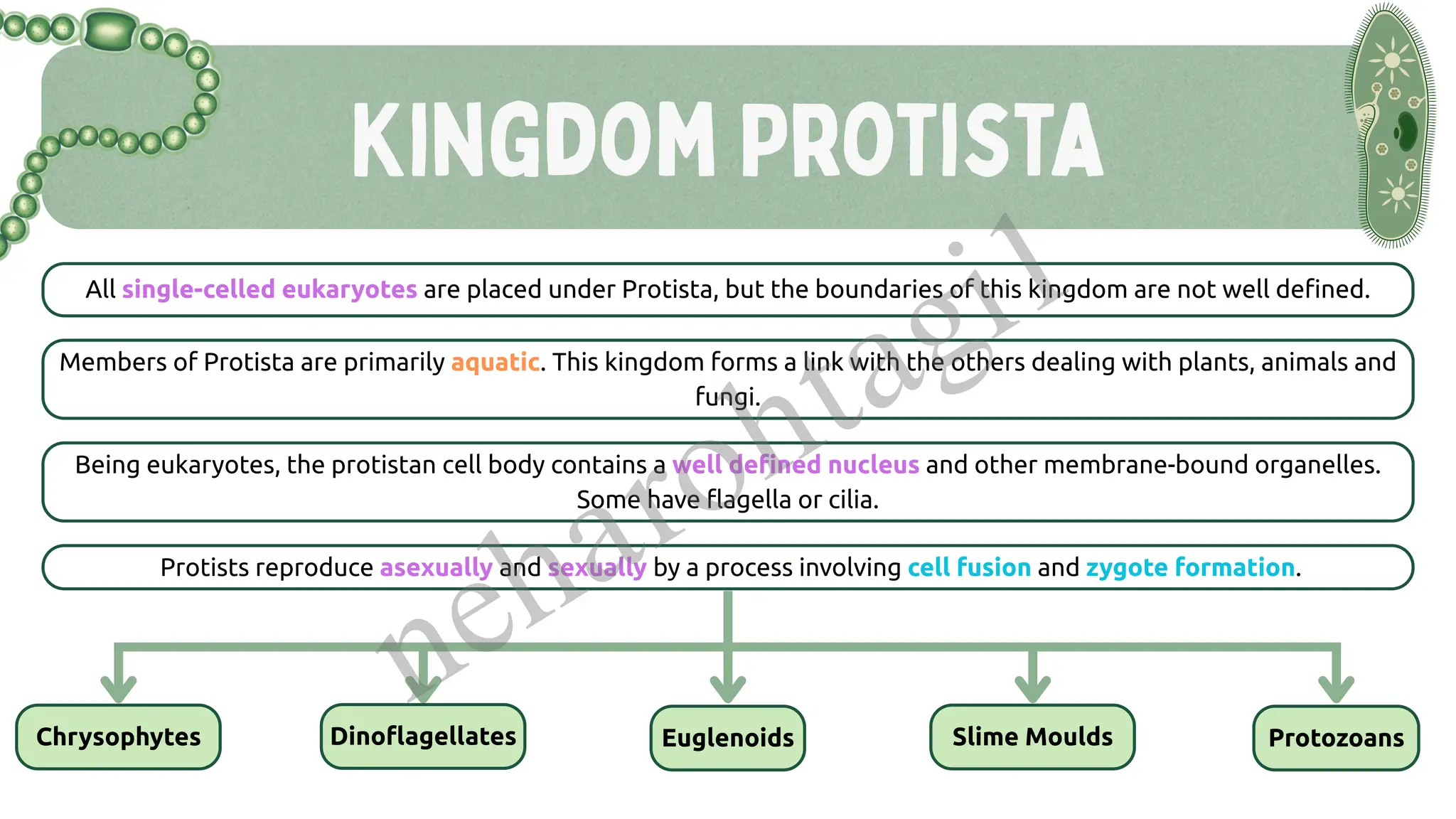
Kingdom Protista
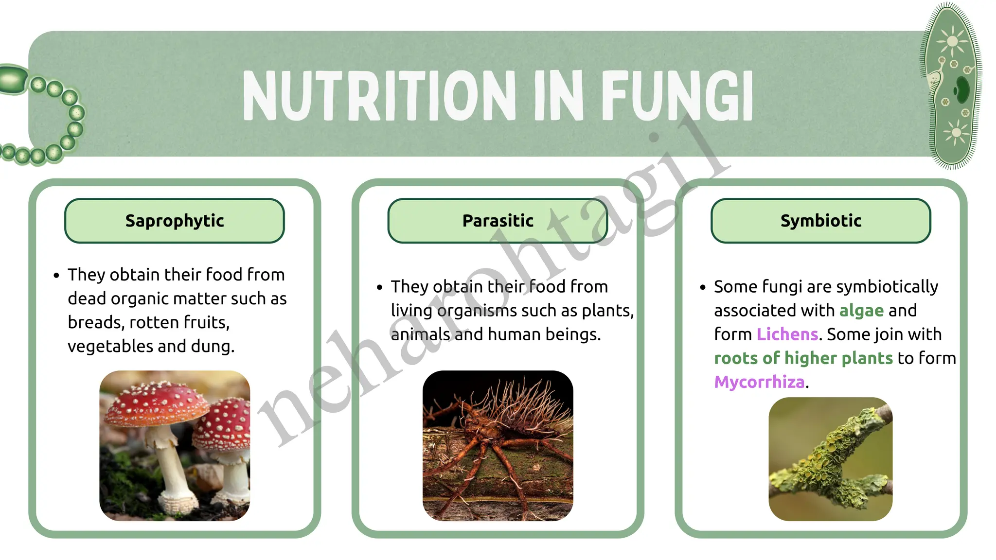
Kingdom Fungi
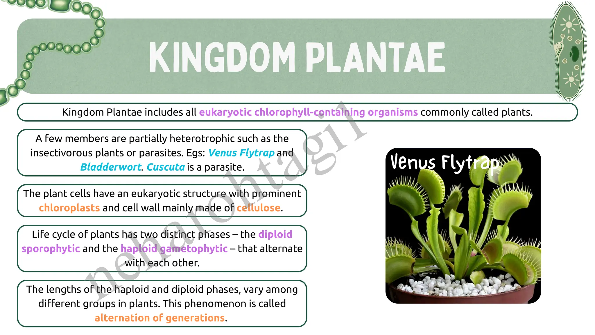
Kingdom Plantae (briefly)
Kingdom Animalia (briefly)
Kingdom Monera
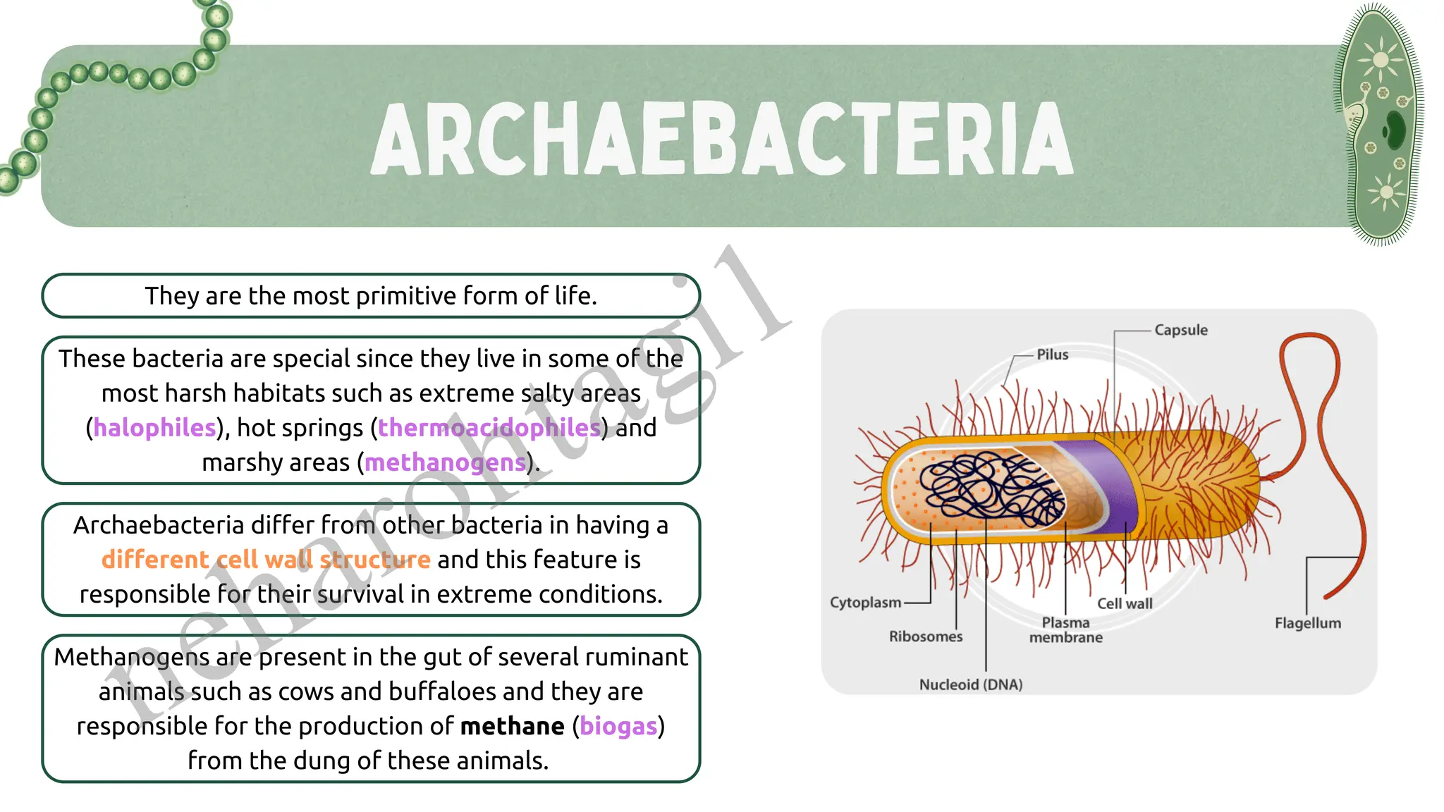
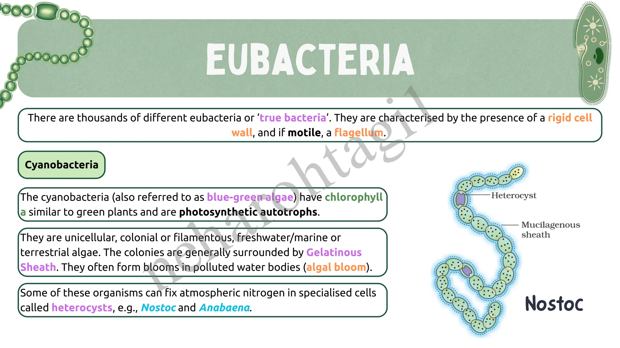
Archaebacteria vs Eubacteria
Cyanobacteria and their role
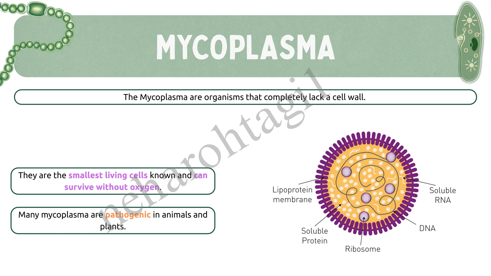
Mycoplasma and other unique features
Kingdom Protista
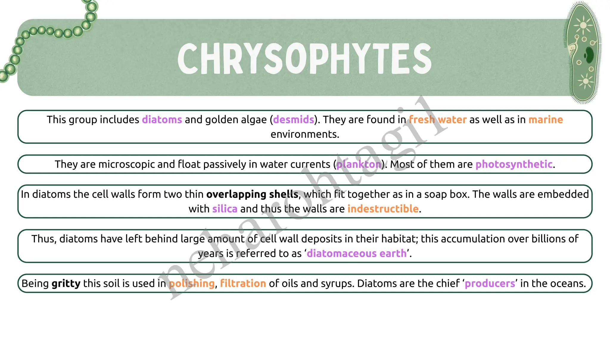
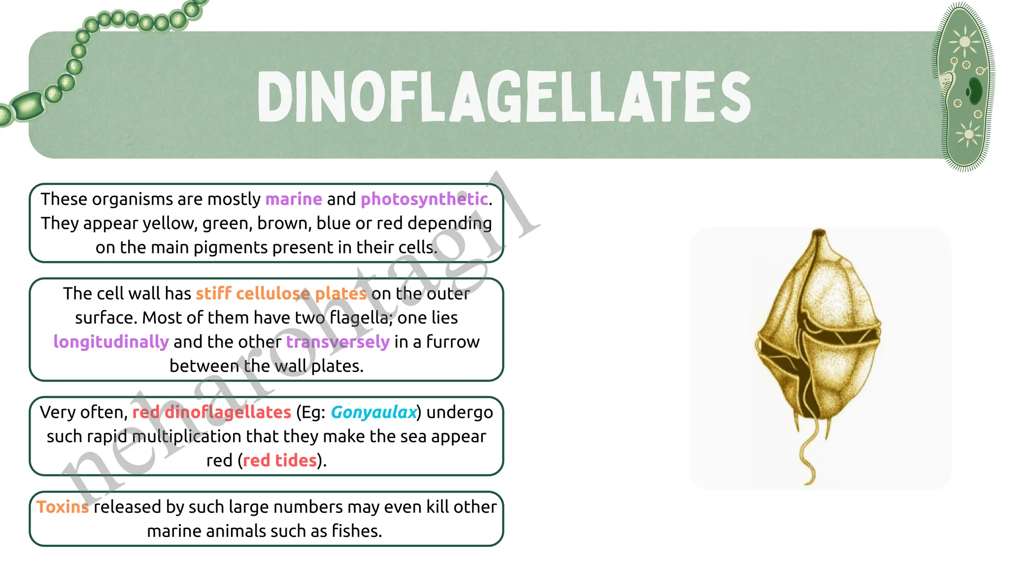
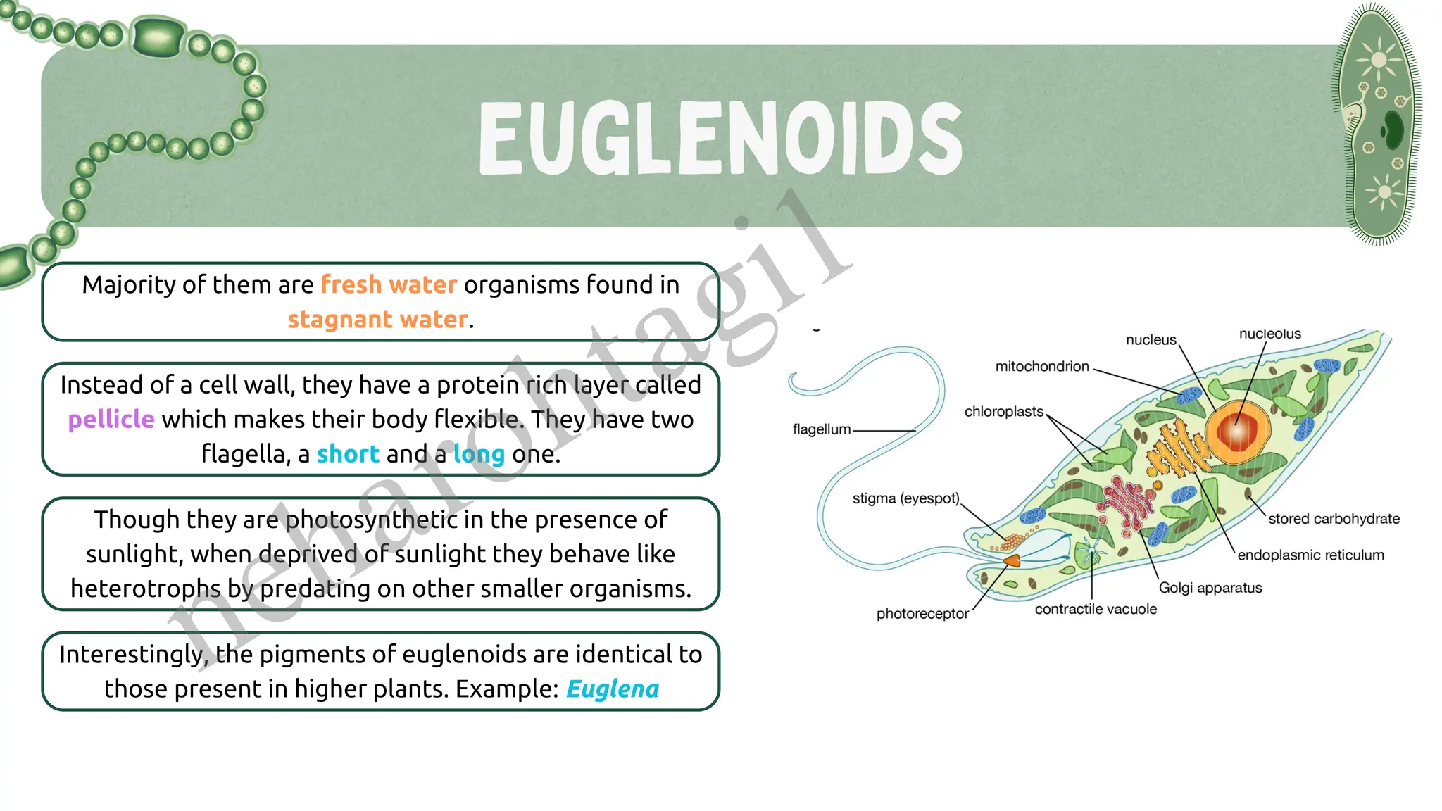
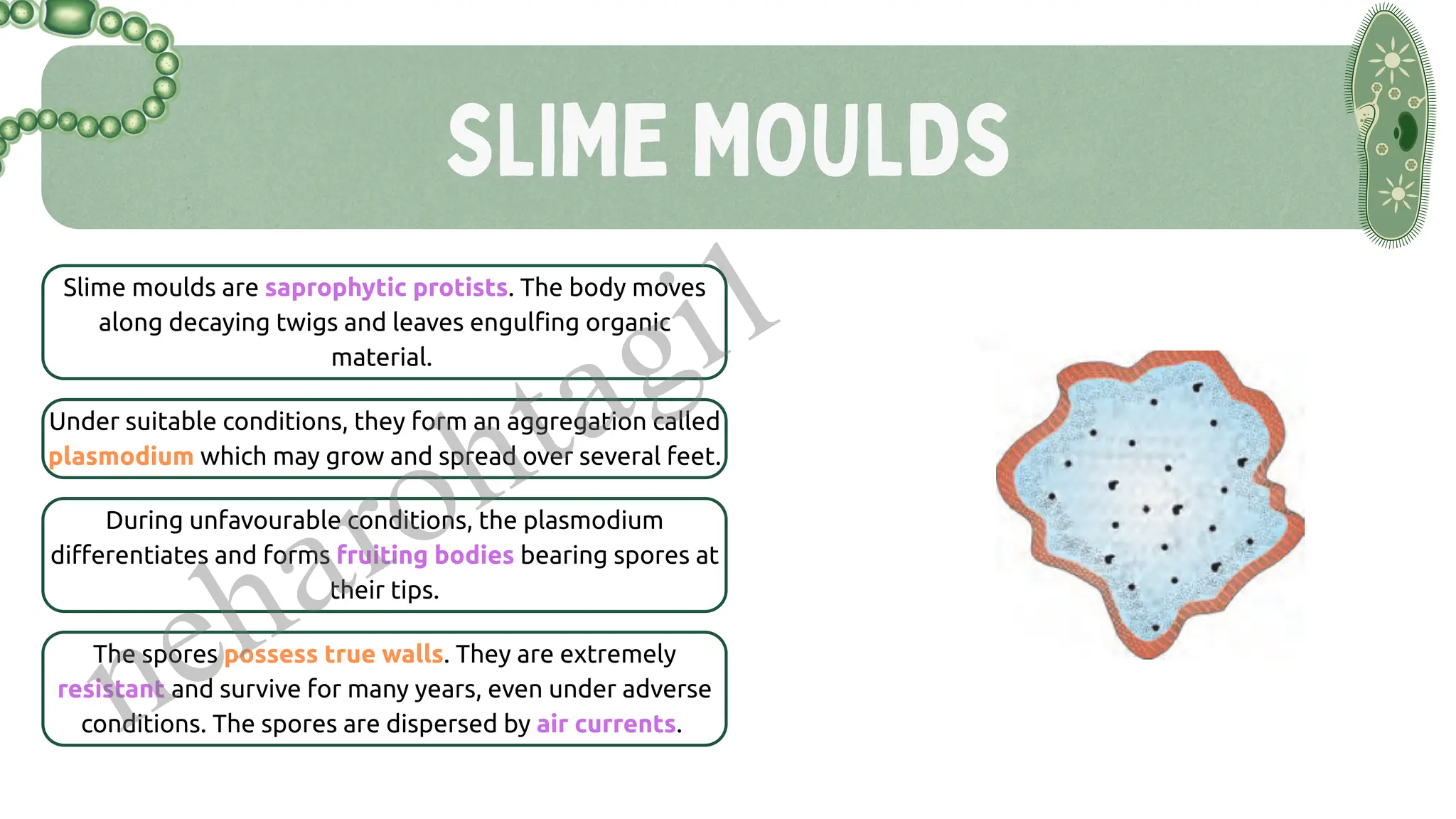
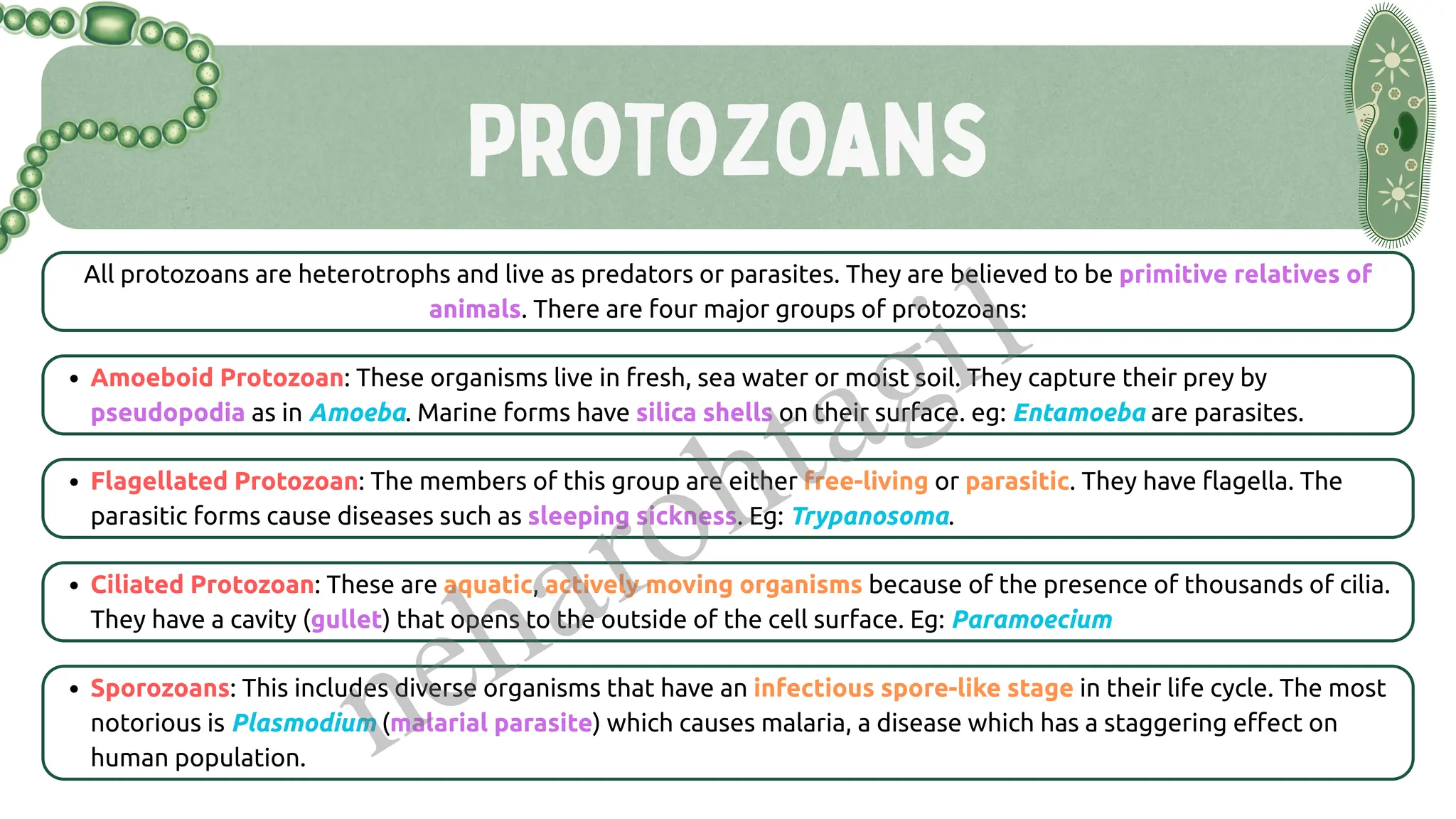
Chrysophytes, Dinoflagellates, Euglenoids, Slime moulds, Protozoans
Characteristics and classification within the kingdom
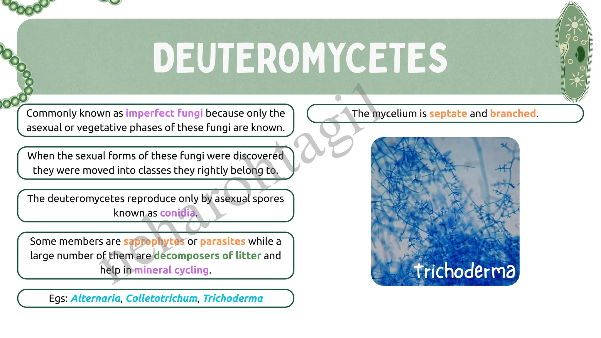
Kingdom Fungi
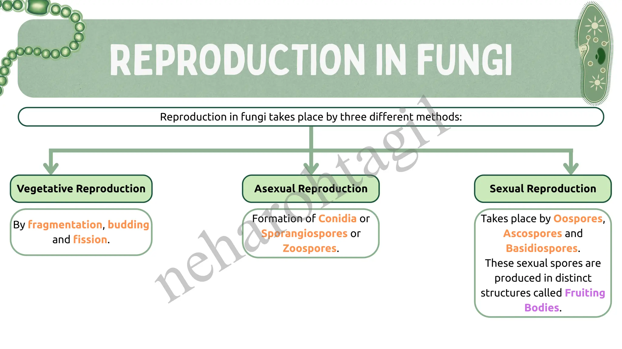
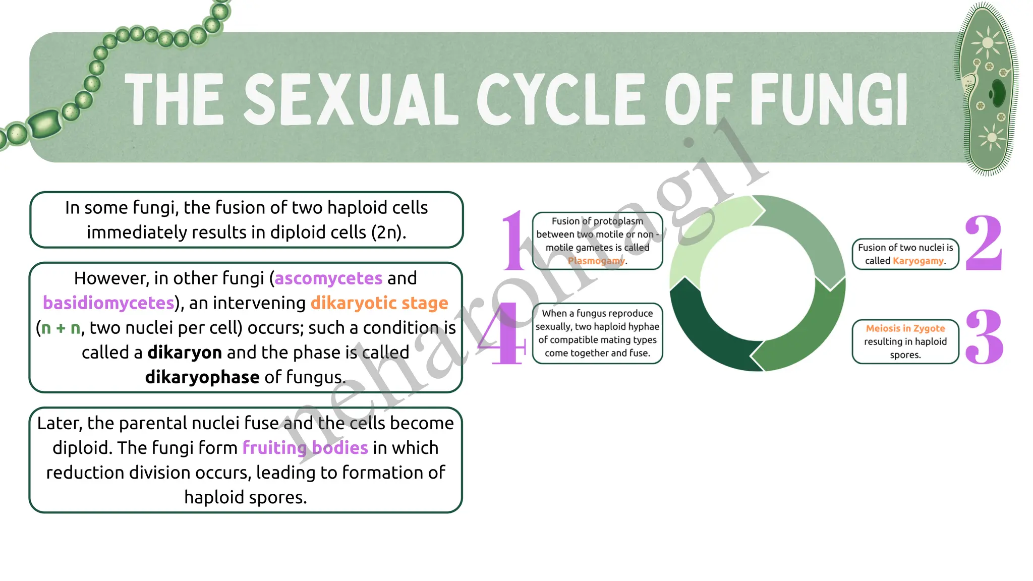
Structure and reproduction
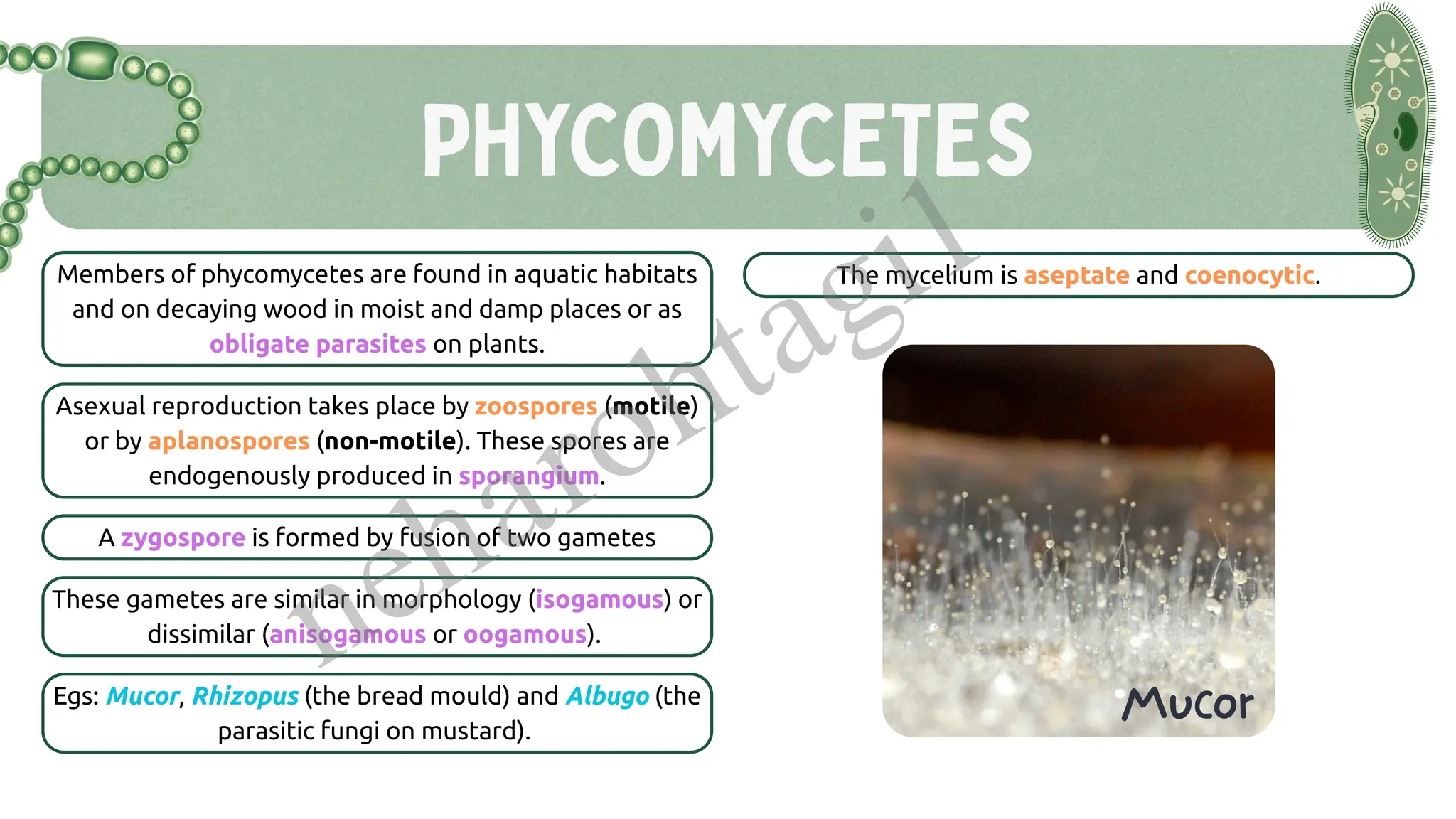
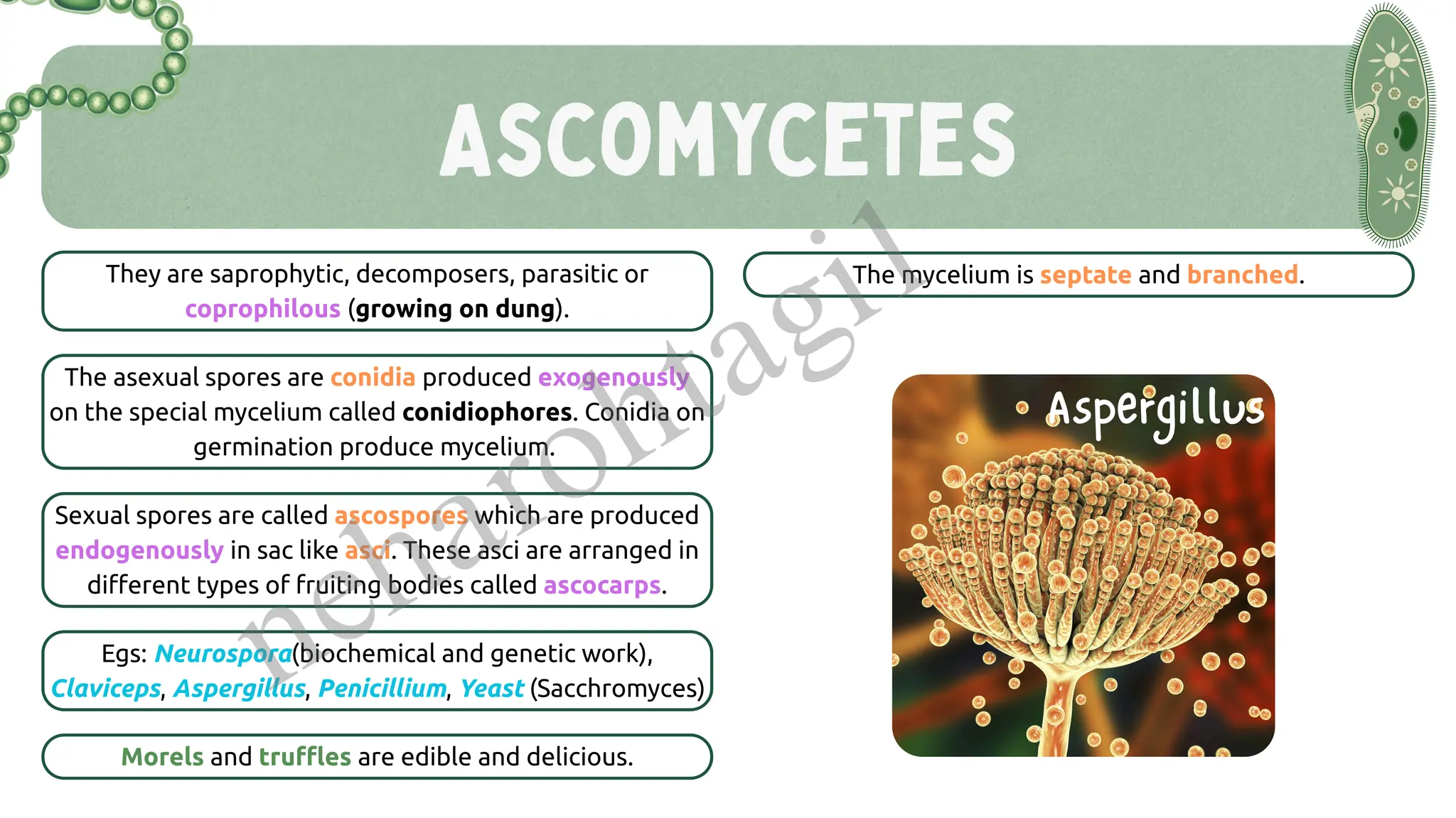
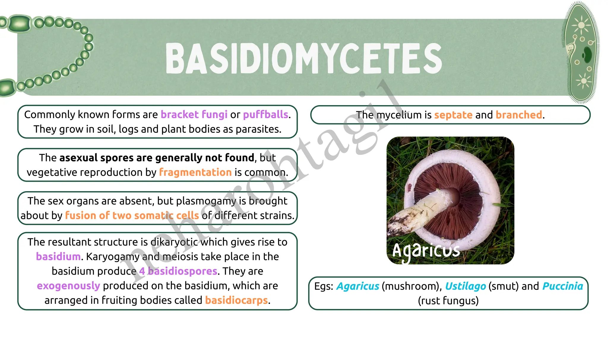
Classes: Phycomycetes, Ascomycetes, Basidiomycetes, Deuteromycetes
Economic and ecological importance
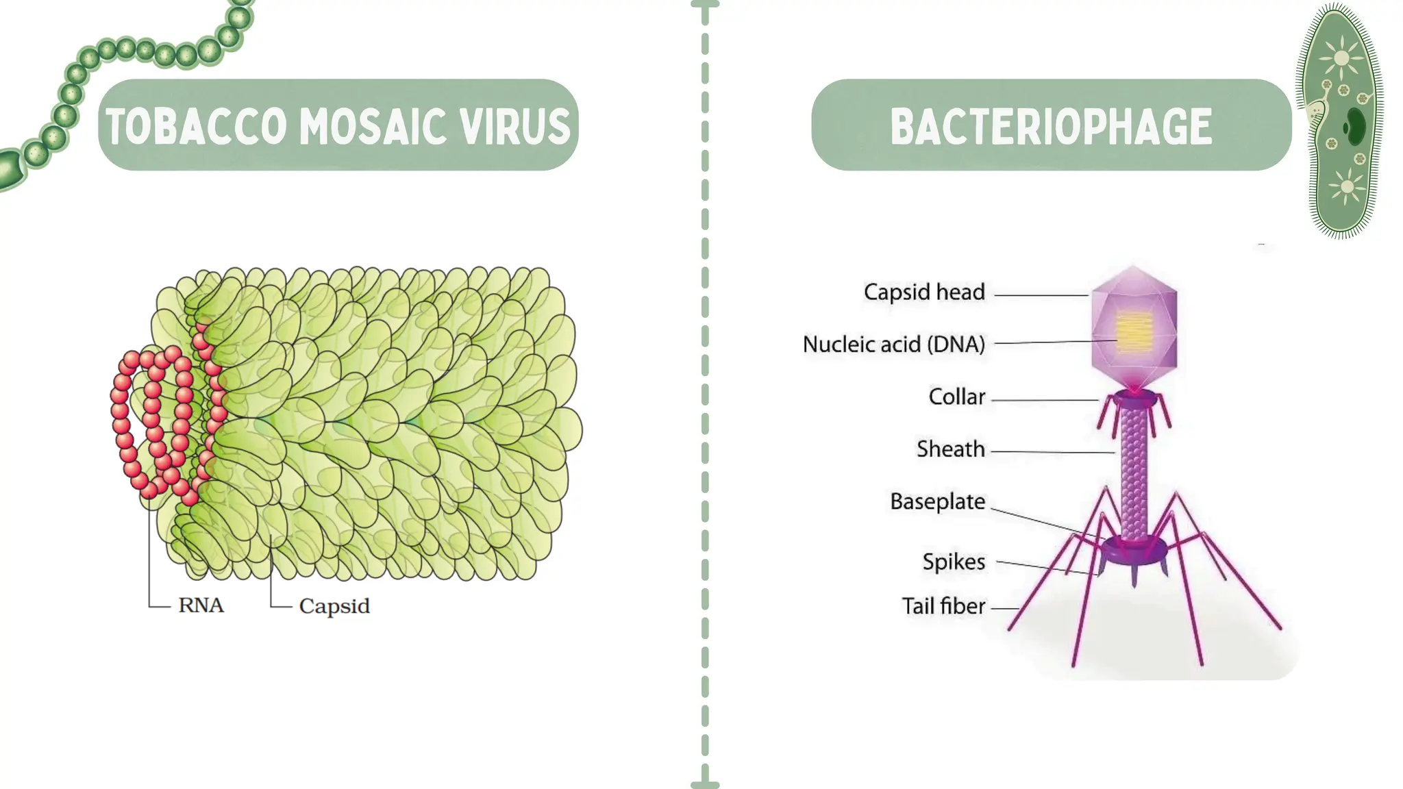
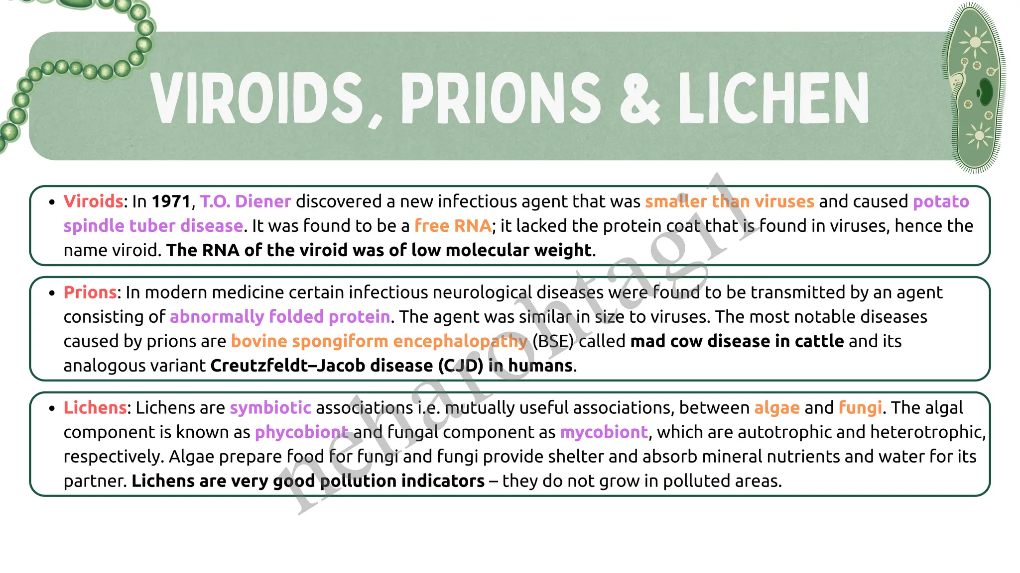
Viruses, Viroids & Lichens
Not included in five-kingdom classification
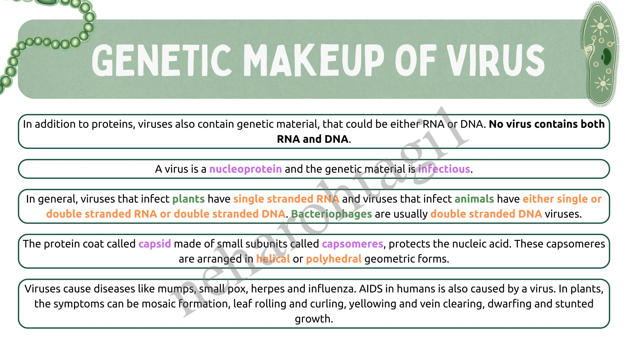
Discovery, structure, characteristics
Virus vs Viroid vs Prion
Symbiotic life of lichens
🔬 Presentation Features:
✅ NCERT-based structured notes
✅ Diagrams & flowcharts for easy recall
✅ Bullet-point summaries for each kingdom
✅ Key NEET MCQ triggers highlighted
✅ PYQ insights and important examples underlined
✅ Revision-friendly layout for quick recap
🎯 Learning Outcomes:
After using this presentation, students will be able to:
Understand the basis and need for biological classification
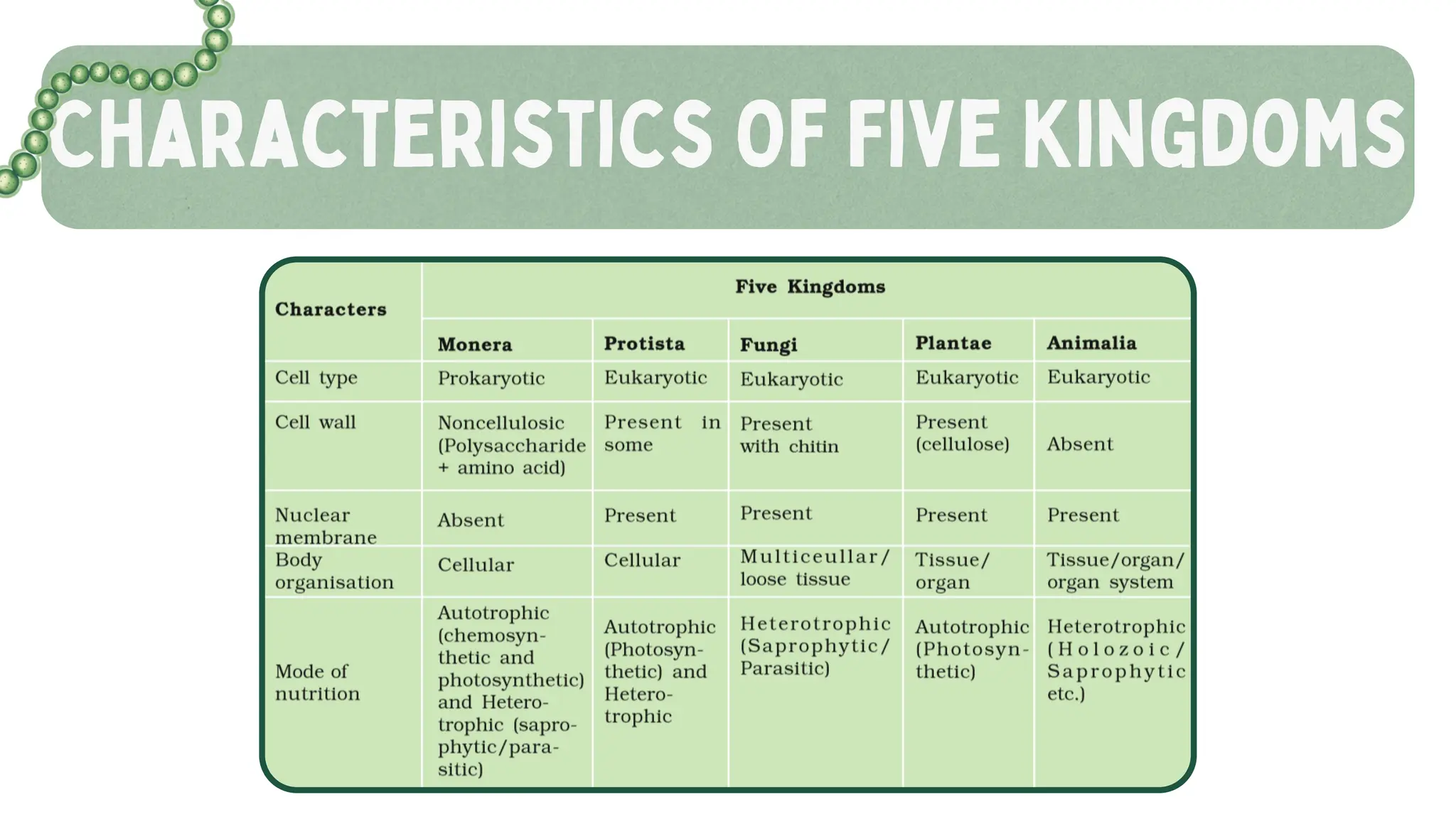
Differentiate among the five kingdoms with key features
Identify examples likely to appear in NEET questions
Recognize differences between living and non-living systems like viruses and viroids
Prepare confidently for both board exams and entrance tests
📌 Best Suited For:
Class 11 CBSE Biology Students
NEET UG Aspirants
Educators and Tutors teaching foundational biology
Students revising NCERT Class 11 Chapter 2
Note: This presentation strictly adheres to the latest NCERT syllabus and includes NEET-focused content, making it a valuable tool for both learning and revision.