
More Related Content
What's hot
What's hot (20)
Similar to Biochemistry reference ranges
Similar to Biochemistry reference ranges (20)
Recently uploaded
Recently uploaded (20)
Biochemistry reference ranges
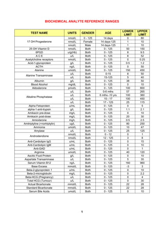
- 1. BIOCHEMICAL ANALYTE REFERENCE RANGES 1 TEST NAME UNITS GENDER AGE LOWER LIMIT UPPER LIMIT nmol/L 0 - 125 14 days 0 80 nmol/L Female 14 days-125 1 1017 OH Progesterone nmol/L Male 14 days-125 1 10 25 OH Vitamin D nmol/L Both 0 - 125 50 150 6PGD u/g(Hb) Both 0 - 125 8 9.5 A C E u/L Both 0 - 125 8 52 Acetylcholine receptors nmol/L Both 0 - 125 0 0.25 Acid-1-glycoprotein g/L Both 0 - 125 0.5 1.2 ACTH ng/L Both 0 - 125 0 50 Alanine umol/L Both 0 - 125 150 450 u/L Both 0-15 8 50 Alanine Transaminase u/L Both 15-125 5 40 Albumin g/L Both 0 - 125 38 50 Blood Alcohol mg/dL Both 0 - 125 0 10 Aldosterone pmol/L Both 0 - 125 100 800 u/L Both 0-6 mths 57 265 u/L Both 6 mths -10 yrs 100 360 u/L Both 10 - 17 100 450 Alkaline Phosphatase u/L Both 17 - 125 25 115 Alpha Fetoprotein u/mL Both 0 - 125 0 5 alpha-1-anti-trypsin g/L Both 0 - 125 1.1 2.1 Amikacin pre-dose: mg/L Both 0 - 125 0 10 Amikacin post-dose: mg/L Both 0 - 125 20 30 Amiodarone mg/L Both 0 - 125 0.5 2.5 Amitriptyline (+nortriptylin) ug/L Both 0 - 125 80 250 Ammonia umol/L Both 0 - 125 10 47 Amylase u/L Both 0 - 125 25 125 nmol/L Both 0 - 12 0 1 Androstendione nmol/L Both 12 - 125 3 8 Anti-Cardiolipin IgG u/mL Both 0 - 125 0 15 Anti-Cardiolipin IgM u/mL Both 0 - 125 0 10 Anti-GAD u/mL Both 0 - 125 0 1 Arginine umol/L Both 0 - 125 40 120 Ascitic Fluid Protein g/L Both 0 - 125 0 10 Aspartate Transaminase u/L Both 0 - 125 5 35 Serum Vitamin B12 ng/L Both 0 - 125 160 960 Base Excess mmol/L Both 0 - 125 -3 3 Beta-2-glycoprotein 1 u/mL Both 0 - 125 0 10 Beta-2-microglobulin mg/L Both 0 - 125 0 2.2 Beta-HCG (Pregnancy) u/L Both 0 - 125 0 4 Total HCG (Tumour) u/L Both 0 - 125 0 30 Actual Bicarbonate mmol/L Both 0 - 125 22 28 Standard Bicarbonate mmol/L Both 0 - 125 22 28 Serum Bile Acids umol/L Both 0 - 125 0 10
- 2. BIOCHEMICAL ANALYTE REFERENCE RANGES 2 TEST NAME UNITS GENDER AGE LOWER LIMIT UPPER LIMIT umol/L Both 0 - 7 days 0 100 Total Bilirubin umol/L Both 0 - 125 0 17 C Reactive Protein mg/L Both 0 - 125 0 10 C1 Esterase Inhibitor g/L Both 0 - 125 0.15 0.35 C1q and C2 levels mg/L Both 0 - 125 118 244 C1r Complement mg/L Both 0 - 125 75 105 C2 Complement mg/L Both 0 - 125 14 25 C22 umol/L Both 0 - 125 30 98 C24 umol/L Both 0 - 125 24 66 C24/C22 ratio Both 0 - 125 0 1 C26 umol/L Both 0 - 125 0.1 1 C26/C22 ratio Both 0 - 125 0 0.03 C3 g/L Both 0 - 125 0.88 2.01 C4 g/L Both 0 - 125 0.16 0.47 C4 Binding Protein mg/L Both 0 - 125 50 150 C5 Complement g/L Both 0 - 125 0.07 0.12 C6 Complement mg/L Both 0 - 125 45 96 C7 Complement mg/L Both 0 - 125 55 85 C8 Complement mg/L Both 0 - 125 112 172 C9 Complement mg/L Both 0 - 125 125 265 CA 125 (Ovarian) u/mL Both 0 - 125 0 21 CA 15-3 (Breast) ku/L Both 0 - 125 0 30 CA 19-9 (Gastro) ku/L Both 0 - 125 0 33 g/L Both 0 - 4 mths 0.08 0.23 g/L Both 4 mths – 1 yr 0.12 0.35 g/L Both 1 - 10 0.2 0.4 g/L Both 10 - 13 0.15 0.23 Caeruloplasmin g/L Both 13 - 125 0.2 0.63 Calcitonin ng/L Both 0 - 125 0 11.5 Calcium mmol/L Both 0 - 125 2.2 2.6 Carbamazepine umol/L Both 0 - 125 20 55 Carboxyhaemoglobin % Both 0 - 125 0 2.5 CEA ug/L Both 0 - 125 0 5 Chloride mmol/L Both 0 - 125 95 108 Cholesterol mmol/L Both 0 - 125 0 5.2 Citrullinated Protein Abs u/mL Both 0 - 125 0 10 Clobazam nmol/L Both 0 - 125 0 670 Clomipramine (+d.m. clomipramine) ug/L Both 0 - 125 200 500 Clonazepam nmol/L Both 0 - 125 80 270 Conjugated (dir) Bili umol/L Both 0 - 125 0 6 Copper umol/L Both 0 - 125 12 25 Corr. Fructosamine umol/L Both 0 - 125 0 395 Corrected Calcium mmol/L Both 0 - 125 2.2 2.6 Correction Ratio Both 0 - 125 1.1 1.3 Cortisol nmol/L Both 0 - 125 140 700
- 3. BIOCHEMICAL ANALYTE REFERENCE RANGES 3 TEST NAME UNITS GENDER AGE LOWER LIMIT UPPER LIMIT C-Peptide mu/L Both 0 - 125 260 650 u/L Female 0 - 125 0 165 Creatine Kinase u/L Male 0 - 125 0 190 umol/L Female 0 - 14 days 40 70 umol/L Female 14 days - 6 mths 35 60 umol/L Female 6 mths – 1 yr 30 55 umol/L Female 1 - 12 30 70 umol/L Female 12 - 17 40 80 umol/L Female 17 - 125 60 100 umol/L Male 14 days - 6 mths 40 70 umol/L Male 6 mths – 1 yr 35 60 umol/L Male 1 - 12 30 70 umol/L Male 12 - 17 40 100 Serum Creatinine umol/L Male 17 - 125 75 125 Creatinine Clearance mL/min Both 0 - 125 80 150 CSF Albumin g/L Both 0 - 125 0 0.24 CSF IgG g/L Both 0 - 125 0 0.03 CSF Protein g/L Both 0 - 125 0.1 0.4 D.M. Clobazam nmol/L Both 0 - 125 0 7000 D.M. Diazepam nmol/L Both 0 - 125 0 5500 Desethylamiodarone mg/L Both 0 - 125 0.5 2.5 Desipramine ug/L Both 0 - 125 50 150 umol/L Female 0 - 125 0.9 11.7 DHEA Sulphate umol/L Male 0 - 125 2.2 15.2 Diazepam nmol/L Both 0 - 125 0 3500 Digoxin nmol/L Both 0 - 125 1 3 DNA antibodies (ELISA) iu/mL Both 0 - 125 0 40 Dothiepin (+d.m. dothiepin) ug/L Both 0 - 125 60 200 Doxepin (+d.m. doxepin) ug/L Both 0 - 125 100 300 Serum Erythropoietin iu/L Both 0 - 125 9.1 29.5 Ethosuximide umol/L Both 0 - 125 0 700 Fasting Glucose mmol/L Both 0 - 125 3.5 6 Felbamate umol/L Both 0 - 125 210 460 ug/L Female 0 - 125 9 120 Ferritin ug/L Male 0 - 125 18 370 Serum Folate ug/L Both 0 - 125 3 17 Free T3 pmol/L Both 0 - 125 3.25 6.21 Free T4 pmol/L Both 0 - 125 9 19 Fructosamine umol/L Both 0 - 125 0 285 u/L Female 0 - 125 3.4 21.6 FSH u/L Male 0 - 125 1.4 13.6 Gabapentin umol/L Both 0 - 125 12 120 u/L Female 0 - 125 0 32 Gamma GT u/L Male 0 - 125 0 50
- 4. BIOCHEMICAL ANALYTE REFERENCE RANGES 4 TEST NAME UNITS GENDER AGE LOWER LIMIT UPPER LIMIT Gastrin pmol/L Both 0 - 125 0 40 GBM ELISA u/mL Both 0 - 125 0 10 Globulin g/L Both 0 - 125 20 35 Glucagon pmol/L Both 0 - 125 0 50 Glutamic acid umol/L Both 0 - 125 25 130 Glutamine umol/L Both 0 - 125 460 800 Glycine umol/L Both 0 - 125 100 330 Growth Hormone mu/L Both 0 - 125 0 13 mg/dL Female 0 - 125 38 210 Serum Haptoglobins mg/dL Male 0 - 125 30 170 Hb A1c % Both 0 - 125 4.1 6.4 mmol/L Female 0 - 125 0.9 2.1 HDL Cholesterol mmol/L Male 0 - 125 0.8 1.4 Histidine umol/L Both 0 - 125 30 150 Histone antibodies Both 0 - 125 0 40 g/L Both 0 - 14 days 0.01 0.08 g/L Both 14 - 42 days 0.02 0.15 g/L Both 1.5 - 3 mths 0.05 0.4 g/L Both 3 - 6 mths 0.1 0.5 g/L Both 6 - 9 mths 0.15 0.7 g/L Both 9 mths – 1 yr 0.2 0.7 g/L Both 1 - 2 0.3 1.2 g/L Both 2 - 3 0.3 1.3 g/L Both 3 - 6 0.4 2 g/L Both 6 - 9 0.5 2.4 g/L Both 9 - 12 0.7 2.5 g/L Both 12 - 15 0.8 2.8 g/L Both 15 - 45 0.8 2.8 IgA g/L Both 45 - 125 0.8 4 ku/L Both 0 - 1 0 29 ku/L Both 1 - 2 0 49 ku/L Both 2 - 3 0 45 ku/L Both 3 - 9 0 52 IgE ku/L Both 9 - 125 0 87 nmol/L Female 0 - 2 2.6 25.6 nmol/L Female 2 - 3 2.6 27.4 nmol/L Female 3 - 4 2.9 31.1 nmol/L Female 4 - 5 3.3 34.8 nmol/L Female 5 - 6 4 40.3 nmol/L Female 6 - 7 5.9 47.6 nmol/L Female 7 - 8 7.3 65.9 nmol/L Female 8 - 9 9.5 73.2 nmol/L Female 9 - 10 11.7 87.8 IGF-1 nmol/L Female 10 - 11 13.9 102.5
- 5. BIOCHEMICAL ANALYTE REFERENCE RANGES 5 TEST NAME UNITS GENDER AGE LOWER LIMIT UPPER LIMIT nmol/L Female 11 - 12 16.1 117.1 nmol/L Female 12 - 13 18.3 120.8 nmol/L Female 13 - 14 19 124.5 nmol/L Female 14 - 15 19.8 120.8 nmol/L Female 15 - 16 19 109.8 nmol/L Female 16 - 20 18.3 98.8 nmol/L Female 20 - 30 14.6 73.2 nmol/L Female 30 - 60 12.8 43.9 nmol/L Female 60 - 125 3.7 32.9 nmol/L Male 0 - 2 2.6 25.6 nmol/L Male 2 - 3 2.6 27.4 nmol/L Male 3 - 4 2.9 31.1 nmol/L Male 4 - 5 3.3 34.8 nmol/L Male 5 - 6 4 40.3 nmol/L Male 7 - 8 4.8 36.6 nmol/L Male 8 - 9 5.9 43.9 nmol/L Male 9 - 10 7.3 54.9 nmol/L Male 10 - 11 9.1 62.2 nmol/L Male 11 - 12 11 73.2 nmol/L Male 12 - 13 13.2 84.2 nmol/L Male 13 - 14 15.4 95.2 nmol/L Male 14 - 15 17.6 102.5 nmol/L Male 15 - 16 10 102.5 nmol/L Male 16 - 20 19 98.8 nmol/L Male 20 - 30 14.6 73.2 nmol/L Male 30 - 60 12.8 43.9 IGF-1 nmol/L Male 60 - 125 3.7 32.9 g/L Both 0 - 14 days 5 17 g/L Both 14 - 42 days 3.9 13 g/L Both 1.5 - 3 mths 2.1 7.7 g/L Both 3 - 6 mths 2.4 8.8 g/L Both 6 - 9 mths 3 9 g/L Both 9 mths – 1 yr 3 10.9 g/L Both 1 - 2 3.1 13.8 g/L Both 2 - 3 3.7 15.8 g/L Both 3 - 6 4.9 16.1 g/L Both 6 - 9 5.4 16.1 g/L Both 9 - 12 5.4 16.1 g/L Both 12 - 15 5.4 16.1 IgG g/L Both 15 - 125 6 16 g/L Both 0 - 10 days 3.8 8.4 g/L Both 10 days - 6 mths 1.5 3 g/L Both 6 mths- 2 yrs 2.3 5.8 g/L Both 2 - 5 2.3 6.4 IgG 1 subclass g/L Both 5 - 10 3.6 7.3
- 6. BIOCHEMICAL ANALYTE REFERENCE RANGES 6 TEST NAME UNITS GENDER AGE LOWER LIMIT UPPER LIMIT g/L Both 10 - 15 3.8 7.7 IgG 1 subclass g/L Both 15 - 125 3.2 10.2 g/L Both 0 - 10 days 1.2 4 g/L Both 10 days - 6 mths 0.3 0.5 g/L Both 6 mths – 2 yrs 0.3 2.9 g/L Both 2 - 5 0.7 4.5 g/L Both 5 - 10 1.4 4.5 g/L Both 10 - 15 1.3 4.5 IgG 2 subclass g/L Both 15 - 125 1.2 6.6 g/L Both 0 - 10 days 0.3 1.5 g/L Both 10 days - 6 mths 0.1 0.6 g/L Both 6 mths – 2 yrs 0.1 0.8 g/L Both 2 - 5 0.1 1.1 g/L Both 5 - 10 0.3 1.1 g/L Both 10 - 15 0.2 1.3 IgG 3 subclass g/L Both 15 - 125 0.2 1.9 g/L Both 0 - 10 days 0 0.5 g/L Both 10 days - 6 mths 0 0.5 g/L Both 6 mths – 2 yrs 0 0.5 g/L Both 2 - 5 0 0.8 g/L Both 5 - 10 0 1 g/L Both 10 - 15 0 1.1 IgG 4 subclass g/L Both 15 - 125 0 1.3 IgG Insulin antibodies mg/L Both 0 - 125 0 5 g/L Both 0 - 14 days 0.05 0.2 g/L Both 14 - 42 days 0.08 0.4 g/L Both 1.5 - 3 mths 0.15 0.7 g/L Both 3 - 6 mths 0.2 1 g/L Both 6 - 9 mths 0.4 1.6 g/L Both 9 mths – 1 yr 0.6 2.1 g/L Both 1 - 2 0.5 2.2 g/L Both 2 - 3 0.5 2.2 g/L Both 3 - 6 0.5 2 g/L Both 6 - 9 0.5 1.8 g/L Both 9 - 12 0.5 1.8 g/L Both 12 - 15 0.5 1.9 g/L Both 15 - 45 0.5 1.9 IgM g/L Both 45 - 125 0.4 2 Imipramine(+desipramine) ug/L Both 0 - 125 150 300 Immuno-Reactive Trypsin (IRT) ug/L Both 0 - 125 0 120 Insulin mu/L Both 0 - 125 3 17 Intrinsic Factor Both 0 - 125 0 1.2 Ionised Calcium mmol/L Both 0 - 125 1.12 1.32 umol/L Female 0 - 125 12 28 Iron umol/L Male 0 - 125 14 30
- 7. BIOCHEMICAL ANALYTE REFERENCE RANGES 7 TEST NAME UNITS GENDER AGE LOWER LIMIT UPPER LIMIT % Female 0 - 125 15 50 Iron Saturation % Male 0 - 125 20 55 Isoleucine umol/L Both 0 - 125 26 100 Lactate mmol/L Both 0 - 125 0.6 2.4 Lamotrigine umol/L Both 0 - 125 4 60 LDH u/L Both 0 - 125 125 243 LDL Cholesterol mmol/L Both 0 - 125 0 4.5 Blood Lead ug/L Both 0 - 125 0 100 Leucine umol/L Both 0 - 125 65 220 u/L Female 0 - 125 2.9 21.7 LH u/L Male 0 - 125 1.8 8.2 Lithium mmol/L Both 0 - 125 0.7 1.2 Lysine umol/L Both 0 - 125 100 300 Magnesium mmol/L Both 0 - 125 0.7 1 Blood Manganese ug/L Both 0 - 125 0.5 1.5 Mannan-binding ligand Both 0 - 125 0.5 4 Maprotiline ug/L Both 0 - 125 100 300 Blood Mercury ug/L Both 0 - 125 0 10 Methaemoglobin % Both 0 - 125 0 2 Methionine umol/L Both 0 - 125 10 60 Mianserin (+d.m.mianserin) ug/L Both 0 - 125 25 100 mg/mmol Female 0 - 125 0 3.5 Microalbumin : Creatinine ratio mg/mmol Male 0 - 125 0 2.5 ug/L Female 0 - 125 0 76 Serum Myoglobin ug/L Male 0 - 125 0 92 Neurotensin pmol/L Both 0 - 125 0 100 Nortriptyline ug/L Both 0 - 125 50 150 pmol/L Female 0 - 125 77 921 Oestradiol pmol/L Male 0 - 125 40 161 Ornithine umol/L Both 0 - 125 25 120 Pancreolauryl Test Ratio % Both 0 - 125 30 100 Paracetamol mg/L Both 0 - 125 0 25 pmol/L Both 0 - 15 1.3 10 Parathyroid Hormone pmol/L Both 15 - 125 1.3 6.8 pCO2 kPa Both 0 - 125 4.6 6 Peritoneal Fluid Protein g/L Both 0 - 125 0 10 pH Both 0 - 125 7.36 7.44 Phenobarbitone umol/L Both 0 - 125 65 172 Phenylalanine umol/L Both 0 - 125 35 100 Phenytoin umol/L Both 0 - 125 40 80 mmol/L Both 0 - 2 days 1.4 2.8 mmol/L Both 2 days – 12 yrs 1.3 2.1 mmol/L Both 12 - 20 1.1 2 Phosphate mmol/L Both 20 - 125 0.8 1.4 Phytanate umol/L Both 0 - 125 0 15
- 8. BIOCHEMICAL ANALYTE REFERENCE RANGES 8 TEST NAME UNITS GENDER AGE LOWER LIMIT UPPER LIMIT Placental Alkaline Phosphatase IU/L Both 0 - 125 0 100 Pleural Fluid Protein g/L Both 0 - 125 0 10 pO2 kPa Both 0 - 125 10.6 14.6 mmol/L Both 0 - 1 mths 3.4 6.6 mmol/L Both 1 - 3 3.4 5.6Potassium mmol/L Both 3 - 125 3.4 4.9 Pancreatic Polypeptide pmol/L Both 0 - 125 0 300 Prealbumin g/L Both 0 - 125 0.2 0.4 Pristanate umol/L Both 0 - 125 0 2 Procollagen III amino-terminal Peptide ug/L Both 0 - 125 1.2 4.2 nmol/L Female 0 - 125 0 1 Progesterone nmol/L Male 0 - 125 0 1 mu/L Female 0 - 125 25 629 Prolactin mu/L Male 0 - 125 54 280 Proline umol/L Both 0 - 125 85 290 Protriptyline ug/L Both 0 - 125 70 200 ug/L Male 0 - 50 0 2.1 ug/L Male 50 - 60 0 3.1 ug/L Male 60 - 70 0 4.1 PSA ug/L Male 70 - 125 0 4.9 Pyruvate Kinase u/g(Hb) Both 0 - 125 11 21 Red Cell Folate ug/L Both 0 - 125 160 713 Plasma Renin Both 0 - 125 0.5 3.1 Rheumatoid Factor u/mL Both 0 - 125 0 50 Salicylate mg/L Both 0 - 125 0 50 Scl-70 antibodies Both 0 - 125 0 10 Blood Selenium ug/L Both 0 - 125 48 128 Serine umol/L Both 0 - 125 90 290 Serum Osmolality mosmol/kg Both 0 - 125 280 300 nmol/L Female 0 - 125 18 114 SHBG nmol/L Male 0 - 125 13 71 Sodium mmol/L Both 0 - 125 135 147 Somatostatin pmol/L Both 0 - 125 0 150 Tacrolimus (FK506) ug/L Both 0 - 125 1.5 15 Taurine umol/L Both 0 - 125 40 140 nmol/L Female 0 - 125 2 4 nmol/L Male 0 - 50 10 52Testosterone nmol/L Male 50 - 125 7 26 nmol/L Female 0 - 125 0 2.8 nmol/L Male 0 - 50 10 52Testosterone (extracted) nmol/L Male 50 - 125 7 26 umol/L Both 1 days 28 56 umol/L Both 1 day – 1 yr 28 56Theophylline (Aminophylline) umol/L Both 1 - 125 55 110 Threonine umol/L Both 0 - 125 70 220 Thiopurine Methyl Transferase (TPMT) Both 0 - 125 35 79
- 9. BIOCHEMICAL ANALYTE REFERENCE RANGES 9 TEST NAME UNITS GENDER AGE LOWER LIMIT UPPER LIMIT Thyroglobulin ng/mL Both 0 - 125 0 30 Thyroid Binding Globulin nmol/L Both 0 - 125 241 722 Thyroid Peroxidase antibodies iu/mL Both 0 - 125 0 9 TIBC umol/L Both 0 - 125 45 80 Tissue transglutaminase (IgA) iu/mL Both 0 - 125 0 5 Topiramate umol/L Both 0 - 125 6 74 g/L Both 0 - 3 59 78 g/L Both 3 - 17 63 83Total Protein g/L Both 17 - 125 60 85 Transferrin g/L Both 0 - 125 2 3.2 Triglycerides mmol/L Both 0 - 125 0.5 2.1 Trimipramin (+d.m. trimipramin) ug/L Both 0 - 125 100 300 Troponin I ug/L Both 0 - 125 0 0.04 Tryptase ug/L Both 0 - 125 2 14 Tryptophan umol/L Both 0 - 125 30 80 TSH mu/L Both 0 - 125 0.3 5 TSH receptor Antibodies (stimulating) Both 0 - 125 0 1 Tyrosine umol/L Both 0 - 125 30 120 mmol/L Both 0 - 13 0.06 0.24 mmol/L Female 0 - 125 0.12 0.36Urate mmol/L Male 0 - 125 0.2 0.42 mmol/L Both 0 - 6 2.2 8.3 mmol/L Both 6 - 13 2.5 7.4 mmol/L Both 13 - 17 2.2 7.6 Urea mmol/L Both 17 - 125 2 6.6 Urine 5 HIAA Excretion umol/d Both 0 - 125 0 50 Urine 5-aminolaevulininc acid (5HVA) umol/mmol Both 0 - 125 0 3.8 Urine Adrenaline nmol/d Both 0 - 125 0 144 Urine Calcium Excretion mmol/d Both 0 - 125 2 7.5 Urine Creatinine mmol/L Both 0 - 125 1.8 50 Urine Creatinine Excretion mmol/d Both 0 - 125 8 17 Urine Dopamine nmol/d Both 0 - 125 0 3194 Urine Free Cortisol Excretion nmol/d Both 0 - 125 80 350 Urine Magnesium Excretion mmol/d Both 0 - 125 2 8 Urine Mercury ug/L Both 0 - 125 0 10 Urine Microalbumin mg/L Both 0 - 125 0 20 Urine Nitrogen Excretion mmol/d Both 0 - 125 8 21 Urine Noradrenaline nmol/d Both 0 - 125 0 560 Urine Osmolality mosmol/kg Both 0 - 125 50 1300 Urine Oxalate excretion umol/d Both 0 - 125 100 460 Urine Oxalate : Creatinine ratio umol/mmol Both 0 - 125 1 38 Urine Phosphate Excretion mmol/d Both 0 - 125 15 50 Urine Porphobilinogen umol/mmol Both 0 - 125 0 1.5 Urine Potassium Excretion mmol/d Both 0 - 125 35 80 Urine Protein g/L Both 0 - 125 0 0.2 Urine Protein Excretion g/d Both 0 - 125 0 0.3
- 10. BIOCHEMICAL ANALYTE REFERENCE RANGES 10 TEST NAME UNITS GENDER AGE LOWER LIMIT UPPER LIMIT Urine Sodium Excretion mmol/d Both 0 - 125 100 240 Urine Total Porphyrin nmol/mmol Both 0 - 125 0 35 Urine Urate Excretion mmol/d Both 0 - 125 2 7 Urine Urea Excretion mmol/d Both 0 - 125 250 600 Valine umol/L Both 0 - 125 90 300 Valproate umol/L Both 0 - 125 350 700 Vigabatrin umol/L Both 0 - 125 6 278 VIP pmol/L Both 0 - 125 0 30 Vitamin E umol/L Both 0 - 125 11.5 35 Voltage Gated Ca channel Both 0 - 125 0 45 Voltage Gated K channel Both 0 - 125 0 100 Zinc umol/L Both 0 - 125 7 18