
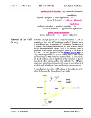
Download to read offline
![SIU School of Medicine BIOCHEMISTRY Carbohydrate Metabolism
Faculty: E.C. Niederhoffer Problem Unit 4 - Page 38
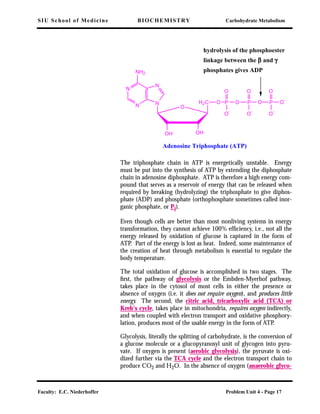
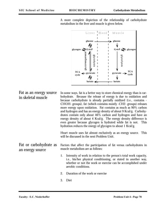
this is not the situation in vivo (see Table 1 in Module I), this is not
the energy of hydrolysis in a cell. The actual free energy is given by
where ∆G° is the standard state free energy, R is the gas constant
(1.99 cal/deg/mol), T is the temperature in Kelvin (i.e. 273.15 plus
the temperature in Celsius), and [ATP], [ADP], and [P] are the con-
centrations of ATP, ADP, and Pi in the cell. In most cells, the con-
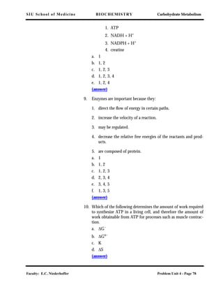
centrations of ATP, ADP, and Pi are such that the ∆G is on the order
of -10 to -13 kcal/mol.
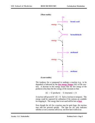
When a chemical change takes place, the maximum energy available
for useful work is the decrease in free energy (∆G) which accompa-
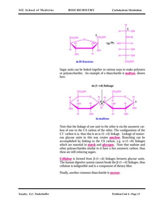
nies the change, under the conditions of temperature and concentra-
tion at the time of the change. The free energy that is released by
ATP hydrolysis may be captured for useful work in the cell, e.g., syn-
thesis of a new bond, active transport, transmission of an impulse
along a nerve, and muscular movement.
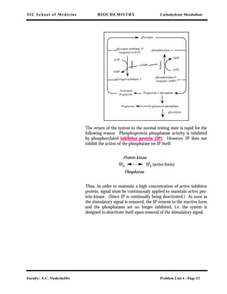
As we have seen, there are many phosphorylated compounds involved
in metabolism. Phosphorylated proteins were discussed in the previ-
ous section under the regulation of glycogenolysis. Monophosphate
esters such as these are low energy compounds and are not important
in energy transfer. The greater the release of free energy generated
during the hydrolytic release of Pi, the greater the free energy stored
in the original bond and the greater the phosphate group transfer
potential, i.e., if X~Pi has a greater phosphate potential than ATP,
then the following reaction is possible: X~Pi + ADP → X + ATP,
while the reverse does not occur to as great an extent. If Y-Pi has a
lower phosphate group transfer potential than ATP, the following
reaction is possible: ATP + Y → ADP + Y-Pi, and the reverse is not
favored.
Thus the phosphate group transfer potential indicates the direction
of flow of phosphate groups from donors of high transfer potential to
an acceptor which forms a compound of lower phosphate group
transfer potential. ATP has an intermediate value in the scale of
phosphate group transfer potential. The ATP-ADP system can there-
fore accept Pi from high potential systems and transfer Pi to acceptor
systems of low potential.
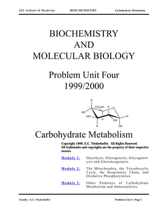
∆G ∆G° RT
ADP[ ] P[ ]
ATP[ ]
--------------------------ln+=](https://image.slidesharecdn.com/biochemistry-150402010123-conversion-gate01-190216190441/85/Biochemistry-150402010123-conversion-gate01-38-320.jpg)
![SIU School of Medicine BIOCHEMISTRY Carbohydrate Metabolism
Faculty: E.C. Niederhoffer Problem Unit 4 - Page 43
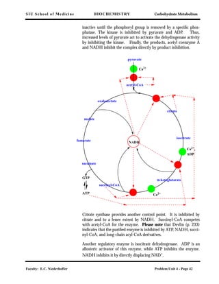
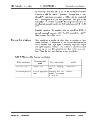
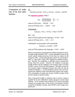
The last point of control is α-ketoglutarate dehydrogenase, which is
inhibited by GTP (ATP), NADH (product inhibition), and succinyl-
CoA. This is another multienzyme complex and some of its compo-
nents are the same as those in the pyruvate dehydrogenase complex.
Figure 6.23 in Devlin (p. 239) illustrates some of these controls. The
cycle, whose main purpose is to produce ATP when coupled with the
respiratory chain and oxidative phosphorylation, is inhibited by the
high-energy compounds GTP and ATP, by the product that feeds
into the respiratory chain (NADH) and by specific intermediates
(acetyl-CoA and succinyl-CoA); it is stimulated by the products of
ATP utilization (ADP and AMP) and by pyruvate which feeds into
the cycle.
Among inhibitors of the TCA cycle is the arsenite ion which inhibits
the pyruvate dehydrogenase and α-ketoglutarate dehydrogenase com-
plexes. (See Clinical Correlation 7.2 in Devlin, p.283)
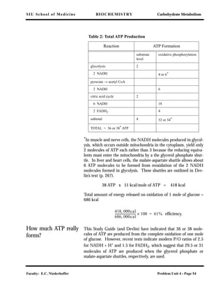
Stoichiometry of the
TCA cycle
The citric acid cycle satisfies stoichiometric requirements for the
complete oxidation of pyruvate by oxygen to carbon dioxide and
water, viz.
Oxidation of pyruvate
CH3-CO-COO- + 5 [O] + H+ ⇔ 3 CO2 + 2 H2O
The uptake of 5 atoms of oxygen corresponds to the removal of 5
pairs of electrons during operation of the cycle. Later the electrons
are transferred to oxygen. Note that oxygen is not directly utilized in
the TCA cycle. Rather, it acts as the electron acceptor at the end of
the electron transport chain. For each atom of oxygen reduced, one
molecule of water is formed. Because 3 molecules of water are taken
up during operation of the cycle, the net change corresponds to the
formation of 2 moles of water per mole of pyruvate oxidized.
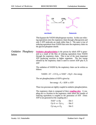
The Respiratory Chain The respiratory chain (or electron transport chain (ETC)) is a
group of proteins located in the inner membrane of the mitochondria
which channel reducing equivalents to oxygen. Reducing equiva-
lents are the electrons (and sometimes protons) which are lost by a
substrate when it is oxidized and, thus, are capable of reducing
another compound.
Electrons flow through the respiratory chain from a high energy level
to a low energy level, much as water flows downhill. The electron
transport chain serves as a conduit, or pipe, through which the reduc-
ing equivalents may flow, and simultaneously capture a portion of the](https://image.slidesharecdn.com/biochemistry-150402010123-conversion-gate01-190216190441/85/Biochemistry-150402010123-conversion-gate01-43-320.jpg)
![SIU School of Medicine BIOCHEMISTRY Carbohydrate Metabolism
Faculty: E.C. Niederhoffer Problem Unit 4 - Page 44
energy, similar to a turbine in a hydroelectric plant which converts
the downhill flow of water to electricity. In this case, the flow of
reducing equivalents is coupled to pumping protons across a mem-
brane to create a proton concentration gradient that can be utilized
elsewhere to do work and synthesize ATP. A pair of electrons and
two protons are removed from a substrate by a dehydrogenase, result-
ing in oxidation of the substrate. The electrons and one proton (from
NADH) are passed together through the first part of the chain to
coenzyme Q (ubiquinone). Then the electrons are passed through
the cytochrome system, finally reducing an atom of oxygen. The
reduction of oxygen by the reducing equivalents results in the forma-
tion of water:
[O] + 2 e- + 2 H+ ⇔ H2O
There are three questions that need to be considered:
a) where do the reducing equivalents which feed the respiratory
chain come from?;
b) what are the members of the respiratory chain which channel the
reducing equivalents?; and
c) what reactions are involved that permit the capture of energy?
The origin of reducing
equivalents
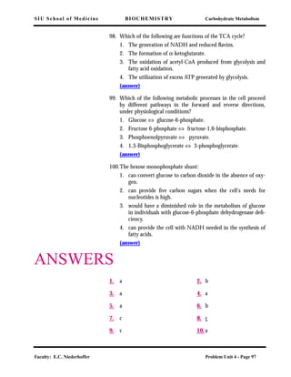
The product of glycolysis is pyruvate which is transported into the
mitochondria. Pyruvate releases energy during its oxidation to CO2
in the tricarboxylic acid cycle. The reducing equivalents then enter
the respiratory chain.
Biological oxidations, like any other oxidations, involve the loss of
electrons. Frequently, protons are lost at the same time, i.e., hydro-
gen atoms are removed. The enzymes that catalyze these reactions
are, therefore, called dehydrogenases. There must be an acceptor for
the electrons. When something is oxidized, something else must be
reduced. The actual electron acceptor in the dehydrogenases is one of
several coenzymes (usually NAD+
or FAD).
Dehydrogenases cata-
lyze the oxidation of
substrates
Many oxidation enzymes use NAD+
as a coenzyme. For example, the
enzyme alcohol dehydrogenase has NAD+ as a coenzyme and medi-
ates the following reaction.
CH3-CH2OH ⇔ CH3-CHO + 2 H+
+ 2 e-
ethanol acetaldehyde
(an alcohol) (an aldehyde)](https://image.slidesharecdn.com/biochemistry-150402010123-conversion-gate01-190216190441/85/Biochemistry-150402010123-conversion-gate01-44-320.jpg)
![SIU School of Medicine BIOCHEMISTRY Carbohydrate Metabolism
Faculty: E.C. Niederhoffer Problem Unit 4 - Page 46
Cyt (Fe2+
) ⇔ Cyt (Fe3+
) + e-
There are several cytochromes in the respiratory chain, viz. b1, c1, c,
a, and a3. Thus, the oxidized form of cytochrome c1 is represented
as Cyt c1(Fe3+) while the reduced form of cytochrome c1 is repre-
sented as Cyt c1(Fe2+
).
Non-heme iron pro-
teins
Non-heme iron proteins contain iron which is not associated with a
porphyrin ring system. The iron is attached to the protein through
iron sulfur clusters, typically [2Fe-2S] or [4Fe-4S] clusters attached to
the protein by coordination with the sulfur of cysteine sulfhydryl
groups. Such proteins are often referred to as iron-sulfur proteins.
The iron atoms can transfer electrons by existing in either oxidized or
reduced states. They are usually closely associated with flavoproteins.
Four complexes in the
Chain
There are four protein complexes which make up the electron trans-
port chain. Complex I is the NADH dehydrogenase complex which
receives the reducing equivalents from NADH (from the TCA cycle).
It is a flavoprotein containing both FMN and non-heme iron (iron-
sulfur) centers. The reducing equivalents exit the complex and are
passed to CoQ, which is freely soluble in the mitochondrial mem-
brane and diffuses to Complex III. Complex II is the succinate dehy-
drogenase discussed below. The reducing equivalents from Complex
II are also passed to CoQ. Complex III is a cytochrome b and c1
complex, which accepts the electrons from ubiquinone (CoQ). The
electrons are passed from Complex III to cytochrome c, a protein
loosely associated with the membrane. Cytochrome c acts as an elec-
tron carrier to transport the electrons to Complex IV, also known as
cytochrome oxidase and contains cytochrome aa3. Cytochrome oxi-
dase catalyzes the reduction of O2 to H2O. This is the only point
where O2 is utilized in oxidative metabolism of sugars.
The respiratory chain
may be entered at two
points
Succinate feeds reducing equivalents directly into the chain via the
following reaction:](https://image.slidesharecdn.com/biochemistry-150402010123-conversion-gate01-190216190441/85/Biochemistry-150402010123-conversion-gate01-46-320.jpg)
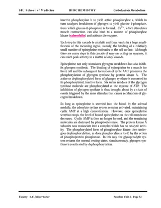
![SIU School of Medicine BIOCHEMISTRY Carbohydrate Metabolism
Faculty: E.C. Niederhoffer Problem Unit 4 - Page 48
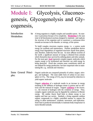
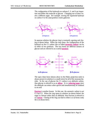
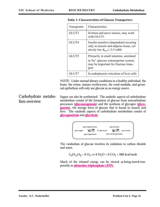
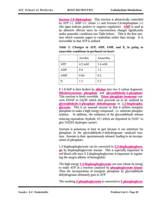
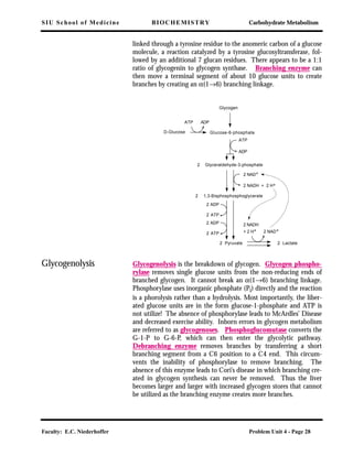
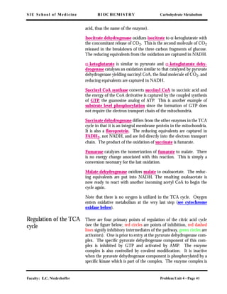
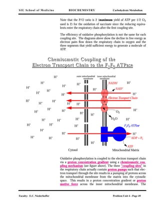
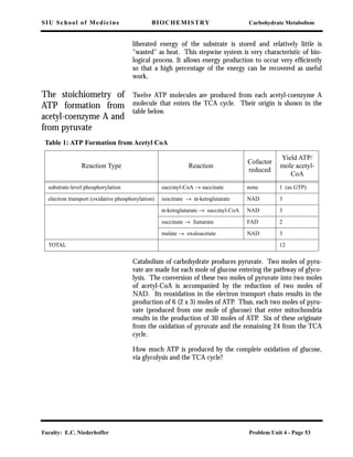
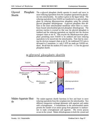
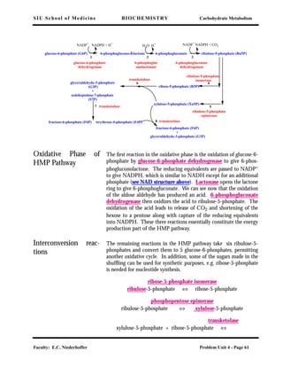
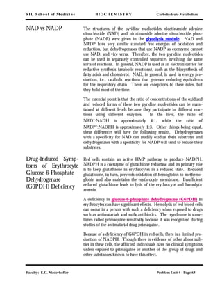
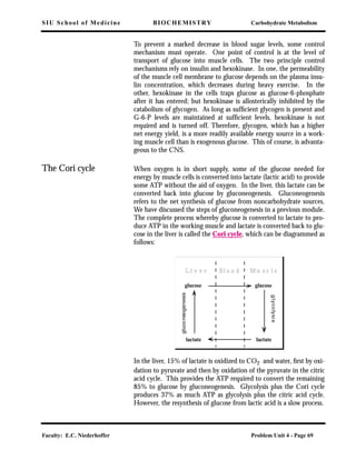
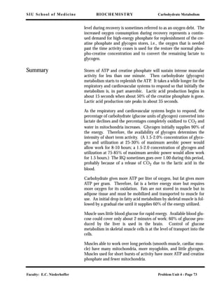
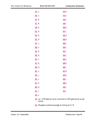
There are two electron carriers that bridge these sites – CoQ between
site 1 and site 2 and cyt c between site 2 and site 3. O2 is the ultimate
electron acceptor after site 3.
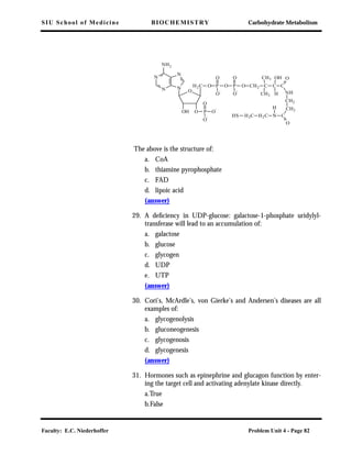
When all three coupling sites are utilized, the maximum yield of
ATP per 1/2 O2 used is 3. The ratio (Pi converted to ATP) / (1/2 O2
[flux of 2e]) is called the P/O ratio.
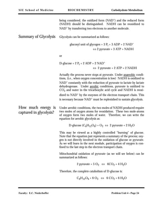
The decline in free energy as electron pairs
flow down the respiratory chain to oxygen.
Each of the three segments yields sufficient
energy to generate a molecule of ATP from
ADP and phosphate
kcal
Q
b
a
c
NAD
50
40
30
20
10
0.27 V
12.2 kcal
0.22 V
9.9 kcal
0.59 V
23.0 kcal
-∆G°'
Substrate
NAD
Flavoprotein
Cytochromes
Oxygen
A schematic view of the
free-energy changes associated
with the oxidative pathway](https://image.slidesharecdn.com/biochemistry-150402010123-conversion-gate01-190216190441/85/Biochemistry-150402010123-conversion-gate01-48-320.jpg)
![SIU School of Medicine BIOCHEMISTRY Carbohydrate Metabolism
Faculty: E.C. Niederhoffer Problem Unit 4 - Page 80
[ATP])
a. 10-6
M glucose
b. 10-4 M glucose
c. 10-2 M glucose
(answer)
19. Lactic dehydrogenase functions most importantly in:
a. aerobic metabolism
b. anaerobic metabolism
(answer)
20. When a cell is switched from aerobic metabolism to anaerobic
metabolism, the ATP concentration changes roughly from 4.2
mM to:
a. 10 mM
b. 4 mM
c. 0.1 mM
(answer)
21.The reaction which irreversibly commits sugar to the glycolytic
pathway is catalyzed by:
a. hexokinase or glucokinase
b. phosphofructokinase
c. phosphoglucomutase
d. glucose phosphate isomerase
e. aldolase
(answer)
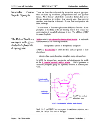
22. NAD+ contains which of the following?
a. thiamine
b. lipoic acid
c. niacin
d. riboflavin
e. CoA
(answer)
23. Phosphoglycerate kinase functions in carbohydrate metabolism
to produce ATP via:
a. oxidative phosphorylation](https://image.slidesharecdn.com/biochemistry-150402010123-conversion-gate01-190216190441/85/Biochemistry-150402010123-conversion-gate01-80-320.jpg)
This document provides an introduction to carbohydrate metabolism. It discusses glycolysis, glycogenesis, glycogenolysis, and gluconeogenesis. Specifically, it covers the basic principles of metabolism, including the roles of enzymes as catalysts and in regulating metabolic pathways. It also provides learning objectives and defines key terms related to carbohydrate metabolism.


![SecurityBoat_Service_Pitch_Deck[24158].pdf](https://cdn.slidesharecdn.com/ss_thumbnails/securityboatservicepitchdeck24158-260121113056-452683e3-thumbnail.jpg?width=640&height=640&fit=bounds)


