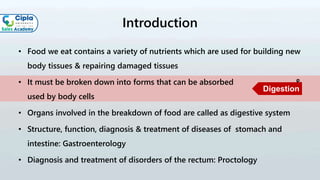
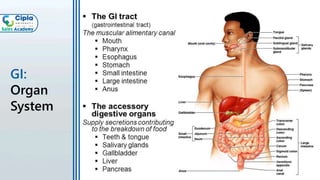
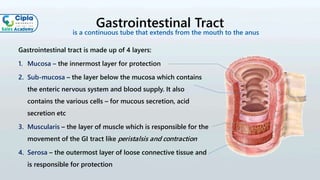
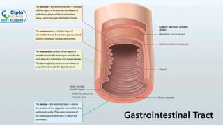
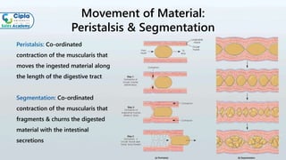
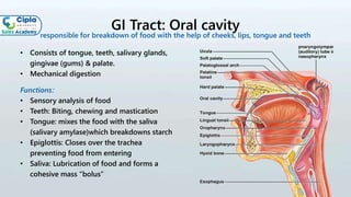
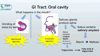
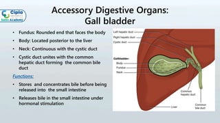
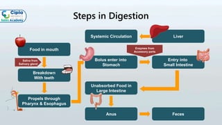
Gastrointestinal System
Basics of
Introduction
• Food we eat contains a variety of nutrients which are used for building new
body tissues & repairing damaged tissues
• It must be broken down into forms that can be absorbed &
used by body cells
• Organs involved in the breakdown of food are called as digestive system
• Structure, function, diagnosis & treatment of diseases of stomach and
intestine: Gastroenterology
• Diagnosis and treatment of disorders of the rectum: Proctology
Digestion
GI:
Organ
System
Gastrointestinal Tract
Gastrointestinal tract is made up of 4 layers:
1. Mucosa – the innermost layer for protection
2. Sub-mucosa – the layer below the mucosa which contains
the enteric nervous system and blood supply. It also
contains the various cells – for mucous secretion, acid
secretion etc
3. Muscularis – the layer of muscle which is responsible for the
movement of the GI tract like peristalsis and contraction
4. Serosa – the outermost layer of loose connective tissue and
is responsible for protection
is a continuous tube that extends from the mouth to the anus
Gastrointestinal Tract
Movement of Material:
Peristalsis & Segmentation
Peristalsis: Co-ordinated
contraction of the muscularis that
moves the ingested material along
the length of the digestive tract
Segmentation: Co-ordinated
contraction of the muscularis that
fragments & churns the digested
material with the intestinal
secretions
GI Tract: Oral cavity
• Consists of tongue, teeth, salivary glands,
gingivae (gums) & palate.
• Mechanical digestion
Functions:
• Sensory analysis of food
• Teeth: Biting, chewing and mastication
• Tongue: mixes the food with the saliva
(salivary amylase)which breakdowns starch
• Epiglottis: Closes over the trachea
preventing food from entering
• Saliva: Lubrication of food and forms a
cohesive mass “bolus”
responsible for breakdown of food with the help of cheeks, lips, tongue and teeth
•
GI Tract: Oral cavity
Saliva
contains
Lingual
lipase
(produced by
lingual glands)
Triglycerides
Fatty acids &
diglycerides
GI Tract: Pharynx
• When food is first swallowed, it passes
from the mouth into the pharynx
• It is composed of skeletal muscle & lined
by mucous membrane
Functions:
• Oropharynx and hypopharynx: Digestive
as well as respiratory functions
• Swallowed food passes from the mouth
into the oropharynx & hypopharynx
• Muscle contraction: Propel food into
esophagus
muscular tube that connects the nasal cavities to the larynx and esophagus
GI Tract: Esophagus
It is lined with a special mucous membrane that protects it from the constant passage of food
Functions:
• Swallowing and passage of food from
pharynx to stomach
• Mucous secretion
• Prevents acid reflux from the stomach
muscular tube connecting pharynx to stomach.
GI Tract: Stomach
Fundus: Above the level of the opening
between the stomach and esophagus.
Cardia: Point where the esophagus joins the
stomach
Body: Main central portion
Pylorus: The pylorus is divided into pyloric
antrum, pyloric canal & pyloric sphincter
• 3 types of muscles: Oblique, circular &
longitudinal
• Rugae: Inner surface thrown into folds
• It is lined with columnar epithelial cells
J-shaped enlargement of the GI tract, that serves as a mixing
and holding reservoir of food
GI Tract: Stomach
Function
GI Tract: Small intestine
Duodenum:
• Short structure in continuation with the
stomach
• Digestion of proteins takes place here
Jejunum:
• Mid section of small intestine connecting
the duodenum to the terminal part of the
small intestine
• Most of the absorption of nutrients takes
place here
Ileum:
• It is about 3 m long, and
contains villi similar to jejunum
• It absorbs mainly vitamin B12 and bile acids
starts at pyloric sphincter of the stomach and ends in the large intestine
GI Tract: Large intestine
• Ilium opens in to the large intestine through
an opening called as ilocecal sphincter
• Cecum: Small pouch attached to it is a
twisted, coiled tube measuring about 8 cm
called appendix
• Colon: Open end of the cecum merges with a
long tube called the colon (Ascending colon-
transverse colon-descending colon-sigmoid
colon)
• Rectum: Last section of the colon
• Anal canal: Opening of the anal canal is
called the anus. It is guarded by the internal
anal sphincter and external anal sphincter
it is the terminal portion of the GI tract
Accessory Digestive Organs:
Salivary glands
Major: Lie completely outside the alimentary tract and connected by an excretory duct
• Parotid, Submandibular, Sublingual
Minor: Lip, cheeks, soft palate
Functions:
• Secrete saliva composed of water, mucus,
proteins, salts, salivary amylase (ptyalin),
immunoglobulins (IgA) & lactoperoxidase
• Serves to moisten food
• Lubricates & moistens oral mucosa and lip
• Initiates the digestion of carbohydrates
Accessory Digestive Organs: Pancreas
gland which lies behind the stomach
Accessory Digestive Organs: Liver
Functional unit of the liver are known as hepatocytes which are involved in absorption and
secretion of certain components
Functions:
• Secretion of bile which helps digest fats
• Involved in synthesis of plasma proteins
• Involved in detoxification drugs and alcohol
• Carbohydrate metabolism and regulation of
blood glucose
• Storage for Vitamins A, D, E,K and B12 and
minerals (iron and copper)
Accessory Digestive Organs:
Gall bladder
• Fundus: Rounded end that faces the body
• Body: Located posterior to the liver
• Neck: Continuous with the cystic duct
• Cystic duct unites with the common
hepatic duct forming the common bile
duct
Functions:
• Stores and concentrates bile before being
released into the small intestine
• Releases bile in the small intestine under
hormonal stimulation
Steps in Digestion
Food in mouth
Breakdown
With teeth
Propels through
Pharynx & Esophagus
Bolus enter into
Stomach
Entry into
Small Intestine
LiverSystemic Circulation
Unabsorbed Food in
Large Intestine
FecesAnus
Saliva from
Salivary gland
Enzymes from
Accessory parts
Blood Supply to the GIT
• Supplied by the celiac artery
• Celiac artery supplies the liver, stomach, spleen and the upper halves of the duodenum and
the pancreas with oxygenated blood
• Most of the blood is returned to the liver via the portal venous system for further
processing and detoxification before returning to the systemic circulation via the hepatic
portal vein
• Three main divisions are : Gastric artery,
Common hepatic artery, splenic artery
• Intestines is primarily supplied by superior
and inferior mesenteric arteries
Let’s learn with
some videos
Digestive System
COMPLETE YOUR ASSESSMENT ON NALANDA
THANK YOU

Basics of GI System

  • 1.
  • 2.
    Introduction • Food weeat contains a variety of nutrients which are used for building new body tissues & repairing damaged tissues • It must be broken down into forms that can be absorbed & used by body cells • Organs involved in the breakdown of food are called as digestive system • Structure, function, diagnosis & treatment of diseases of stomach and intestine: Gastroenterology • Diagnosis and treatment of disorders of the rectum: Proctology Digestion
  • 3.
  • 4.
    Gastrointestinal Tract Gastrointestinal tractis made up of 4 layers: 1. Mucosa – the innermost layer for protection 2. Sub-mucosa – the layer below the mucosa which contains the enteric nervous system and blood supply. It also contains the various cells – for mucous secretion, acid secretion etc 3. Muscularis – the layer of muscle which is responsible for the movement of the GI tract like peristalsis and contraction 4. Serosa – the outermost layer of loose connective tissue and is responsible for protection is a continuous tube that extends from the mouth to the anus
  • 5.
  • 6.
    Movement of Material: Peristalsis& Segmentation Peristalsis: Co-ordinated contraction of the muscularis that moves the ingested material along the length of the digestive tract Segmentation: Co-ordinated contraction of the muscularis that fragments & churns the digested material with the intestinal secretions
  • 7.
    GI Tract: Oralcavity • Consists of tongue, teeth, salivary glands, gingivae (gums) & palate. • Mechanical digestion Functions: • Sensory analysis of food • Teeth: Biting, chewing and mastication • Tongue: mixes the food with the saliva (salivary amylase)which breakdowns starch • Epiglottis: Closes over the trachea preventing food from entering • Saliva: Lubrication of food and forms a cohesive mass “bolus” responsible for breakdown of food with the help of cheeks, lips, tongue and teeth •
  • 8.
    GI Tract: Oralcavity Saliva contains Lingual lipase (produced by lingual glands) Triglycerides Fatty acids & diglycerides
  • 9.
    GI Tract: Pharynx •When food is first swallowed, it passes from the mouth into the pharynx • It is composed of skeletal muscle & lined by mucous membrane Functions: • Oropharynx and hypopharynx: Digestive as well as respiratory functions • Swallowed food passes from the mouth into the oropharynx & hypopharynx • Muscle contraction: Propel food into esophagus muscular tube that connects the nasal cavities to the larynx and esophagus
  • 10.
    GI Tract: Esophagus Itis lined with a special mucous membrane that protects it from the constant passage of food Functions: • Swallowing and passage of food from pharynx to stomach • Mucous secretion • Prevents acid reflux from the stomach muscular tube connecting pharynx to stomach.
  • 11.
    GI Tract: Stomach Fundus:Above the level of the opening between the stomach and esophagus. Cardia: Point where the esophagus joins the stomach Body: Main central portion Pylorus: The pylorus is divided into pyloric antrum, pyloric canal & pyloric sphincter • 3 types of muscles: Oblique, circular & longitudinal • Rugae: Inner surface thrown into folds • It is lined with columnar epithelial cells J-shaped enlargement of the GI tract, that serves as a mixing and holding reservoir of food
  • 12.
  • 13.
    GI Tract: Smallintestine Duodenum: • Short structure in continuation with the stomach • Digestion of proteins takes place here Jejunum: • Mid section of small intestine connecting the duodenum to the terminal part of the small intestine • Most of the absorption of nutrients takes place here Ileum: • It is about 3 m long, and contains villi similar to jejunum • It absorbs mainly vitamin B12 and bile acids starts at pyloric sphincter of the stomach and ends in the large intestine
  • 14.
    GI Tract: Largeintestine • Ilium opens in to the large intestine through an opening called as ilocecal sphincter • Cecum: Small pouch attached to it is a twisted, coiled tube measuring about 8 cm called appendix • Colon: Open end of the cecum merges with a long tube called the colon (Ascending colon- transverse colon-descending colon-sigmoid colon) • Rectum: Last section of the colon • Anal canal: Opening of the anal canal is called the anus. It is guarded by the internal anal sphincter and external anal sphincter it is the terminal portion of the GI tract
  • 15.
    Accessory Digestive Organs: Salivaryglands Major: Lie completely outside the alimentary tract and connected by an excretory duct • Parotid, Submandibular, Sublingual Minor: Lip, cheeks, soft palate Functions: • Secrete saliva composed of water, mucus, proteins, salts, salivary amylase (ptyalin), immunoglobulins (IgA) & lactoperoxidase • Serves to moisten food • Lubricates & moistens oral mucosa and lip • Initiates the digestion of carbohydrates
  • 16.
    Accessory Digestive Organs:Pancreas gland which lies behind the stomach
  • 17.
    Accessory Digestive Organs:Liver Functional unit of the liver are known as hepatocytes which are involved in absorption and secretion of certain components Functions: • Secretion of bile which helps digest fats • Involved in synthesis of plasma proteins • Involved in detoxification drugs and alcohol • Carbohydrate metabolism and regulation of blood glucose • Storage for Vitamins A, D, E,K and B12 and minerals (iron and copper)
  • 18.
    Accessory Digestive Organs: Gallbladder • Fundus: Rounded end that faces the body • Body: Located posterior to the liver • Neck: Continuous with the cystic duct • Cystic duct unites with the common hepatic duct forming the common bile duct Functions: • Stores and concentrates bile before being released into the small intestine • Releases bile in the small intestine under hormonal stimulation
  • 19.
    Steps in Digestion Foodin mouth Breakdown With teeth Propels through Pharynx & Esophagus Bolus enter into Stomach Entry into Small Intestine LiverSystemic Circulation Unabsorbed Food in Large Intestine FecesAnus Saliva from Salivary gland Enzymes from Accessory parts
  • 20.
    Blood Supply tothe GIT • Supplied by the celiac artery • Celiac artery supplies the liver, stomach, spleen and the upper halves of the duodenum and the pancreas with oxygenated blood • Most of the blood is returned to the liver via the portal venous system for further processing and detoxification before returning to the systemic circulation via the hepatic portal vein • Three main divisions are : Gastric artery, Common hepatic artery, splenic artery • Intestines is primarily supplied by superior and inferior mesenteric arteries
  • 21.
  • 22.
  • 23.
    COMPLETE YOUR ASSESSMENTON NALANDA THANK YOU