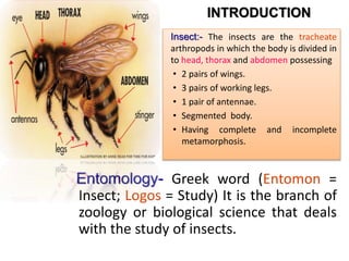
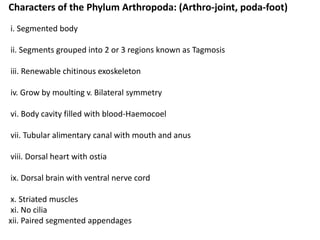
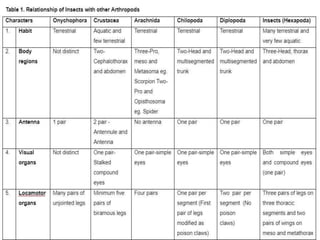
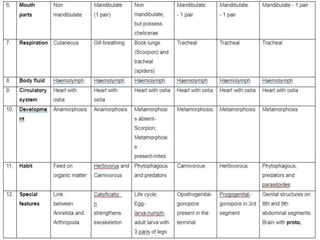
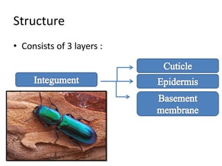
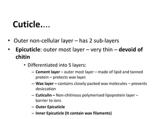
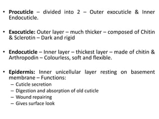
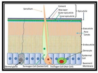
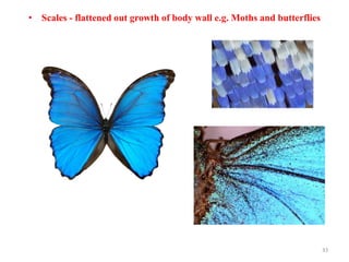
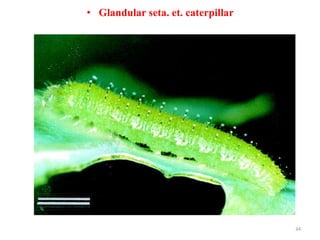
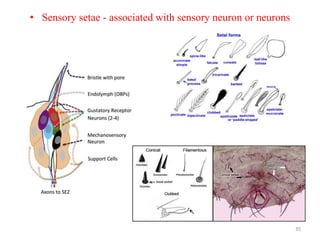
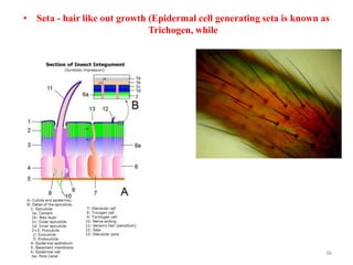
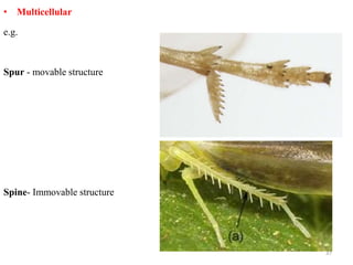
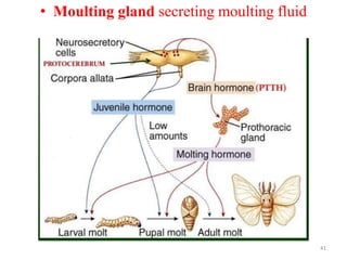
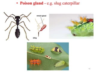
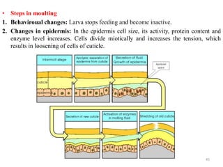
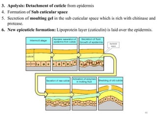
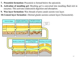
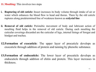
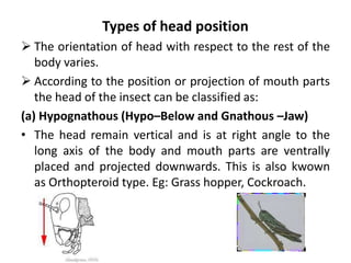
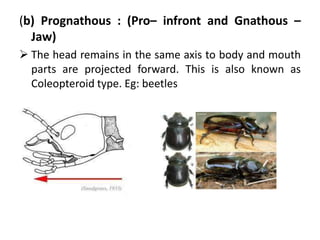
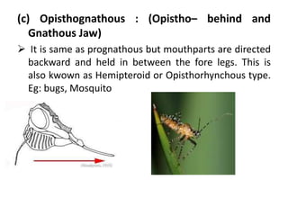
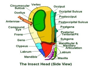
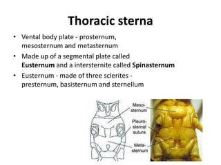
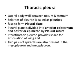
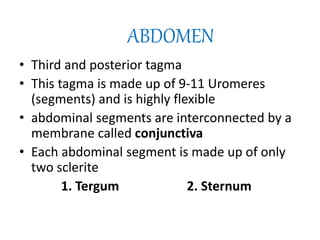
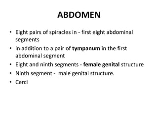
1. Course No. – ENTO-121
TITLE-FUNDAMENTALS OF
ENTOMOLOGY
Dr. Anita Sharma
Asstt. Professor (Entomology)
2. Entomology- Greek word (Entomon =
Insect; Logos = Study) It is the branch of
zoology or biological science that deals
with the study of insects.
INTRODUCTION
Insect:- The insects are the tracheate
arthropods in which the body is divided in
to head, thorax and abdomen possessing
• 2 pairs of wings.
• 3 pairs of working legs.
• 1 pair of antennae.
• Segmented body.
• Having complete and incomplete
metamorphosis.
3. • Insect is Greek word = Cut in pieces or
segmented.
• Insects belong to the Phylum Arthropoda (Artho=
jointed, Poda= Legs) which is the biggest phylum
of kingdom Animalia. More than three quarters
of the animals on earth are arthropods, and most
of these are insects.
• Kingdom Animalia is classified into twelve phyla.
INTRODUCTION
4. • Study and use of insects in crime investigations is known
as Forensic Entomology.
• Study of insects related to live stock and veterinary
animals is known as Veterinary Entomology.
• Study of insects in relation to Human beings is known as
Medical Entomology.
• Study of insects in relation to Agriculture is known as
Agriculture Entomology
Branches of Entomology
5. Topic -1, HISTORY OF ENTOMOLOGY IN INDIA
Aristotle (384-322 B.C.)– Father of biological classification. First person grouped insects in
winged and wing less groups. He gave the terms like Coleoptera and Diptera.
Carolus Linnaeus (1758)– Father of Taxonomy.
William Kirby considered as “Founder of Entomology” because of his significant role in
Entomology in the world. He published a book “An Introduction to Entomology” (1815-
1826).
Snodgrass R. E. (1875)– referred as a Father of Insect Morphology. He wrote book -
Principles of Insect Morphology.
Mithan Lal Runwal (1908)– Outstanding work on termites/ white ants. Contributions to
ecology, embryology and locust.
1940 - Dr. T.V. Ramakrishna Ayyar published the book "Handbook of Economic Entomology"
which met the long felt need of the students of Agriculture and agricultural scientists as well
and also known as “Father of Indian Entomology”.
Dr.S.Pradhan (1969) - Wrote a "Insect Pests of Crops" and Father of Modern Applied
Entomology in India.
6. History of Entomology in India
1758 - 10th edition of Systema Naturae by Linnaeus with only 12 Indian insects which was
the earliest record.
1779 - Dr. J.G. Koenig - Medical Officer initiated the work on Indian insects on scientific
lines. He also published a special account of the termites of Thanjavur District.
1782 - Dr. Kerr Published on account of lac insect.
1785 - Asiatic Society of Bengal started in Calcutta and many papers were published in the
Societys publications.
1790 - Roxburgh (Botanist) published a detailed account of lac insect.
1791 - Dr. J. Anderson issued a monograph on Cochineal scale insects
1800 - Buchanan (Traveller) wrote on the cultivation of lac in India and on sericulture in
some parts of South India. Denovan published Natural History of Insects which was the first
contribution on the insects of Asia and was revised in 1842 by West Wood.
1875 - Foundation of the Indian Museum at Calcutta
7. 1883 - Bombay Natural History Society was started. After the foundation of these two
organisations scientific studies received greater attention in India. Numerous contributions
of Indian insects were published in the Journal of the Bombay Natural History.
1893 - Rothney published on Indian Ants (earliest record of biological pest control in
India) i.e. White ants attach on stationary items was kept free by red ants.
1897 - Bingham's issued volumes on "Hymenoptera' (Ants, bees and wasps). Since than
volumes on other groups of insects like Coleoptera (beetles), Hemiptera (bugs), Odonata
(dragenfly and damselfly), etc., were published.
1889 - Indian Museum, Calcutta published the Indian Museum Notes in five volumes.
1901 - (Lionel de Nicevelle) posting of the first entomologist to the Government of India.
1905 - Establishment of Imperial Agricultural Research Institute at Pusa (Bihar). Maxwell
Lefroy became first Imperial Entomologist of Govt. of India.
1906 - “Indian Insect Pests” & “Indian Insect Life” Books by Professor Maxwell.
Subsequently State Governments also took up entomological work.
1914 - T.B. Fletcher, the first Government Entomologist of Madras State, published his
book "Some South Indian Insects".
8. 1916 –”Indian Forest of Economic Importance: Coleoptera; was published by the first
Imperial Forest Entomologist E.P. Stebbing”.
1921 - Indian Central Cotton Committee to investigate on pests of cotton.
1925 - Indian Lac Research Institute.
1934- Hem Singh Pruthi as Imperial Entomologist, start ‘Entomological Society of India’ in
1938. Afzal Hussain was the first president of the society and VC were HS Pruthi and Ayyar.
1940 - Dr. T.V. Ramakrishna Ayyar published the book "Handbook of Economic
Entomology" which met the long felt need of the students of Agriculture and agricultural
scientists as well and also known as “Father of Indian Entomology”.
1968 - Dr. M.S. Mani's "General Entomology"
1969 - Dr. H.s. Pruthi's "Textbook of Agricultural Entomology". Dr. Pradhan's "Insect Pests
of Crops“
1946 - Government of India started the "Directorate of plant protection, quarentine and
storage.".
1960 - "The Desert Locust in India" monograph by Y.R. Rao.
1969 - "The monograph on Indian Thysanoptera" by Dr. T.N. Ananthakrishnan
9. o 1912– Plant Quarantine Act.
o 1914–Destructive Insects and Pests Act (DIPA).
o 1916–Imperial Forest Research Institute at Dehradun.
o 1925–Indian Lac Research Institute started at Ranchi.
o 1937–A laboratory for storage pests was started at Hapur, U.P.
o 1937- Establishment of Entomology division at IARI New Delhi.
o 1939–Locust Warning Organization established at Jodhpur.
o 1946–‘Directorate of Plant Protection, Quarantine & Storage at Faridabaad.
o 1968– ‘Central Insecticide Act’.
Institutes/Organizations
10. • NCIPM, New Delhi-(National Centre for Integrated Pest
Management - 1988).
• PDBC, Bangalore-(Project Directorate of Biological Control -1993).
• NBAIR-National Bureau of Agricultural Insect Resources and
formerly it is a NBAII, Bengalore in 1957.
• CIB, Faridabad – Central Insecticide Board.
• NPPTI, Hyderabad–National Plant Protection Training Institute.
Entomological Institutes
11. FACTORS FOR INSECTS ABUNDANCE
Measures of dominance
1. More number of species: In the animal kingdom more than 85 per cent of the
species belongs to insect group. Total number of insects described so far is more than
9 lakhs.
2. Large number of individuals in a single species: e.g., Locust swarm comprising of
109 number of individuals, occupying large area.
3. Great variety of habitats: Insects thrive well under varied conditions.
4. Long geological history: Insects were known to occupy this earth for more than
350 million years, which is a good track record. This has given the insects great
variety of adoptions under different conditions.
12. Reasons for dominance
1. Capacity for flight: Insects posess wings, which is the lateral extension of
exoskeleton. Insects are the earliest animals and the only flying invertebrates.
Flight is used for the following purpose-
i. To seek food, mate, shelter and oviposition sites
ii. To colonize in a new habitat and also to exchange habitat.
iii. To escape from enemies and unfavourable conditions.
iv. To migrate (i.e. for long distance travel e.g. Locusts)
2. Adaptability or Universality: Insects are the earliest groups to make their life
on the earth and to occupy vast habitats of soil and water.
i. Found in wide range of climatic conditions, from -50ñC to 40ñC.
ii. Psilopa petroli found in crude petroleum well.
iii. Ephydra fly living in great salt lake.
iv. Every flowering plant providing food for one or many Phytophagous insects.
v. Even the decomposing materials serving as food for many Saprophagous
insects.
vi. Many Carnivorous insects are parasitic on other animals and insects.
13. 3. Size: Majority of insects are small conferring the following physiological and
ecological advantages.
i. Less space, food, time and energy requirements for development and sustaining
life.
ii. Energy Utilization maximum.
iii. Less gravitational effect.
iv. Muscular action and tracheal respiration more effective.
v. Easy escape from enemies.
4. Exoskeleton: Insect body is covered with an outer cuticle called exoskeleton which
is made up of a cuticular protein called Chitin. This is light in weight and gives
strength, rigidity and flexibility to the insect body.
Uses:
i.Act as external armour
ii.Provides space for muscle attachment
iii.Prevents water loss
14. 5. Resistance to desiccation: Insects minimise the water loss from their body
surface through the following processes.
I. Prevention of water loss:
i. Lipids and polyphenols present in the Epicuticle acts as water proofing.
ii. Was layer with closely packed wax molecules prevents escape of water.
iii. Spiracles are closed to prevent water loss.
iv. In the egg stage shell development prevents water loss and desication of inner
embryos.
II. Conservation of water
i. Capable of utilizing metabolic water
ii. Rectal resorption of water from faeces.
iii. Terrestrial insects use less quantity of water to remove the nitrogenous waste
(Uric acid) which is water insoluble.
6. Tracheal system of respiration: This ensures direct transfer of adequate oxygen
to actively breathing tissues. Spiracles through their closing mechanism admit air
and restrict water loss.
15. 7. Reproductive potential: Reproductive potential of insect is high due to the
following reasons:
i Egg laying capacity (fecundity) is high. e.g., Queen termite lays 6000 - 7000 eggs per
day for 15 long years.
ii. Development period is short. e.g., Corn aphid produces 16 nymphs per female
which reaches the adulthood within 16 days. There by one generation is completed
within a short period of 16 days, which favours greater genetic changes in the insect
population, like quicker development of insecticide resistant strains.
iii. Careful selection of egg lying sites and protection of eggs.
iv. Exhibits parental care like progressive provisioning (e.g. bees) and mass
provisioning (e.g. Wasps)
v. Presence of special types of reproduction other than oviparity and viviparity.
* Polyembryony: Development of many individuals from a single egg. e.g. parasitic
wasps.
* Parthenogenesis: Reproduction without male or without fertilization, e.g. aphids
* Paedogenesis: Reproduction by immature stages. e.g. certain flies.
16. 8. Complete metamorphosis: More than 82 per cent of insects undergo complete
metamorphosis (Holometabolous insects) with the following four stages.
i. Egg: Inactive, inexpensive, inconspicuous and embryo develops inside.
ii. Larva: Active, feeds, digests, grows and store food.
iii. Pupa: Inactive, internal reorganisation and resist adverse conditions.
iv. Adult: Active, reproduce and disperse.
As the larval and adult food sources are different, competition for food is less.
9. Defense mechanisms: By using the following defense mechanisms, insects escape
from the enemies to increase their survival rate.
i. Behavioural: Thanatosis - insects pretends as if dead. e.g. some beetles.
ii. Structural e.g. hardened forewings of beetles known as elytra protect the beetles
from predation of birds.
iii. Colourational: Presence of protective colours. e.g.Stick insects
iv. Chemical: Presence of defensive chemicals. e.g. Bees producing venom
10. Hexapod locomotion: Insects uses 3 legs at a time during locomotion, while the
remaining 3 legs are static, which gives greater stability.
19. POSITION OF INSECTS IN ANIMAL KINGDOM AND ITS
RELATIONSHIP WITH OTHER ARTHROPODA
Classification: 7 classes.
Phylum : Arthropoda
Classes :
1. Onychophora (claw bearing)e.g. Peripatus, has similarities with arthropoda like
antenae, open circulatory system, walking legs with claw and tracheal system.
2. Crustacea (Crusta - shell)e.g. Prawn, crab, wood louse
3. Arachnida (Arachne - spider)e.g. Scorpion, spider, tick, mite
4. Chilopoda (Chilo - lip; poda - appendage)e.g. Centipedes (carnivorous)
5. Diplopoda (Diplo - two; poda- - appendage)e.g. Millipede (scavengers)
6. Trilobita (an extinct group)- The dominant arthropodes in the early Paleozoic
seas (many million ago)
7. Hexapoda or Insectae.g. Insects.
20. Characters of the Phylum Arthropoda: (Arthro-joint, poda-foot)
i. Segmented body
ii. Segments grouped into 2 or 3 regions known as Tagmosis
iii. Renewable chitinous exoskeleton
iv. Grow by moulting v. Bilateral symmetry
vi. Body cavity filled with blood-Haemocoel
vii. Tubular alimentary canal with mouth and anus
viii. Dorsal heart with ostia
ix. Dorsal brain with ventral nerve cord
x. Striated muscles
xi. No cilia
xii. Paired segmented appendages
21.
22.
23. • Insect body wall – Integument/Exoskeleton
• External covering – ectodermal in origin
• Rigid, flexible, lighter, stronger and variously
modified
Insect Integument: Structure and Function
25. • Outer non-cellular layer – has 2 sub-layers
• Epicuticle: outer most layer – very thin – devoid of
chitin
• Differentiated into 5 layers:
– Cement layer – outer most layer – made of lipid and tanned
protein – protects wax layer.
– Wax layer – contains closely packed wax molecules – prevents
desiccation
– Cuticulin – Non-chitinous polymerised lipoprotein layer –
barrier to ions
– Outer Epicuticle
– Inner Epicuticle (It contain wax filaments)
Cuticle....
26. • Procuticle – divided into 2 – Outer exocuticle & Inner
Endocuticle.
• Exocuticle: Outer layer – much thicker – composed of Chitin
& Sclerotin – Dark and rigid
• Endocuticle – Inner layer – thickest layer – made of chitin &
Arthropodin – Colourless, soft and flexible.
• Epidermis: Inner unicellular layer resting on basement
membrane – Functions:
– Cuticle secretion
– Digestion and absorption of old cuticle
– Wound repairing
– Gives surface look
27.
28. Composition....
Chitin: Main constituent of cuticle
– Nitrogenous polysaccharide and polymer of N-
acetylglucosamine.
– (water insoluble and soluble in acids, alkalies and
organic solvents)
Arthropodin: Untanned cuticular protein (water soluble).
Sclerotin : Tanned cuticular protein (water insoluble).
Resilin: Elastic cuticular protein – for flexibility of sclerites.
29. • Cuticular in growth of body wall – provide
space for muscle attachment. 2 types:
– Apodeme – hollow invagination of body wall
(ridge like)
– Apophysis – Solid invagination of body wall (spine
or fingure like)
Endoskeleton...
38. • GLANDS
• Cuticular glands are either unicellular or multicellular.
• Following are some of the examples.
• i. Wax gland - e.g. Honey bee and mealy bug
• ii. Lac gland - e.g. Lac insects
• iii. Moulting gland secreting moulting fluid.
• iv. Androconia or scent scale - e.g.moth
• v. Poison gland - e.g. slug caterpillar
38
43. Functions of Body wall
i. Acts as external armour and strengthen external organs like jaws and ovipositor
ii. Protects the organs against physical aberation, injurious chemicals, parasites,
predators and pathogen.
iii. Internally protects the vital organs, foregut, hindgut and trachea.
iv. Provides space for muscle attachment and gives shape to the body.
v. Prevents water loss from the body.
vi. Cuticular sensory organs helps in sensing the environment.
vii. Cuticular pigments give colour.
43
44. MOULTING (Ecdysis)
• Ecdysis
• Periodical process of shedding the old cuticle accompanied by the formation of
new cuticle is known as moulting or ecdysis.
• The cuticular parts discarded during moulting is known as Exuvia.
• Moulting occurs many times in an insect during the immatured stages before
attaining the adult-hood.
• The time interval between the two subsequent moulting is called as Stadium and
the form assumed by the insect in any stadium is called as Instar.
44
45. • Steps in moulting
1. Behaviroual changes: Larva stops feeding and become inactive.
2. Changes in epidermis: In the epidermis cell size, its activity, protein content and
enzyme level increases. Cells divide miotically and increases the tension, which
results in loosening of cells of cuticle.
45
46. 3. Apolysis: Detachment of cuticle from epidermis
4. Formation of Sub cuticular space
5. Secretion of moulting gel in the sub cuticular space which is rich with chitinase and
protease.
6. New epicuticle formation: Lipoprotein layer (cuticulin) is laid over the epidermis.
46
47. 7. Procuticle formation: Procuticle is formed below the epicuticle.
8. Activation of moulting gel: Moulting gel is converted into moulting fluid rich in
enzymes. This activates endocuticle digestion and absorption.
9. Wax layer formation: Wax threads of pore canals secrete wax layer.
10.Cement layer formation : Dermal glands secretes cement layer (Tectocuticle).
47
48. 11.Moulting: This involves two steps
i. Rupturing of old cuticle: Insect increases its body volume through intake of air or
water which enhances the blood flow to head and thorax. There by the old cuticle
ruptures along predetermined line of weakness known as ecdysial line
ii. Removal of old cuticle: Peristaltic movement of body and lubricant action of
moulting fluid helps in the removal of old cuticle. During each moulting the
cuticular coverings discarded are the cuticular of legs, internal linings of foregut and
hindgut and trachea.
12.Formation of exocuticle: The upper layer of procuticle develops as
exocuticle through addition of protein and tanning by phenolic substance.
13.Formation of endocuticle: The lower layer of procuticle develops as
endocuticle through addition of chitin and protein. This layer increases in
thickness.
48
49. • Control of Moulting: It is controlled by endocrine gland like prothoracic gland
which secrete moulting hormone. Endocrine glands are activated by prothoracico-
tropic hormones produced by neurosecretory cells of brain.
49
50. • Moulting: Shedding of old cuticle accompanied by formation of new cuticle is
called Moulting or Ecdysis.
• Detachment of cuticle from the epidermis is apolysis
• The time interval between two moults is called a stadium
• The formation of an insect during a stadium is called an instar
• The cast off skin by an insect is called as exuviae
• Chitin which makes up the exo-and endocuticle is a nitrogenous polysaccharide
formed from long chains N-acetyl D-glucosamine units (C8H13O5N)n
50
52. Cockroach and Grasshopper is a typical insect as it
possesses all important characters of class insect.
In general, insect body is divided in to a series of
rings or segments are known as “somites” or
“metameres”.
The type of arrangement of these body segments in
embryonic stage is known as primary segmentation
while in adult insects is known as the secondary
segmentation.
53. Insect body is divided in to three regions or tagmata
namely head, thorax and abdomen.
This grouping of body segments in to regions is known as
tagmosis.
Head consists of 6 segments i.e., mouthparts, compound
eyes, simple eyes (ocelli) and a pair of antennae.
Thorax consists of 3 segments i.e. prothorax,
mesothorax and metathorax. Meso and metathorax are
together known as pterothorax.
All the three thoracic segments possess a pair of legs and
meso and meta thorax possess one pair of wings.
Abdomen has 11 segments with genital appendages on
8th and 9th segments.
The insect body generally consists of 20 segments.
56. It is the foremost part in insect body consisting of 6 segments that
are fused to form a head capsule. Labral, antennary, ocular,
mandibular, maxillary and labial segments.
The head segments can be divided in to two regions i.e.
procephalon and gnathocephalon (mouth).
Head is attached or articulated to the thorax through neck or cervix.
Head capsule is sclerotized and the head capsule excluding
appendages formed by the fusion of several sclerites is known as
cranium.
Inside the head, an endoskeletal structure called the tentorium
which give supports to the brain, and provides a rigid origin for
muscles of the mandibles and other mouthparts.
Head is concerned with feeding and sensory perception.
57. Types of head position
The orientation of head with respect to the rest of the
body varies.
According to the position or projection of mouth parts
the head of the insect can be classified as:
(a) Hypognathous (Hypo–Below and Gnathous –Jaw)
• The head remain vertical and is at right angle to the
long axis of the body and mouth parts are ventrally
placed and projected downwards. This is also kwown
as Orthopteroid type. Eg: Grass hopper, Cockroach.
58. (b) Prognathous : (Pro– infront and Gnathous –
Jaw)
The head remains in the same axis to body and mouth
parts are projected forward. This is also known as
Coleopteroid type. Eg: beetles
59. (c) Opisthognathous : (Opistho– behind and
Gnathous Jaw)
It is same as prognathous but mouthparts are directed
backward and held in between the fore legs. This is
also kwown as Hemipteroid or Opisthorhynchous type.
Eg: bugs, Mosquito
60. SCLERITES AND SUTURES OF HEAD
The head capsule is formed by the union of number of
sclerites or cuticular plates or areas which are joined
together by means of cuticular lines or ridges known as
sutures or any of the large or small sclerotized/harden
areas of the body wall.
These sutures provide mechanical support to the
cranial wall.
Suture
The sclerites separated from each other by means of
thin impressed line called suture. (Sometimes referred
as a sulcus).
61. Generally insect possess the following sclerites:
1. Vertex: It is the top portion of epicranium which lies behind the
frons or the area between the two compound eyes.
2. Clypeus: It is situated above the labrum, separated by fronto-clypeal
suture & also separated from gena by clypogenal suture.
3. Frons: It is unpaired, facial part of the head capsule lying between the
arms of epicranial suture. Facial area below the vertex and above
clypeus
4. Gena: It is the area extending from below the compound eyes to just
above the mandibles. It is separated from frons by frontoganal suture
and from clypeus clypogenal suture.
62. 5. Occiput : Cranial area between occipital and post
occipital suture.
6. Post occiput: It is the extreme posterior part of the
insect head that remains before the neck region.
7. Occular sclerites: These are cuticular ring like
structures present around each compound eye.
8. Antennal sclerites: These form the basis for the
antennae and present around the scape.
64. The common sutures present in head are:
1. Clypeolabral suture: It is the suture present
between clypeus and labrum.
2. Clypeofrontal suture or epistomal suture: The
suture present between clypeus and frons.
3. Epicranial suture: It is an inverted ‘Y’ shaped suture
distributed above the facial region extending up to
the epicranial part of the head.
4. Occipital suture: It is ‘U’ shaped or horseshoe
shaped suture between epicranium and occiput.
65. 5. Genal suture: It is the sutures present on the lateral
side of the head i.e. gena.
6. Post occipital suture: It is the only real suture in
insect head. Posterior end of the head is marked by
the post occipital suture to which the sclerites are
attached. As this suture separates the head from the
neck, hence named as real suture.
7. Occular suture: It is circular suture present around
each compound eye.
8. Antennal suture: It is a marginal depressed ring
around the antennal socket.
68. Thorax
• Middle tagma
• Three segmented - pro, meso and meta
• Meso and meta thorax with wing are called as Pterothorax
• Thorax is made of three scleritic plates
1. dorsal body plate - Tergum or Nota
2. ventral body plate (Sterna)
3. lateral plate (Pleura)
69. Thoracic nota
• 3 segments - pronotum, mesonotum & metanotum
respectively
• Pronotum - undivided & Saddle shaped in grass hopper,
Shield like in cockroach
• Pterothoracic notum - have 3 transverse sutures (Antecostal,
Pre scutal and Scuto-scutellar)
&5 tergites(Acrotergite, Prescutum, Scutum, Scutellum and
Post-scutellum)
70. Thoracic sterna
• Vental body plate - prosternum,
mesosternum and metasternum
• Made up of a segmental plate called
Eusternum and a intersternite called Spinasternum
• Eusternum - made of three sclerites -
presternum, basisternum and sternellum
71. Thoracic pleura
• Lateral body wall between notum & sternum
• Selerites of pleuron is called as pleurites
• fuse to form Pleural plate
• Pleural plate is divided into anterior episternum
and posterior epimeron by Pleural suture
• Pterothoracic pleuron provides space for
articulation of wing and
• Two pairs of spiracles are also present in the
mesopleuron and metapleuron.
72. ABDOMEN
• Third and posterior tagma
• This tagma is made up of 9-11 Uromeres
(segments) and is highly flexible
• abdominal segments are interconnected by a
membrane called conjunctiva
• Each abdominal segment is made up of only
two sclerite
1. Tergum 2. Sternum
73. ABDOMEN
• Eight pairs of spiracles in - first eight abdominal
segments
• in addition to a pair of tympanum in the first
abdominal segment
• Eight and ninth segments - female genital structure
• Ninth segment - male genital structure.
• Cerci