Web & Social Media Analytics Previous Year Question Paper.pdf
Basic anatomy pdf
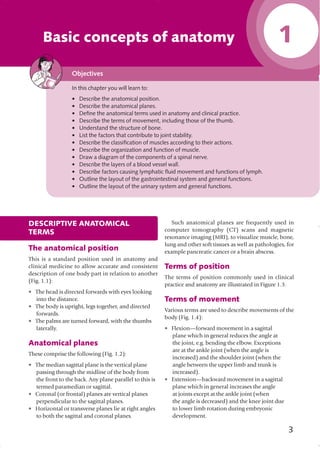
1. Objectives
In this chapter you will learn to:
• Describe the anatomical position.
• Describe the anatomical planes.
• Define the anatomical terms used in anatomy and clinical practice.
• Describe the terms of movement, including those of the thumb.
• Understand the structure of bone.
• List the factors that contribute to joint stability.
• Describe the classification of muscles according to their actions.
• Describe the organization and function of muscle.
• Draw a diagram of the components of a spinal nerve.
• Describe the layers of a blood vessel wall.
• Describe factors causing lymphatic fluid movement and functions of lymph.
• Outline the layout of the gastrointestinal system and general functions.
• Outline the layout of the urinary system and general functions.
Basic concepts of anatomy 1
3
Such anatomical planes are frequently used in
computer tomography (CT) scans and magnetic
resonance imaging (MRI), to visualize muscle, bone,
lung and other soft tissues as well as pathologies, for
example pancreatic cancer or a brain abscess.
Terms of position
The terms of position commonly used in clinical
practice and anatomy are illustrated in Figure 1.3.
Terms of movement
Various terms are used to describe movements of the
body (Fig. 1.4):
• Flexion—forward movement in a sagittal
plane which in general reduces the angle at
the joint, e.g. bending the elbow. Exceptions
are at the ankle joint (when the angle is
increased) and the shoulder joint (when the
angle between the upper limb and trunk is
increased).
• Extension—backward movement in a sagittal
plane which in general increases the angle
at joints except at the ankle joint (when
the angle is decreased) and the knee joint due
to lower limb rotation during embryonic
development.
DESCRIPTIVE ANATOMICAL
TERMS
The anatomical position
This is a standard position used in anatomy and
clinical medicine to allow accurate and consistent
description of one body part in relation to another
(Fig. 1.1):
• The head is directed forwards with eyes looking
into the distance.
• The body is upright, legs together, and directed
forwards.
• The palms are turned forward, with the thumbs
laterally.
Anatomical planes
These comprise the following (Fig. 1.2):
• The median sagittal plane is the vertical plane
passing through the midline of the body from
the front to the back. Any plane parallel to this is
termed paramedian or sagittal.
• Coronal (or frontal) planes are vertical planes
perpendicular to the sagittal planes.
• Horizontal or transverse planes lie at right angles
to both the sagittal and coronal planes.
Ch01-M3417.qxd 3/19/07 3:22 PM Page 3
2. Basic Concepts of Anatomy
• Abduction—movement away from the median
plane.
• Adduction—movement towards the median
plane.
• Supination—lateral rotation of the forearm,
causing the palm to face anteriorly.
• Pronation—medial rotation of the forearm,
causing the palm to face posteriorly.
• Eversion—turning the sole of the foot outwards.
• Inversion—turning the sole of the foot inwards.
• Rotation—movement of part of the body around
its long axis.
• Circumduction—a combination of flexion,
extension, abduction, and adduction.
The terms used to describe movements of the thumb
are perpendicular to the movements of the body, e.g.
flexion of the thumb is at 90° to that of flexion of the
fingers (Fig. 1.5).
BASIC STRUCTURES OF ANATOMY
Skin
The skin completely covers the body surface and is the
largest organ of the body. The functions of the skin
include:
• Protection from ultraviolet light and mechanical,
chemical, and thermal insults.
• Sensations including pain, temperature, touch
and pressure.
• Thermoregulation.
• Metabolic functions, e.g. vitamin D synthesis.
The skin is composed of the following (Fig. 1.6):
• The epidermis forms a protective waterproof
barrier. It consists of keratinized stratified
squamous epithelium, which is continuously
being shed and replaced. It is avascular.
• The dermis supports the epidermis and it has a
rich network of vessels and nerves. It is composed
mainly of collagen fibres with elastic fibres giving
the skin its elasticity.
• The hypodermis or superficial fascia. It consists of
fatty tissue which provides thermal insulation
and protection for underlying structures.
4
neck
Posterior view
head
scapular
region
back
loin
buttock
Anterior view
face
arm
upper limb
forearm
hand
thigh
lower limb
leg
foot
elbow
wrist
flank
groin
heel
knee
hip
ankle
abdomen
shoulder
breast
thorax
Fig. 1.1 Anatomical position and
regions of the body.
To differentiate supination from
pronation remember that you hold a
bowl of soup with a supinated forearm.
Ch01-M3417.qxd 3/19/07 3:22 PM Page 4
3. The skin appendages include:
• Hairs—highly modified, keratinized structures.
• Sweat glands—produce sweat, which plays a role
in thermoregulation.
• Sebaceous glands—produce sebum, which
lubricates the skin and hair.
5
Basic Structures of Anatomy 1
median
plane
coronal
plane
inferior
superior
horizontal
plane
posterior
(dorsal)
medial
lateral
anterior
(ventral)
Fig. 1.2 Anatomical planes.
median
plane
posterior anterior
Superior
Inferior
medial
proximal
distal
distal
proximal
lateral
Superior
Inferior
A
Dermatology
A genetic mutation in collagen synthesis affects the
protein’s function. Dermal collagen is normally
resistant to stretch, preventing excessive elasticity.
However, this is lost in Ehlers–Danlos syndrome
where individuals have very elastic skin as well as
other features due to collagen in joints (are
hyperextendable) or heart valves (mitral valve
regurgitation).
Fig. 1.3 Relationship and comparison (A) and classification
(B) of terms of position commonly used in anatomy and
clinical practice.
Position Description
Anterior In front of another structure
Posterior Behind another structure
Superior Above another structure
Inferior Below another structure
Deep Further away from body surface
Superficial Closer to body surface
Medial Closer to median plane
Lateral Further away from median plane
Proximal Closer to the trunk or origin
Distal Further away from the trunk or origin
Ipsilateral The same side of the body
Contralateral The opposite side of the body
Ch01-M3417.qxd 3/19/07 3:23 PM Page 5
4. Basic Concepts of Anatomy
• Nails—highly specialized appendages found on
the dorsal surface of each digit.
Fascia
The fascia of the body may be divided into superficial
and deep layers.
The superficial fascia (subcutaneous fatty tissue)
consists of loose areolar tissue that unites the dermis
to the deep fascia. It contains cutaneous nerves, blood
vessels and lymphatics that supply to the dermis. Its
thickness varies at different sites within the body and
women have a thicker layer than men.
In some places sheets of muscle lie in the fascia,
e.g. muscles of facial expression.
The deep fascia forms a layer of fibrous tissue
around the limbs and body and the deep structures.
Intermuscular septa extend from the deep fascia,
attach to bone, and divide limb musculature into
compartments. The fascia has a rich nerve supply and
it is, therefore, very sensitive. The thickness of the
fascia varies widely: e.g. it is thickened in the iliotibial
tract but very thin over the rectus abdominis muscle
and absent over the face. The arrangement of the
fascia determines the pattern of spread of infection as
well as blood due to haemorrhaging into tissues.
Bone
Bone is a specialized form of connective tissue with a
mineralized extracellular component.
The functions of bone include:
• Locomotion (by serving as a rigid lever).
• Support (giving soft tissue permanent shape).
• Attachment of muscles.
• Calcium homeostasis and storage of other
inorganic ions.
• Production of blood cells (haematopoiesis).
6
extension extension
flexion flexion
A B
abduction
adduction
medial
rotation
lateral
rotation
plantarflexion = flexion
dorsiflexion = extension
circumduction
pronation
supination
abduction
adduction
medial
rotation
lateral
rotation
inversion
eversion
G
E F
D
C
Fig. 1.4 Terms of movement.
(A) Flexion and extension of forearm at elbow joint.
(B) Flexion and extension of leg at knee joint.
(C) Dorsiflexion and plantarflexion of foot at ankle joint.
(D) Abduction and adduction of right limbs and rotation of left
limbs at shoulder and hip joints, respectively.
(E) Pronation and supination of forearm at radioulnar joints.
(F) Circumduction (circular movement) of lower limb at hip
joint.
(G) Inversion and eversion of foot at subtalar and transverse
tarsal joints.
Ch01-M3417.qxd 3/19/07 3:23 PM Page 6
5. Classification of bone
Bones are classified according to their position and
shape.
The position can be described as:
• Axial skeleton, consists of the skull, vertebral
column including the sacrum, ribs, and sternum.
• Appendicular skeleton, consists of the pelvic
girdle, pectoral girdle, and bones of the upper
and lower limbs.
Types of shape include:
• Long bones, e.g. femur, humerus.
• Short bones, e.g. carpal bones.
• Flat bones, e.g. skull vault.
• Irregular bones, e.g. vertebrae.
General structure of bone
Bone is surrounded by a connective tissue membrane
called the periosteum (Fig. 1.7). This is continuous
with muscle attachments, joint capsules and the
Fig. 1.5 Terms of movement for the thumb.
(Adapted from Crash Course:
Musculoskeletal System by SV Biswas and R
Iqbal. Mosby.)
(A) Neutral hand position.
(B) Extension (radial abduction).
(C) Flexion (transpalmar adduction).
(D) Abduction (palmar abduction).
(E) Opposition.
(F) Adduction.
A B
C
E
D
F
blood vessel
sebaceous
gland
sweat gland
hair follicle
nerve
fat
hair
epidermis
dermis
arrector
pili muscle
skeletal
muscle
deep fascia
superficial
fascia/
hypodermis
Fig. 1.6 Structure of skin and subcutaneous tissue.
7
Basic Structures of Anatomy 1
Ch01-M3417.qxd 3/19/07 3:23 PM Page 7
6. Basic Concepts of Anatomy
deep fascia. There is an outer fibrous layer and an
inner cellular layer. The inner layer is vascular, and it
provides the underlying bone with nutrition. The
periosteum is an osteogenic layer consisting of
osteoproginator cells that can differentiate into
osteoblasts, e.g. at a fracture site and cause forma-
tion of a bone cuff (callus) which stabilizes the
fracture.
Bone includes the following components:
• The outer compact layer or cortical bone provides
great strength and rigidity.
• The cancellous or spongy bone consists of a
network of trabeculae arranged to resist
external forces.
• The medullary cavity of long bones and the
interstices of cancellous bone are filled with
bone marrow. At birth virtually all the bone
marrow is red (haematopoietic), but this
is replaced by yellow (fatty) marrow—only
the ribs, sternum, vertebrae, clavicle,
pelvis, and skull bones contain red marrow in
adult life.
• The endosteum is a single layer of osteogenic cells
lining the inner surface of bone.
Blood supply of bones
There are two main sources of blood supply to bone:
• A major nutrient artery that supplies the marrow.
• Vessels from the periosteum.
The periosteal supply to bone assumes greater
importance in the elderly. Extensive stripping of the
periosteum, e.g. during surgery or following trauma,
may result in bone death.
Joints
These are unions between bones of which there are
three major types (Fig. 1.8).
Synovial joints
These are moveable joints and have the following
features:
• The bone ends are covered by hyaline articular
cartilage.
• The joint is surrounded by a fibrous capsule.
• A synovial membrane lines the inner aspect of the
joint and its capsule, except where there is
cartilage and it secretes synovial fluid. This
lubricates the joint and transports nutrients,
especially to the cartilage.
• Some synovial joints, e.g. the
temporomandibular joints, are divided into two
cavities by an articular disc.
Blood supply of joints
A vascular plexus around the epiphysis provides the
joint with a very good blood supply.
8
epiphyseal plate
epiphyseal plate
compact bone
medullary cavity
periosteum
cancellous bone
articular cartilage
diaphysis
metaphysis
epiphysis
metaphysis
epiphysis
Fig. 1.7 Long bone and its components.
Orthopaedics
As an individual ages their bone density is reduced
(osteopenia). The cortical bone becomes thinner and
the trabeculae decrease in number. As a result, bone
structure is weaker and predisposes to fractures,
especially in osteoporotic postmenopausal women.
Fractures tend to occur where, in normality, there is a
greater amount of trabecular bone to cortical bone,
e.g. radius (Colles fracture), femoral neck and
vertebral body. Fractures occurring secondary to
another process, e.g. osteoporosis, are known as
pathological fractures.
Ch01-M3417.qxd 3/19/07 3:23 PM Page 8
7. sensation of joint position and it is necessary for
motor control and posture.
Stability of joints
Stability is achieved by the following components:
• Bony—e.g. in a firm ball-and-socket joint such as
the hip joint, bony contours contribute to
stability.
• Ligaments—these are important in most joints,
and they act mainly to prevent excessive
movement.
• Muscles—these are an important stabilizing
factor in most joints.
Muscles and tendons
Skeletal muscles are aggregations of contractile fibres
that move the joints of the skeleton.
Muscles are usually joined to bone by tendons at
their origin and insertion.
Muscle action
Muscles can be classified according to their action:
• Prime mover—the muscle is the major muscle
responsible for a particular movement, e.g.
brachialis is the prime mover in flexing the
elbow.
• Antagonist—any muscle that opposes the action
of the prime mover: as the primer mover
contracts the antagonist relaxes, e.g. triceps
brachii relaxes during elbow flexion.
• Fixator—prime mover and antagonist acting
together to ‘fix’ a joint, e.g. muscles holding
the scapula steady when deltoid moves the
humerus.
• Synergist—prevents unwanted movement in
an intermediate joint, e.g. extensors of the
carpus contract to fix the wrist joint, allowing
the long flexors of the fingers to function
effectively.
Nerve supply of joints
According to Hilton’s law, the motor nerve to a
muscle tends also to give a sensory branch to the joint
that the muscle moves and another branch to the skin
over the joint. The capsule and ligaments are supplied
by afferent nerve endings, including pain fibres.
Innervation of a joint and the muscles that move that
joint allow proprioception to occur. This is the
9
Basic Structures of Anatomy 1
In general, if a joint is very stable it has a
reduced range of movement, e.g. the
stable hip joint compared with the less
stable shoulder joint; the latter has a
greater range of movement.
head
of femur
diploë
coronal suture with
collagen fibres
A Fibrous joint – suture
C Primary cartilaginous joint
B Fibrous joint – syndesmosis
E Synovial joint
epiphyseal
(growth) plate
neck of
femur
ulna
synovial
membrane
intervertebral
disc
lateral view
vertebral body
joint
cavity
articular
cartilage
fibrous
capsule
D Secondary
cartilaginous joint
radius
interosseous
membrane
compact
bone
Fig. 1.8 Types of joints.
(A) Fibrous joint—sutural (bones are united by fibrous tissue,
as in sutures of the skull).
(B) Fibrous joint—syndesmosis (bones are joined by a sheet of
fibrous tissue).
(C) Primary cartilaginous joint (where bone and hyaline
cartilage meet).
(D) Secondary cartilaginous joint (articular surfaces are
covered by a thin lamina of hyaline cartilage; the hyaline
laminae are united by fibrocartilage).
(E) Synovial joint.
Ch01-M3417.qxd 3/19/07 3:23 PM Page 9
8. Basic Concepts of Anatomy
Muscle design
Muscle fibres may be either parallel or oblique to the
line of pull of the whole muscle.
Parallel fibres allow maximal range of movement.
These muscles may be quadrangular, fusiform, or
strap shaped, e.g. sartorius and sternocleidomastoid.
Oblique fibres increase the force generated at the
expense of a reduced range of movement. These
muscles may be unipennate (e.g. flexor pollicis longus),
bipennate (e.g. dorsal interossei), multipennate (e.g.
deltoid) or triangular (e.g. deltoid).
Muscle organization and function
Motor nerves control the contraction of skeletal
muscle. Each motor neuron together with the muscle
fibres it supplies constitutes a motor unit.
The size of motor units varies considerably: where
fine precise movements are required, a single neuron
may supply only a few muscle fibres, e.g. the extrinsic
eye muscles; conversely, in the large gluteus maximus
muscle, a single neuron may supply several hundred
muscle fibres. The smaller the size of the motor
unit, the more precise are the possible movements. If
powerful contractions are required then larger motor
units are recruited (activated) which cause contraction
of larger muscles.
The force generated by a skeletal muscle is related
to the cross-sectional area of its fibres. For a fixed
volume of muscle, shorter fibres produce more force
but less shortening.
In muscles, there is an optimum length of muscle
filaments, which produces optimum tension and
contraction. Optimum tension is reduced if the
muscle becomes stretched beyond this length or
is compressed. This is a property of the muscle
length–tension relationship.
Muscle attachments
The ends of muscles are attached to bone, cartilage
and ligaments by tendons. Some flat muscles are
attached by a flattened tendon, an aponeurosis or
fascia.
When symmetrical halves of a muscle fuse to form
a seam like intersection, e.g. in mylohyoid muscle, a
raphe is formed.
When tendons cross joints they are often enclosed
and protected by a synovial sheath, a layer of con-
nective tissue lined by a synovial membrane and
lubricated by synovial fluid.
Bursae are sacs of connective tissue filled with
synovial fluid, which lie between tendons and bony
areas, acting as cushioning devices.
Nerves
The nervous system is divided into the central nervous
system and the peripheral nervous system: the central
nervous system is composed of the brain and spinal
cord; the peripheral nervous system consists of the
cranial and spinal nerves, and their distribution. The
nervous system may also be divided into the somatic
and autonomic nervous systems.
The conducting cells of the nervous system are
termed neurons. A typical motor neuron consists of a
cell body which contains the nucleus and gives off a
single axon and numerous dendrites (Fig. 1.9). The
cell bodies of most neurons are located within the
central nervous system, where they aggregate to form
nuclei. Cell bodies in the peripheral nervous system
aggregate in ganglia.
Axons are nerve fibres that conduct action poten-
tials generated in the cell body to influence other
neurons or affect organs. They may be myelinated or
non-myelinated.
Most nerves in the peripheral nervous system are
bundles of motor, sensory and autonomic axons. The
head is largely supplied by the 12 cranial nerves. The
10
Clinical examination
During a neurological and musculoskeletal
examination muscle power is assessed by asking the
patient to perform movements against resistance,
e.g. asking the patient to flex the elbow while the
examiner tries to prevent this by holding the wrist
and supporting the patient’s elbow. The power is
graded (5 to 0) by the UK Medical Research Council
(MRC) scale:
Grade 5: Full power
Grade 4: Contraction against resistance
Grade 3: Contraction against gravity
Grade 2: Contraction with gravity eliminated
Grade 1: Flicker of muscle contraction
Grade 0: No muscle contraction
Muscle weakness is seen in myasthenia gravis when
autoantibodies are produced that attack the receptors
on the neuromuscular junction (NMJ). Rapid
repeated movements cause muscle fatigue.
Ch01-M3417.qxd 3/19/07 3:23 PM Page 10
9. the structural basis of a reflex arc (Fig. 1.10). The reflex
arc is an involuntary protective mechanism that
occurs unconsciously although higher centres can
influence its activity, i.e. increase or decrease activity.
In a stroke the inhibitory input of higher centres that
dampens the reflex arc activity is lost and hyper-
reflexia (exaggerated limb reflexes) occurs.
Autonomic nerves are either sympathetic or
parasympathetic. Sympathetic preganglionic fibres
arise from the thoracic and upper two lumbar seg-
ments of the spinal cord. The preganglionic fibres
synapse in a ganglion of the sympathetic chain which
runs either side of the vertebral column. The post-
ganglionic fibres that arise from the sympathetic
chain ganglia can either enter a spinal nerve to supply
the limbs or body wall. Some preganglionic fibres do
not synapse in the sympathetic chain. Instead they
pass through the chain and synapse in a prevertebral
ganglion, e.g. coeliac ganglion. Postganglionic fibres
arise from prevertebral ganglia and supply viscera, e.g.
stomach. Parasympathetic preganglionic fibres arise
from cranial nerves and sacral nerves (S2–S4). They
synapse in ganglia associated with organs, e.g. a
pulmonary ganglion, to form postganglionic fibres
that innervate an organ, e.g. lung.
Spinal nerves
There are 31 pairs of spinal nerves: 8 cervical, 12
thoracic, 5 lumbar, 5 sacral, and the coccygeal nerve.
trunk and the limbs are supplied by the segmental
spinal nerves.
Motor nerves originate in the ventral (anterior)
horn of the spinal cord (Fig. 1.10) and synapse with
the sarcolemma (plasma membrane) of muscle to
form a structure called the motor endplate. A nerve
impulse reaches the end of the nerve fibre causing the
release of neurotransmitter. This leads to depo-
larization of the sarcolemma and initiation of muscle
contraction.
Sensory nerves carry impulses from receptors in
skin, muscle or viscera to the dorsal (posterior) horn
of the spinal cord. Receptors respond to specific
stimuli, e.g. stretch, noxious substances or pressure.
Sensory neurons synapse with neurons, which ascend
in the spinal cord and travel to higher centres, e.g.
cerebral cortex or cerebellum. They also synapse with
motor neurons directly or via an interneuron. This is
11
Basic Structures of Anatomy 1
Clinical examination/neurology
When testing reflexes the reflex arc is being assessed
at a particular spinal cord level. On striking a tendon
with a hammer it stretches the tendon and a receptor
within the muscle (a muscle spindle). This receptor
monitors muscle length and prevents over-stretching
by initiating a reflex arc and causing muscle
contraction to counter the stretching. This is
witnessed as a jerk of the limb; for example, on
striking the patella tendon the quadriceps muscle
contracts, causing knee extension. The common limb
reflexes and their spinal cord segment levels, which
are tested, are:
• Biceps brachii (C5–6)
• Triceps brachii (C7–8)
• Brachioradialis (C6–7)
• Quadriceps femoris (L3–4)
• Gastrocnemius (S1–2)
dendrites
nucleus
nerve cell body
Nissl bodies
axon
Ranvier's node
axon terminal
myelin
collateral
branch
neurolemma
(nerve cell
membrane)
Fig. 1.9 Structure of a typical neuron.
Ch01-M3417.qxd 3/19/07 3:23 PM Page 11
10. Basic Concepts of Anatomy
The spinal cord ends at the lower border of the first
lumbar vertebra in the adult. Below this, the nerve
roots of the cord form a vertical bundle: the cauda
equina.
Each spinal nerve is formed by the union of the
anterior and posterior roots (Fig. 1.10):
• The anterior root contains motor fibres for skeletal
muscles. Those from T1 to L2 also contain
preganglionic sympathetic fibres; S2 to S4 also
contain preganglionic parasympathetic fibres.
• The posterior root contains sensory fibres whose
cell bodies are in the posterior root ganglion.
Immediately after formation, the spinal nerve
divides into anterior and posterior rami. The great
nerve plexuses, e.g. the brachial, lumbar and sacral,
are formed by anterior rami. The posterior rami
supply the erector spinae muscles and skin that cover
them.
The spinal nerves each supply an area of skin called
a dermatome (except the face, which is supplied by
the fifth cranial nerve). The nerve supply of each
dermatome overlaps above and below with adjacent
dermatomes. Testing for loss of sensation over a
dermatome indicates the level of a lesion within the
12
skin
splanchic nerve
prevertebral
ganglion
Somatic nerves
sensory
motor
Sympathetic nerves
presynaptic
postsynaptic
stomach
sensory
motor
skeletal
muscle
blood
vessel
skin
heart
grey ramus
white ramus
sympathetic
chain ganglion
diaphragm
anterior
root
posterior
root
lateral horn of
grey matter
anterior
ramus
posterior
ramus
posterior root ganglion
Fig. 1.10 Components of a typical
spinal nerve.
Ch01-M3417.qxd 3/19/07 3:23 PM Page 12
11. where gaseous exchange occurs. Deoxygenated blood
is eventually returned to the heart first by venules then
by veins (Fig. 1.11A). Valves in the low-pressure
venous system are required to prevent back-flow of
blood. However, some veins have no true valves, e.g.
venae cavae, vertebral, pelvic, head and neck veins.
The general structure of the blood vessel wall
consists of three layers or tunicas (Fig. 1.11B). The
contents of each vary with vessel type and its function.
Arteries have a well-developed tunica media of
smooth muscle. The walls of the largest arteries
contain numerous elastic tissue layers; however, veins
have relatively little smooth muscle and elastic tissue.
Capillaries consist of an endothelial tube.
The larger vessels, e.g. aorta, also contain an
additional external layer of blood vessels (vasa
spinal cord. Dermatomes of the limbs and trunk are
illustrated in the relevant chapters.
Cardiovascular system
The cardiovascular system functions principally to
transport oxygen and nutrients to the tissues and
carbon dioxide and other metabolic waste products
away from the tissues.
The right side of the heart pumps blood to the
lungs via the pulmonary circulation. The left side of
the heart pumps oxygenated blood through the aorta
to the rest of the body via the systemic circulation
(Fig. 1.11A).
Blood is distributed to the organs via the arteries
and then arterioles, which branch to form capillaries
13
Basic Structures of Anatomy 1
superficial
temporal
common carotid
subclavian
axillary
brachial
ulnar
radial
deep arch
superficial
arch
left
brachiocephalic
subclavian
internal jugular
external jugular
azygos
facial
vertebral
aortic arch
internal thoracic
aorta
common iliac
external iliac
axillary
superior
vena cava
cephalic
basilic
inferior
vena cava
median
forearm
dorsal
venous
arch
femoral
great
saphenous
popliteal
small
saphenous
femoral
popliteal
anterior tibial
posterior tibial
Fig. 1.11(A) The arterial tree (A) and venous tree (B) of the cardiovascular system.
Ch01-M3417.qxd 3/19/07 3:23 PM Page 13
12. Basic Concepts of Anatomy
vasorum) and nerves (vasa nervosa) that supplies the
wall.
Anastomosis
Not all blood traverses a capillary bed. Direct
connections (anastomoses) between arterioles and
venules (arteriovenous shunts) exist. Pre-capillary
sphincters regulate flow through the capillary bed
under sympathetic nerve control. In the skin such
shunts are involved in thermoregulation. Capillary
beds can be opened up or closed off depending on
metabolic requirements, e.g. during exercise.
Direct communication between larger vessels can
be advantageous. If an artery becomes occluded
anastomoses maintain the circulation to an organ.
When an artery is slowly occluded by disease, new
vessels may develop (collaterals), forming an
alternative pathway, e.g. coronary arteries.
When such communications are absent (e.g. the
central artery of the retina) between arteries the vessel
is known as an end artery. Occlusion in these vessels
causes necrosis.
Lymphatics
Figure 1.12 illustrates the lymphatic system in man.
Fluid moves out of capillaries into tissues at the
arterial end due to hydrostatic pressure, which is created
by blood pressure. At the venous end of the capillary
oncotic pressure acts to draw fluid back into the vessel.
Oncotic pressure is created by proteins, e.g. albumin
and cations (sodium ions). However, not all fluid is
returned to the blood and excess within the tissues
drains into the lymphatic system. Movement of
lymphatic fluid through the vessels is the result of
(i) muscle contraction, (ii) pulsation of an adjacent
artery, (iii) a suction action by the negative intrathoracic
pressure, and (iv) pressure within the lymphatic vessels.
The lymphatics on the right side of the head, neck,
upper limb and thorax drain into the right lymphatic
duct which enters the venous circulation at the
junction of the right subclavian and right internal
jugular veins. The rest of the body drains into the
thoracic duct, which enters the venous circulation at
the junction of the left subclavian and left internal
jugular veins (Fig. 1.12).
14
smooth
muscle
tunica
media
tunica
adventitia
tunica
intima
external
elastic
lamina
internal
elastic
lamina
endothelium
fibrocollagenous layer
(in larger vessels contains blood
vessels and is known as vasa vasorum)
Fig. 1.11(C) Cross section of vessel wall
showing basic layers.
Ch01-M3417.qxd 3/19/07 3:23 PM Page 14
13. the intestinal villi contain chyle (a milky lymph
fluid), which drains into larger lymphatic vessels and
eventually into the thoracic duct.
Lymphatics are found in all tissues except the
central nervous system, eyeball, internal ear, cartilage,
bone, and the epidermis of the skin.
Lymph carries foreign material (not recognized as
self), which may be presented by special cells in the
lymph nodes (antigen-presenting cells) to cells of the
immune system to mount an immune response. The
lymphatics also are involved in the absorption and
transport of fats. Lacteals (end lymphatic vessels) of
15
Basic Structures of Anatomy 1
internal
jugular vein
thoracic duct
aorta
posterior
mediastinal
nodes
superficial
lymphatic
vessels
lumbar nodes
iliac nodes
superficial
inguinal nodes
superficial
lymphatic
vessels
Anterior view
cervical nodes
right
lymphatic
duct
subclavian vein
brachiocephalic
vein
axillary nodes
cubital nodes
cisterna chyli
deep lymphatic
vessels
deep inguinal
nodes
popliteal nodes
deep lymphatic
vessels
supraclavicular nodes
Fig. 1.12 The lymphatic system (shaded area drains into the right lymphatic duct; unshaded area drains into the thoracic duct).
Ch01-M3417.qxd 3/19/07 3:23 PM Page 15
14. Respiratory system
The upper part of the respiratory tract, consisting of
the nasal and oral cavities, pharynx, larynx and
trachea, is responsible for conditioning the air by
(i) humidifying and warming e.g. blood vessels in the
nasal cavity, and on conchae that increase the surface
area avaliable, (ii) trapping of foreign material e.g.
hair in the nasal vestibule and mucus secretion. The
lower respiratory tract consists of a series of branching
tubes that form the bronchial tree (see Chapter 3),
which ends in the alveolar sacs where gaseous
exchange occurs.
The general structure of the respiratory tree wall
changes with function, e.g. the bronchi walls contain
cartilage whereas the bronchioles lack cartilage. The
alveoli consist of a sphere of epithelium surrounded
by a network of capillaries.
Respiratory epithelium of the trachea, bronchi and
bronchioles consists of cells which contain cilia
(small hairs) that beat rhythmically and propel
trapped foreign particles (within mucus) towards
the pharynx. Moreover, the alveoli consist of thin
epithelial cells (pneumocytes) which reduce the
distance that gases have to diffuse across between it
and the capillaries of the lung. This increases gaseous
exchange efficiency.
The functions of the respiratory system include:
• Gaseous exchange.
• Metabolism and activation or inactivation of
some proteins, e.g. angiotensin-converting
enzyme.
• Acting as a reservoir for blood.
• Phonation (vocal sound production).
• Olfactory function.
Urinary system
The urinary system is composed of the kidneys,
ureters, bladder and urethra (Fig. 1.14). The kidneys
filter the blood at the glomerulus, and along the
length of the nephron unit selective absorption and
secretion occurs. The urine that is formed from these
processes enters the renal pelvis and the ureters. The
latter empty into the bladder, which stores urea until
such time that it may be voided (micturation). The
functions of the kidneys are:
• Excretion of waste products, e.g. urea (produced
in the liver).
• Absorption of filtered substances, e.g. glucose,
ions, proteins.
Basic Concepts of Anatomy
Gastrointestinal system
The gastrointestinal system has three functions:
• Digestion of food material starting with
mastication and continuing in stomach and
duodenum.
• Absorption of the products of digestion in the
small intestine.
• Absorption of fluid and formation of solid faeces
in the large intestine.
The process of digestion begins in the mouth with
enzyme secretion by the salivary glands and chewing
(mastication). In the stomach, acid and enzyme
secretion continue the process; then, in the second
part of the duodenum, pancreatic enzymes, along
with bile from the liver, complete this process. The
majority of absorption occurs in the jejunum, which
has an increased surface area due to plicae circularis
(folds), villi (finger-like projections) and microvilli
(microscopic projections on individual cells). Carbo-
hydrates and proteins enter the portal circulation (see
below) via the intestinal villi capillaries and fats enter
the lacteals of the lymphatic system.
The portal circulation is a circulation consisting of
two capillary beds. Capillaries originating in the
intestine enter veins that eventually drain into the
hepatic portal vein and this drains into the liver
capillaries (sinusoids). Hepatic veins drain blood
from the liver into the systemic circulation and it
returns to the heart. The portal vein also receives
tributaries from the stomach, spleen and pancreas.
There are anastomoses with the systemic venous
circulation at the gastro-oesophageal and recto-anal
junctions (portosystemic anastomoses).
The general structure of the gastrointestinal tract
wall is illustrated in Figure 1.13. Modifications to this
denote its underlying function, e.g. there are more
folds and villi in the jejunum than in the ileum or
colon.
16
Oncology
Lymphatic drainage of organs provides one of the
routes by which a cancer can spread to other
anatomical sites (metastasis). In breast carcinoma,
metastasis can be to the lymph nodes of the armpit
(axilla), or in gastric carcinoma spread can be to the
left supraclavicular nodes only and this is known as
Troissier’s sign.
Ch01-M3417.qxd 3/19/07 3:23 PM Page 16
15. stomach
Ao
B
C
D
F
E
non keratinized stratified
squamous epithelium
A1
B
C
D
F
E
gastric pits
A2
B
C
D
F
E
villi
A3
B
C
D
F
E
rectum
oesophagus
large intestine
– caecum
– ascending colon
– transverse colon
– descending colon
– sigmoid colon
key
Ao
A1
A2
A3
B
C
D
E
F
small intestine
– jejunum
– ileum
duodenum
oesophageal epithelium
stomach epithelium
small intestine epithelium
large intestine epithelium
muscularis mucosae layer
submucosa layer
circular smooth muscle layer
longitudinal smooth
muscle layer
serosa
Fig. 1.13 The gastrointestinal system. The illustration shows the basic layers of the gastrointestinal tract wall with epithelial
adaptions, which dictate function.
• Metabolism of vitamin D.
• Blood pressure and sodium regulation (renin
secretion).
• Rate of red blood cell production, e.g.
erythropoietin secretion.
The ureters and bladder have a muscular wall and are
lined by urothelium (transitional epithelium). This
is a specialized stratified epithelium allowing
distension, especially of the bladder to accommodate
large volumes of fluid.
RADIOLOGICAL ANATOMY
Introduction
The use of plain radiography is frequently requested
to detect and aid the diagnosis of disease within the
thorax, abdomen or in bones. Using contrast studies
to distinguish adjacent structures of similar lucency
on a film can enhance the clinical usefulness of this
investigation, especially in the gastrointestinal tract to
detect a perforation of the bowel wall or a lesion. A
contrast study uses a substance, e.g. barium, which
appears radio-opaque (white) on an X-ray film and
allows internal anatomical structures not normally
seen to be visualized. The contrast study can be single
17
Radiological Anatomy 1
Ch01-M3417.qxd 3/19/07 3:23 PM Page 17
16. Basic Concepts of Anatomy
(when only barium is used) or double when both
barium and air are introduced into the intestines.
Angiography is a procedure in which a contrast
medium is injected into an artery or vein via a
percutaneous catheter. It is used to assess vascular
disease such as atherosclerosis (fatty plaques) in the
coronary arteries or an aneurysm (a balloon-like
swelling) in the abdominal aorta.
The following chapters will introduce normal
radiographic anatomical structures and give a method
for reading X-rays because they will be presented
to you not only in your anatomy studies but also
in the clinical years. The pre-registration house
officer will usually be expected to perform the initial
interpretation of an X-ray.
18
ureter
A and B
C
D
E
bladder
key
A
B
C
D
E
urothelium
submucosa
longitudinal smooth muscle
circular smooth muscle
serosa (connective tissue)
inferior vena cava
kidney
adrenal gland
E C D C B A
C
B
6
5 9
7
1
3
4
2
8
A
key
1.
2.
3.
4.
5.
6.
7.
8.
9.
afferent arteriole
efferent arteriole
glomerulus
Bowmanís capsule
proximal convoluted
tubule
thin descending limb of
Loop of Henle
thick ascending limb of
Loop of Henle
distal convoluted
tubule
collecting duct
Fig. 1.14 Components of the urinary tract. Inset A shows the structure of a nephron, inset B shows the structure of the ureter
and inset C shows the structure of the bladder wall.
Ch01-M3417.qxd 3/19/07 3:23 PM Page 18