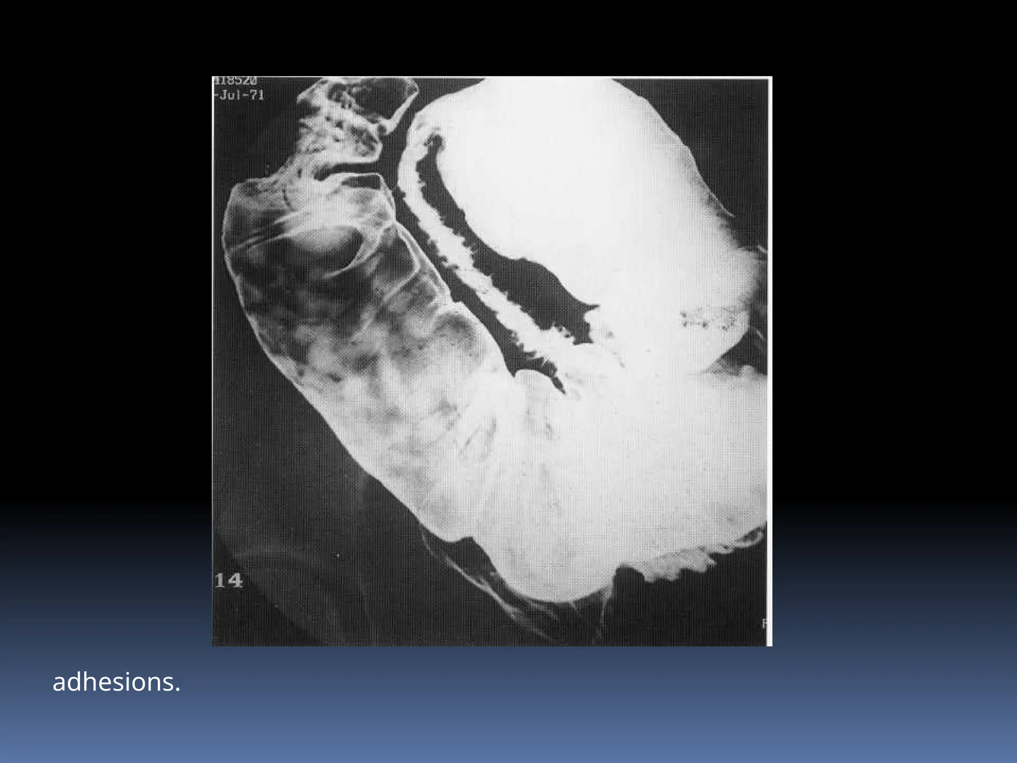
BARIUM IMAGING IN SMALL
BOWEL
MODERATOR : DR M M PATIL
PRESENTER : DR NARENDRA G S
Introduction :
 The small intestine measures approximately 5 m in length
and extends from the duodenojejunal flexure to the
ileocaecal valve. It is attached by its mesentery to the
posterior abdominal wall and this allows its mobility.
 The proximal two-fifths constitute the jejunum and the
distal three-fifths the ileum. The jejunum lies mainly in the
left upper and lower quadrants and the ileum in the lower
abdomen and the right iliac fossa.
 The valvulae conniventes have a circular configuration
and are about 2 mm thick in the distended jejunum,
becoming more spiral shaped and about 1 mm thick in
the ileum.
 They may be absent in the distended terminal ileum,
resulting in a rather featureless outline.
 Jejunal diameter should not exceed 3.5 cm on barium
Follow-through & 4.5 cm on enteroclysis.
BARIUM STUDIES
1. Single/double contrast - BMFT
2. Enteroclysis.
3. Peroral pneumocolon.
4. Ileostomy enema.
Indications
 Pain
 Diarrhoea
 Anaemia/gastrointestinal bleeding
 Partial obstruction
 Malabsorption
 Abdominal mass
 Failed small bowel enema.
Contraindications
 Complete obstruction
 Suspected perforation (unless a water-
soluble contrast medium is used).
Contrast medium
 100% w/v 300 ml (150 ml if performed immediately
after a barium meal) barium. The transit time through
the small bowel is reduced by the addition of 10 ml of
Gastrografin to the barium. In children, 3-4 ml kg-1
is a
suitable volume.
 In situations where barium is contraindicated, non-ionic
water-soluble solutions have been used .
Patient preparation
 Metoclopramide 20 mg orally may be given 20
min before the examination.
Preliminary film :Plain abdominal film.
Technique
 The aim is to deliver a single column of barium into the small
bowel. This is achieved by lying the patient on the right side after
the barium has been ingested.
 Metoclopramide enhances the rate of gastric emptying. If the
transit time through the small bowel is found to be slow, a dry
meal may help to speed it up.
 If a follow-through examination is combined with a barium
meal, glucagon is used for the duodenal cap views rather than
Buscopan because it has a short length of action and does not
interfere with the small-bowel transit time.
Films
 Prone PA films of the abdomen are taken every 20 min
during the first hour, and subsequently every 30 min
until the colon is reached. The prone position is used
because the pressure on the abdomen helps to separate
the loops of small bowel.
 Spot films of the terminal ileum are taken supine. A
compression pad is used to displace any overlying loops
of small bowel that are obscuring the terminal ileum.
Additional films
 1. To separate loops of small bowel:
a. obliques
b. with X-ray tube angled into the pelvis
c. with the patient tilted head down.
 2. To demonstrate diverticula:
a. erect - this position will reveal any fluid levels caused
by contrast medium retained within the diverticula.
Complications :
 Leakage of barium from an unsuspected perforation.
 Aspiration of stomach contents due to the Buscopan.
 Conversion of a partial large bowel obstruction into a
complete obstruction by the impaction of barium.
 Barium appendicitis, if barium impacts in the
appendix.
Small bowel
enema/enteroclysis :
 Advantages :This procedure gives better visualization of the small
bowel because rapid infusion of a large, continuous column of
contrast medium directly into the jejunum which avoids
segmentation of the barium column and does not allow time for
flocculation to occur.
 Disadvantages :
1. Intubation may be unpleasant for the patient, and may occasionally
prove difficult.
2. It is more time-consuming for the radiologist.
3. There is a higher radiation dose to the patient (screening the tube
into position)
Contrast medium
 70% w/v barium.
 This is diluted with water to give a 20% solution (total volume 1500
ml). The reduced viscosity produces better mucosal coating, and the
reduced density permits the visualization of bowel loops which may
have been obscured by a denser contrast medium in an overlying
loop.
 An alternative way to gain a double contrast effect is to use 600 ml of
0.5% methylcellulose after 500 ml of 70% w/v barium. Even with these
modifications, it may still be difficult to obtain good distension and
double contrast effect of the distal small bowel and terminal ileum.
Equipment
1. Bilbao-Dotter tube with a guide-wire (the tube is
longer than the wire so that there is reduced risk
of perforation when introducing the wire).
2. Silk tube: 140-cm long tube with a tungsten-filled
guide-tip. It is made of polyurethane and the stylet
and the internal lumen of the tube are coated with
a water-activated lubricant to facilitate the smooth
removal of the stylet after insertion.
Patient preparation:
1. A low-residue diet for 2 days prior to the examination.
2. If the patient is taking any antispasmodic drugs, they must
be stopped 1 day prior to the examination.
3. Amethocaine lozenge 30 mg, 30 min before the
examination.
Immediately before the examination ,
4.the pharynx is anaesthetized with lignocaine spray.
Technique
 The patient sits on the edge of the X-ray table. The pharynx is
thoroughly anaesthetized with lignocaine spray. patency of the nasal
passages is checked by asking the patient to sniff with one nostril
occluded. The Silk tube should be passed with the guide-wire pre-
lubricatcd and fully within the tube, whereas for the Bilbao-Dotter tube
it may be more comfortable to introduce the guide-wire after the tube
tip is in the stomach.
 The tube is then passed through the nose or the mouth, and brief
lateral screening of the neck may be helpful in negotiating the
epiglottic region. The patient is asked to swallow as the tube is passed
through the pharynx. The tube is then advanced into the antrum.
 The patient then lies down and the tube is passed into the duodenum.
Various manoeuvres may be used alone or in combination, to help this
part of the procedure, which may be difficult.
a. Lie the patient on- the left side so that the gastric air bubble rises to the
antrum, thus straightening out the stomach.
b. Advance the tube whilst applying clockwise rotational motion .
c. In the case of the Bilbao-Dotter tube, introduce the guide-wire.
d. In the case of the Silk tube, lie the patient on the right side, as the tube
has a tungsten-weighted guide-tip which will then tend to fall towards
the antrum.
e. Get the patient to sit up, to try to overcome the tendency of the tube to
coil in the fundus of the stomach.
 When the tip of the tube has been passed through the pylorus, the
guide-wire tip is maintained at the pylorus as the tube is passed over
it along the duodenum to the level of the ligament of Treitz.
Clockwise torque applied to the tube may again help in getting past
the junction of the first and second parts of the duodenum. The tube
is passed as far as the duodenojejunal flexure to diminish the risk of
aspiration due to reflux of barium into the stomach.
 Barium is then run in quickly, and spot films are taken of the barium
column and its leading edge at the regions of interest, until the
colon is reached. If methylcellulose is used, it is infused
continuously, after an initial bolus of 500 ml of barium, until the
barium has reached the colon.
 The tube is then withdrawn, aspirating any residual fluid in the
stomach. Again, this is to decrease the risk of aspiration.
 Finally, prone and supine abdominal films are taken
 Aftercare
1. Nil orally for 5 h after the procedure.
2. The patient should be warned that diarrhoea may occur as a
result of the large volume of fluid given.
 Complications
1. Aspiration
2. Perforation of the bowel owing to manipulation of the guide-wire.
Barium-Enteroclysis
ENTEROCLYSIS v/s BMFT
 Contrast administered at desired
rate,
 Pylorus bypassed
 Distension of bowel can be
assessed
 Bowel proximal to stenosis
dilates-stands out
 Time taken 20 to 30 min
 Direct rapid infusion produce
hypotonia
 Reliability high superior
 Critically ill,elderly
 No discomfort
 Transit time assesed
 Overlapping & poor
distension of bowel loops
 Prolonged study
PERORAL PNEUMOCOLON
Mainly to evaluate distal ileum and IC junciton
Air insufflated after barium reaches transverse colon
Ileostomy enema :
 Symptoms following ileostomy may be due to recurrent
disease, for example Crohn's, adhesions related to the
procedure, or a stomal hernia.
 A Foley catheter is inserted into the stoma, its balloon
inflated just deep to the anterior abdominal wall, and
barium suspension injected via a syringe, followed by
some air for a double-contrast effect.
 The procedure is the same but water-soluble contrast is
used.
Normal Findings
 Small bowel gradually tapers in diameter from the
duodenojejunal junction to the terminal ileum, so
the jejunum (up to 3 cm in luminal diameter) 4-
7folds per inch & the ileum (up to 2cm) (2-4 folds
per inch) , changes in enteroclysis.
 The folds are composed of mucosa and
submucosa, whereas individual villi lining the folds
are composed only of mucosa and lamina propria.
Frontal spot image from enteroclysis shows normal folds in distal jejunum
as thin (1–2- mm-thick) delicate structures perpendicular to longitudinal
axis of bowel. There are normally four to seven folds per inch of jejunum.
CONGENITAL LESIONS :
 Malrotation: Intestinal malrotation can be broadly defined as any
deviation from the normal 270° counterclockwise rotation of the
midgut during embryologic development.
 Duplication: Cystic or tubular. Intramural duplications may cause
obstruction,
 The typical triradiate fold configuration of the diverticulum is
infrequently visualised on contrast studies.
 Inverted Meckel‘s diverticulum is a rare but well-recognised cause of
intestinal obstruction. Congenital stenoses and atresias also occur,
usually because of incomplete canalisation, with the duodenum the
most common site.
Meckel diverticulum as
tubular
outpouching from distal
ileum. No folds are seen in
the diverticulum.
BMFT revealing
large meckel’s
diverticulum
Small bowel obstruction:
 Mechanical intestinal obstruction accounts for approximately 20% of
surgical admissions.
 Causes may be generally divided into extrinsic and intrinsic groups.
 Extrinsic causes include
 Adhesions (following surgery or peritoneal inflammation),
 Hernias (inguinal, femoral or internal, particularly paraduodenal)
 Masses, most notably disseminated peritoneal malignancy.
 Congenital malrotation or peritoneal (Ladd's) bands are rarer extrinsic
causes.
.
adhesive band
small bowel
adhesion
 Intrinsic mural disease may be due to inflammatory strictures,
notably due to Crohn's disease or radiation enteritis,
ischaemia, or rarely primary small-bowel tumours (which
may also he accompanied by intussusception).
 Intraluminal obstruction may be due to gallstones or foreign
bodies.
 Non-steroidal tablets may cause intestinal membranes,
resulting in obstruction.
).
adhesions.
 Water-soluble studies are often requested by surgeons to
diagnose acute obstruction. These are likely to he less useful
than CT for diagnosis of the level and cause, predominantly
because of slow transit & coupled with distal contrast dilution.
 Patients settled on conservative treatment if contrast had
entered the colon but laparotomy was likely if it had not
 These features are best sought using enteroclysis, although
compression during follow-through techniques can be useful by
demonstrating loop fixity and abrupt angulation
 Prestenotic dilatation suggests a degree of functional
obstruction but the distensibility of a strictured
segment is best assessed using enteroclysis due to
infusion pressure.
 Massive small-bowel dilatation secondary to chronic
strictures can occur and may be complicated by
bacterial overgrowth.
ILEUS & PSEUDO OBSTRUCTION
 Causes of paralytic ileus, are often needs to be differentiated from
mechanical obstruction,
 Both small and large bowel may be dilated. The commonest etiologies
are laparotomy ,peritonitis , electrolyte imbalance , may also be
implicated.
 Constitutional disease, for example scleroderma (systemic sclerosis)
Some may be associated with a gut myopathy or neuropathy which
gives rise to the clinical picture of intestinal pseudo-obstruction.
 The cardinal radiological feature of scleroderma is duodenal and
jejunal dilatation associated with fold crowding and slow transit due to
collagen replacement of intestinal smooth muscle.
Primary visceral myopathy.
Scleroderma
CROHN’S DISEASE
 Idiopathic inflammatory disease which may affect any
part of the luminal gastrointestinal tract from mouth to
anus.
 Characterised by discontinuous transmural ulceration,
fistulation and spontaneous abscess formation.
 Most patients (60-80 %)will have small bowel disease,
with the terminal ileum most commonly affected (55 % of
all patients). Approximately 25% overall will have colonic
disease only.
 Villous oedema and blunting are the earliest detectable
radiological change, manifest as a granular pattern on high-
quality contrast studies ,the `grains' are due to individual filling
defects produced by the enlarged and inflamed vili and are best
appreciated on compression studies.
 Ulceration becomes linear and deeper, with typical transmural
penetration accompanied by mural thickening.
 Mucosal oedema and inflammation intervenes between these
ulcers to cause the characteristic 'cobblestone' appearance.
Crohn's disease. intense mucosal
granularity,
Crohn’s disease. Fold thickenin
Crohn's disease. Fat suppressed T2-weighed MR scan shows
thickened ileal loops (curved arrows) and also reveals a parastomal
abscess
 Ulceration is frequently discontinuous and patchy and
also asymmetrical along the bowel circumference;
indrawing at the site of ulceration may he accompanied
by ballooning of the contralateral wall, creating a
characteristic pseudodiverticlar appearance.
 Advanced disease may also he complicated by strictures,
fistulation, abscess formation and, rarely, by tumour.
Strictures are generally easy to demonstrate using
contrast studies
multiple aphthoid ulcers as punctate collections of barium surrounded by
radiolucent mounds of edema
Cobblestoning of the
terminal ileum,
thickening of the wall of
the terminal ileum, and
an enlarged ileocaecal
valve in Crohn's disease
Crohn disease shows
cobblestoning in distal
ileum due to intersecting
linear
and transverse ulcers.
separation of diseased
loops from adjacent small
bowel by fibrofatty
proliferation
in mesentery
pseudodiverticulae
aphthous ulcers
background granularity
caused by villous oedema.
ileocecal fistulas with
narrowing of
terminal ileum near ileocecal
valve.
barium enema examination
(with reflux into terminal
ileum) in patient with Crohn
disease shows classic string
sign with
marked narrowing of terminal
ileum due to severe edema
and spasm.
Long stricture in Crohn's disease. A
long segment of narrowing is seen in
the ileum just proximal to the site of
an ileocolic anastomosis in a patient
who had undergone a previous
resection for Crohn's disease.
PRIMARY SMALL BOWEL TUMORS
 Primary small-bowel tumours are rare
and frequently difficult to diagnose
because findings are non-specific and
the diagnosis is often not considered,
which often leading to late presentation
and possibly poor prognosis.
BENIGN TUMORS
 Adenomas & stromal tumors.
 Benign stromal tumours (leiomyomas), the commonest
benign small-bowel tumour, arise from the smooth
muscle of the muscularis propria. They are usually jejunal
and may have endoluminal and exolmninal components.
 They are usually easy to demonstrate on contrast studies
once large enough to cause obstruction or
intussusception
 Adenomas are similar to their colonic counterparts both
morphologically and histologically and are classified in a
similar fashion: tubular, villous, tubulovillous.
 Lipomas may be recognized by their characteristic low attenuation
on CT. Most are ileal) and asymptomatic. When seen on
contrast studies they are smooth and easily compressible.
 Haemangiomas may be capillary or cavernous. Most are too
small to produce a filling defect but frequently present with
anaemia due to haemorrhage.
Benign stromal tumour, Barium follow-
through reveals an intraluminal
mass .,,The tumour is also visible on CT
MALIGNANT TUMORS.
 In contrast to the large bowel, adenocarcinoma is
remarkably uncommon outside of a polyposis syndrome.
 There are well-documented associations with Crohn's and
coeliac disease and the morphology is essentially similar to
that seen in the colon.
 an annular, shouldered, apple-core-type lesion
Small bowel adenocarcinoma
B-cell non-Hodgkin
lymphoma as a
giant cavitated lesion in
distal ileum, with
displacement of adjacent
small-bowel loops by the
surrounding mass.
 Lymphoma is non-Hodgkin's in origin and is the commonest
primary small bowel malignant tumour in some series. Again,
there is an association with coeliac and Crohn's disease.
 There may be diffuse, regular fold thickening without any
obvious, localised tumour mass .In contrast, other cases
exhibit marked focal mural thickening with fistulation, Non-
obstructing stricturing is common.
 Aneurysmal dilatation, which is highly characteristic and due
to cavitating necrosis, often following effective treatment.
cavitated lesion in mid–
small bowel with displacement
of adjacent smallbowel
loops by mass. This patient had
malignant gastrointestinal
stromal tumor.
irregular-segmental
thickening of folds in loop
of mid–small bowel,
with markedly thickened,
lobulated folds
This patient had primary
non-Hodgkin small bowel
lymphoma.
innumerable nodules in distal ileum due to smallbowel lymphoma.
nodules are less uniform in size and larger than typical lymphoid follicles.
In subtotal colectomy with ileosigmoid anastomosis .
Lymphoma. Diffuse fold
thickening and nodularity.
 Carcinoid the majority are in the distal ileum. Tumours larger
than 2 cm are frequently malignant, defined by metastasis.
 An intense desmoplastic response to the primary tumour is
highly characteristic and is well demonstrated by CT.
 Carcinoid syndrome may occur when significant liver metastasis
prevents metabolism of secreted vasoactive serotonin and
bradykinin, allowing them to reach the systemic circulation, and
is characterised by episodic flushing and diarrhoea.
barium enema
examination (with
reflux
into terminal ileum)
shows carcinoid tumor
as
smooth, sessile, 1.5-
cm-diameter polyp
(black
arrows) in terminal
ileum. Also note
multiple ileal diverticula
(white arrows).
patient with carcinoid tumor
shows mass effect,
angulation, and tethering of
ileal loops due to marked
desmoplastic reaction
incited by
tumor in mesentery.
Polypoid Lesions
MUCOSAL SUB MUCOSAL
Adenomas
Hamartomas
Familial adenomatous
polyposis
Peutz jegher’s syndrome
Carcinoids.
Hematogenous
metastases
Malignant melanoma
Lymphoma
Multiple carcinoid tumors
Neurofibromas
Kapossi’s sarcoma
Lipoma.
Inverted meckel’s
diverticulum
POLYPOSIS SYNDROME
 Adenomas in familial adenomatous polyposis (FAP) tend to
cluster around the duodenal ampulla.
 The larger the polyp, the greater the possibility of malignancy
and there is also an association with ampullary carcinoma.
 FAP is also strongly associated with desmoid disease.
 Peutz-Jeghers syndrome is an autosomal dominant disease
characterized by mucocutaneous pigmentation, often
perioral, and gastrointestinal hamartomas.
 Cowden’s disease also describes small intestinal hamartomas
(and also adenomas, hyperplastic polyps & adenomas.)
 Diffuse intestinal inflammatory polyposis cronkhite-canada
syndrome is associated with neuroectodermal change,
manifests as nail dystrophy , alopecia & mal absorption.
Barium follow
through reveal
ileal hamartoma in
peutz jeghers
syndrome
barium enema examination (with
reflux into terminal ileum) shows
lipoma as smooth,
ovoid, submucosal mass in distal
ileum.
multiple smooth-
surfaced
hemispheric
submucosal
masses in small
bowel;
other lesions have
bull’s-eye
appearance
due to central
ulceration.
The patient
had malignant
melanoma with
hematogenous
metastases to
small bowel.
metastatic melanoma shows intussuscepting mass (black arrows) with
telescoping of small bowel (intussusceptum,) into adjacent loop
(intussuscipiens).
A “coil spring” appearance results from barium coating folds of
smooth elongated
mass in distal ileum
with telescoping of
small bowel
(intussusceptum,
white arrow) into
adjacent lumen
(intussuscipiens),
producing coil spring
appearance
This patient
had inverted Meckel
diverticulum acting as
lead point for
intussusception.
OUTPOUCHINGS :
 JEJUNAL DIVERTICULOSIS
 SACCULATIONS
diverticula in
duodenum and
jejunum
as smooth rounded
outpouchings of
varying sizes
Frontal overhead
radiograph from
enteroclysis shows
massive jejunal
diverticulosis
Paradoxically, this
degree of
diverticulosis
can be more
difficult to detect
on barium studies,
because
diverticula are
easily mistaken for
overlapping loops
of small bowel.
patient with scleroderma
shows markedly
dilated duodenum and
proximal jejunum, with
increased number of small-
bowel folds crowded
together ,producing
the “hidebound” sign.
Also note multiple
outpouchings
(arrows) due to asymmetric
fibrosis with sacculation of
opposite wall of bowel.
INFECTIOUS ENTERITIS
 Salmonella, Campylobacter and Staphylococcus are all possible
causative agents. Radiology has no role to play but appearances
may be dramatic if patients are examined during an attack, with
dilatation, ulceration and nodularity.
 In case of chronic infection fold thickening & mild dilatation may
occur.
 Intestinal tuberculosis usually affects the ileocaecal area. Terminal
ileal ulceration in association with a funnelled, contracted
caecum are characteristic. Ulcers tend to be discrete and
transverse or star-shaped.
FLEISCHNER’S SIGN (INVERTED UMBRELLA SIGN)
TB Most common in
terminal ileum
because of… The
increased
physiological stasis,
Increased rate of fluid
and electrolyte
absorption, Minimal
digestive activity and
An abundance of
Stierlin’s sign
Conical caecum
 Ascaris lumbricoides is a large roundworm which is extremely common.
Cause small-bowel obstruction. Their appearance on contrast
studies is characteristic once the worms have swallowed contrast
themselves; barium is seen within their intestinal tract.
 non-specific findings of fold thickening, nodularity, mild dilatation
and flocculation on contrast studies can be elicited.
 Whipple's disease may also be considered an intestinal infection
because of its association with the bacilli Tropheryrna whippelii.
 Contrast studies typically reveal a micronodular mucosal pattern.
SBFT in patient with Whipple
disease shows thickened
irregular folds in jejunum
and proximal ileum due to
accumulation of Whipple
bacilli.
patient with AIDS shows
thickened irregular folds
in jejunum due to
opportunistic infection
by
cryptosporidiosis.
NON INFECTIOUS ENTERITIS
 The small bowel is often unavoidably irradiated as a
consequence of radiotherapy to abdominopelvic tumours.
 An acute radiation enteritis is followed by fibrotic healing which
may precipitate an strictures, notably colonic, and often suffer
from short-bowel endarteritis obliterans. This causes ischaemia
and the subsequent fibrosis & stricture that is characteristic of
chronic radiation enteritis.
Pt on warfarin sodium
shows straightsegmental
thickening of folds (arrows)
due to localized
submucosal hemorrhage
from anticoagulation.
Small-bowel ischemia may
produce similar
findings
straight-segmental
thickening of folds in loop of
ileum due to localized
submucosal edema and
hemorrhage.
 There may he abrupt margination between affected bowel and
normal adjacent bowel excluded from the radiation field.
 Initially the valvulae are thickened but may eventually become
completely effaced. Extensive adhesions between the anti
mesenteric aspects of adjacent loops results in the
phenomenon of `mucosal tacking' and a 'picketfence’
appearance.
 Superficial ulceration, stenosis and obstructive dilatation are
common .
patient with prior radiation
therapy to pelvis shows
straight-segmental
thickening of folds in pelvic
loops of ileum with
narrowing, angulation and
lowgrade obstruction due
to radiation serositis
Barium follow-through in
a patient with extensive
radiation enteritis reveals
strictures, dilatation &
picket fence appearance
 Eosinophillic gastroenteritis is a rare condition caused by widespread
eosinophillic infiltration, which may be revealed on endoscopic biopsy.
Peripheral blood eosinophilia may also be associated. The gastric
antrum and small bowel are most frequently affected and nodular
antral fold thickening is characteristic. Nodular forms also exist.
 Necrotising enteritis affects premature infants, especially those with
additional problems such as respiratory distress. Plain films reveal
gastric and small-bowel dilatation. Intramural pneumatosis is a
characteristic but late finding, as is portal vein gas
Patient with
eosinophilic
gastroenteritis
shows straight-
and irregular-
diffuse thickening
of
folds in small
bowel due to
infiltration of small
bowel
wall by
eosinophils.
MALABSORPTION :
 Impaired absorption of normal dietary constituents, namely
protein, carbohydrates, fats, minerals and proteins.
 Due to luminal disease, mucosal disease, bowel wall disease,
and diseases outside the gastrointestinal tract, including drugs.
many of the infective and non-infective enteritides may cause
malabsorption, as can extensive tumours and endocrine
disorders (diabetes, Zollinger-Ellison syndrome).
 Many findings are non-specific and dilatation, oedematous fold
thickening and impaired motility generally occur.
patient with advanced
gastric carcinoma shows
areas of mass effect,
angulation, and
tethering
on concave border of
distal ileum due to
intraperitoneal-seeded
metastases.
 Coeliac disease: The classical radiological feature is
ileal `jejunisation'. Jejunal folds are either widely
separated or absent altogether and this feature is
accompanied by a paradoxical increase in ileal folds.
 Iuminal dilatation ,Fold thickening may also occur,
because of oedema secondary to hypoalbuminaemia .
 Transient intusception .
 Complications such as ulcerations , T cell lymphoma.
 Tropical sprue : non specific.
patient with celiac
disease
shows markedly
decreased number of
folds per inch of
jejunum with
increased number of
ileal folds ,
producing a “flip-
flop” pattern.
patient with bone
marrow transplant
shows thickened
and effaced folds
(arrows) in several loops
of
distal ileum due to
graft-versus-host
disease.
 Amyloidosis:non-specific dilatation, fold thickening and
impaired motility, suggesting pseudo-obstruction ,
Localised deposition is less common but results in filling
defects, either macro- or micronodular
 Cystic fibrosis :non-specific small-bowel dilatation and fold
thickening, duodenal sacculation is said to be characteristic
and viscid secretions adhering to villi may produce a coarse
reticular pattern.
VASCULAR DISEASE :
 Acute superior mesenteric artery (SMA) occlusion, usually
due to atheromatous thrombus or embolus, will result in
small bowel and right colonic ischaemia.
 Small-bowel collaterals are more developed than in the
colon and healing will ensue if these are adequate,
sometimes with subsequent fibrotic stricture.
 Multiple, gas-filled, dilated small-bowel loops.
 Pain is intermittent and classically follows eating in cases of
chronic angina .
tubular narrowing of multiple loops of distal ileum (with complete obliteration of
folds) due to chronic small-bowel ischemia.
 Mesenteric vein thrombosis most often follows abdominal surgery but is
associated with trauma, portal hypertension and hypercoagulative states.
 There is bleeding into affected loops, with associated oedema, gas-filled
loops with associated mural thickening (thumb-printing if marked),
 Intra mural hemorrhage : isolated segment of mural thickening with high
attenuation. Occurs in cases of trauma.
 Vasculitides :There is small bowel mucosal and submucosal haemorrhage
in approximately 50%. Contrast examinations will reveal fold thickening in
affected areas and CT will show the extent of mural haemorrhage
Small-bowel thickening, causing a
'target' sign, in a young woman with
Henoch-Schonlein purpura
Gross intramural jejunal haemorrhage
revealed by CT in a young man taking
oral anticoagulants
 Nodular lymphoid hyperplasia:
 Nodular filling defects 2-3 mm in size.
 Pneumatosis intestinalis describes gas in the bowel wall.
This may be primary or secondary, due to infection,
ischaemia or trauma.
Terminal ileum nodular lymphoid
hyperplasia.
enlarged lymphoid follicles as small
round nodules (arrows) separated by
normal mucosa in terminal ileum.
Plain films showing pneumatosis
intestinalis evidenced by(arrows).
innumerable air-filled cysts.
Thank you 
ntestinal lymphangiectasia
shows tiny nodules in jejunum (arrows) due to dilated lacteal vessels in lamina
propria

BARIUM IMAGING IN SMALL BOWEL ILEUM.pptx

  • 1.
    BARIUM IMAGING INSMALL BOWEL MODERATOR : DR M M PATIL PRESENTER : DR NARENDRA G S
  • 2.
    Introduction :  Thesmall intestine measures approximately 5 m in length and extends from the duodenojejunal flexure to the ileocaecal valve. It is attached by its mesentery to the posterior abdominal wall and this allows its mobility.  The proximal two-fifths constitute the jejunum and the distal three-fifths the ileum. The jejunum lies mainly in the left upper and lower quadrants and the ileum in the lower abdomen and the right iliac fossa.
  • 3.
     The valvulaeconniventes have a circular configuration and are about 2 mm thick in the distended jejunum, becoming more spiral shaped and about 1 mm thick in the ileum.  They may be absent in the distended terminal ileum, resulting in a rather featureless outline.  Jejunal diameter should not exceed 3.5 cm on barium Follow-through & 4.5 cm on enteroclysis.
  • 4.
    BARIUM STUDIES 1. Single/doublecontrast - BMFT 2. Enteroclysis. 3. Peroral pneumocolon. 4. Ileostomy enema.
  • 5.
    Indications  Pain  Diarrhoea Anaemia/gastrointestinal bleeding  Partial obstruction  Malabsorption  Abdominal mass  Failed small bowel enema.
  • 6.
    Contraindications  Complete obstruction Suspected perforation (unless a water- soluble contrast medium is used).
  • 7.
    Contrast medium  100%w/v 300 ml (150 ml if performed immediately after a barium meal) barium. The transit time through the small bowel is reduced by the addition of 10 ml of Gastrografin to the barium. In children, 3-4 ml kg-1 is a suitable volume.  In situations where barium is contraindicated, non-ionic water-soluble solutions have been used .
  • 8.
    Patient preparation  Metoclopramide20 mg orally may be given 20 min before the examination. Preliminary film :Plain abdominal film.
  • 9.
    Technique  The aimis to deliver a single column of barium into the small bowel. This is achieved by lying the patient on the right side after the barium has been ingested.  Metoclopramide enhances the rate of gastric emptying. If the transit time through the small bowel is found to be slow, a dry meal may help to speed it up.  If a follow-through examination is combined with a barium meal, glucagon is used for the duodenal cap views rather than Buscopan because it has a short length of action and does not interfere with the small-bowel transit time.
  • 10.
    Films  Prone PAfilms of the abdomen are taken every 20 min during the first hour, and subsequently every 30 min until the colon is reached. The prone position is used because the pressure on the abdomen helps to separate the loops of small bowel.  Spot films of the terminal ileum are taken supine. A compression pad is used to displace any overlying loops of small bowel that are obscuring the terminal ileum.
  • 11.
    Additional films  1.To separate loops of small bowel: a. obliques b. with X-ray tube angled into the pelvis c. with the patient tilted head down.  2. To demonstrate diverticula: a. erect - this position will reveal any fluid levels caused by contrast medium retained within the diverticula.
  • 12.
    Complications :  Leakageof barium from an unsuspected perforation.  Aspiration of stomach contents due to the Buscopan.  Conversion of a partial large bowel obstruction into a complete obstruction by the impaction of barium.  Barium appendicitis, if barium impacts in the appendix.
  • 13.
    Small bowel enema/enteroclysis : Advantages :This procedure gives better visualization of the small bowel because rapid infusion of a large, continuous column of contrast medium directly into the jejunum which avoids segmentation of the barium column and does not allow time for flocculation to occur.  Disadvantages : 1. Intubation may be unpleasant for the patient, and may occasionally prove difficult. 2. It is more time-consuming for the radiologist. 3. There is a higher radiation dose to the patient (screening the tube into position)
  • 14.
    Contrast medium  70%w/v barium.  This is diluted with water to give a 20% solution (total volume 1500 ml). The reduced viscosity produces better mucosal coating, and the reduced density permits the visualization of bowel loops which may have been obscured by a denser contrast medium in an overlying loop.  An alternative way to gain a double contrast effect is to use 600 ml of 0.5% methylcellulose after 500 ml of 70% w/v barium. Even with these modifications, it may still be difficult to obtain good distension and double contrast effect of the distal small bowel and terminal ileum.
  • 15.
    Equipment 1. Bilbao-Dotter tubewith a guide-wire (the tube is longer than the wire so that there is reduced risk of perforation when introducing the wire). 2. Silk tube: 140-cm long tube with a tungsten-filled guide-tip. It is made of polyurethane and the stylet and the internal lumen of the tube are coated with a water-activated lubricant to facilitate the smooth removal of the stylet after insertion.
  • 16.
    Patient preparation: 1. Alow-residue diet for 2 days prior to the examination. 2. If the patient is taking any antispasmodic drugs, they must be stopped 1 day prior to the examination. 3. Amethocaine lozenge 30 mg, 30 min before the examination. Immediately before the examination , 4.the pharynx is anaesthetized with lignocaine spray.
  • 17.
    Technique  The patientsits on the edge of the X-ray table. The pharynx is thoroughly anaesthetized with lignocaine spray. patency of the nasal passages is checked by asking the patient to sniff with one nostril occluded. The Silk tube should be passed with the guide-wire pre- lubricatcd and fully within the tube, whereas for the Bilbao-Dotter tube it may be more comfortable to introduce the guide-wire after the tube tip is in the stomach.  The tube is then passed through the nose or the mouth, and brief lateral screening of the neck may be helpful in negotiating the epiglottic region. The patient is asked to swallow as the tube is passed through the pharynx. The tube is then advanced into the antrum.
  • 18.
     The patientthen lies down and the tube is passed into the duodenum. Various manoeuvres may be used alone or in combination, to help this part of the procedure, which may be difficult. a. Lie the patient on- the left side so that the gastric air bubble rises to the antrum, thus straightening out the stomach. b. Advance the tube whilst applying clockwise rotational motion . c. In the case of the Bilbao-Dotter tube, introduce the guide-wire. d. In the case of the Silk tube, lie the patient on the right side, as the tube has a tungsten-weighted guide-tip which will then tend to fall towards the antrum. e. Get the patient to sit up, to try to overcome the tendency of the tube to coil in the fundus of the stomach.
  • 19.
     When thetip of the tube has been passed through the pylorus, the guide-wire tip is maintained at the pylorus as the tube is passed over it along the duodenum to the level of the ligament of Treitz. Clockwise torque applied to the tube may again help in getting past the junction of the first and second parts of the duodenum. The tube is passed as far as the duodenojejunal flexure to diminish the risk of aspiration due to reflux of barium into the stomach.  Barium is then run in quickly, and spot films are taken of the barium column and its leading edge at the regions of interest, until the colon is reached. If methylcellulose is used, it is infused continuously, after an initial bolus of 500 ml of barium, until the barium has reached the colon.
  • 20.
     The tubeis then withdrawn, aspirating any residual fluid in the stomach. Again, this is to decrease the risk of aspiration.  Finally, prone and supine abdominal films are taken  Aftercare 1. Nil orally for 5 h after the procedure. 2. The patient should be warned that diarrhoea may occur as a result of the large volume of fluid given.  Complications 1. Aspiration 2. Perforation of the bowel owing to manipulation of the guide-wire.
  • 21.
  • 22.
    ENTEROCLYSIS v/s BMFT Contrast administered at desired rate,  Pylorus bypassed  Distension of bowel can be assessed  Bowel proximal to stenosis dilates-stands out  Time taken 20 to 30 min  Direct rapid infusion produce hypotonia  Reliability high superior  Critically ill,elderly  No discomfort  Transit time assesed  Overlapping & poor distension of bowel loops  Prolonged study
  • 23.
    PERORAL PNEUMOCOLON Mainly toevaluate distal ileum and IC junciton Air insufflated after barium reaches transverse colon
  • 24.
    Ileostomy enema : Symptoms following ileostomy may be due to recurrent disease, for example Crohn's, adhesions related to the procedure, or a stomal hernia.  A Foley catheter is inserted into the stoma, its balloon inflated just deep to the anterior abdominal wall, and barium suspension injected via a syringe, followed by some air for a double-contrast effect.  The procedure is the same but water-soluble contrast is used.
  • 25.
    Normal Findings  Smallbowel gradually tapers in diameter from the duodenojejunal junction to the terminal ileum, so the jejunum (up to 3 cm in luminal diameter) 4- 7folds per inch & the ileum (up to 2cm) (2-4 folds per inch) , changes in enteroclysis.  The folds are composed of mucosa and submucosa, whereas individual villi lining the folds are composed only of mucosa and lamina propria.
  • 26.
    Frontal spot imagefrom enteroclysis shows normal folds in distal jejunum as thin (1–2- mm-thick) delicate structures perpendicular to longitudinal axis of bowel. There are normally four to seven folds per inch of jejunum.
  • 27.
    CONGENITAL LESIONS : Malrotation: Intestinal malrotation can be broadly defined as any deviation from the normal 270° counterclockwise rotation of the midgut during embryologic development.  Duplication: Cystic or tubular. Intramural duplications may cause obstruction,  The typical triradiate fold configuration of the diverticulum is infrequently visualised on contrast studies.  Inverted Meckel‘s diverticulum is a rare but well-recognised cause of intestinal obstruction. Congenital stenoses and atresias also occur, usually because of incomplete canalisation, with the duodenum the most common site.
  • 29.
    Meckel diverticulum as tubular outpouchingfrom distal ileum. No folds are seen in the diverticulum.
  • 30.
  • 31.
    Small bowel obstruction: Mechanical intestinal obstruction accounts for approximately 20% of surgical admissions.  Causes may be generally divided into extrinsic and intrinsic groups.  Extrinsic causes include  Adhesions (following surgery or peritoneal inflammation),  Hernias (inguinal, femoral or internal, particularly paraduodenal)  Masses, most notably disseminated peritoneal malignancy.  Congenital malrotation or peritoneal (Ladd's) bands are rarer extrinsic causes.
  • 32.
  • 33.
  • 34.
     Intrinsic muraldisease may be due to inflammatory strictures, notably due to Crohn's disease or radiation enteritis, ischaemia, or rarely primary small-bowel tumours (which may also he accompanied by intussusception).  Intraluminal obstruction may be due to gallstones or foreign bodies.  Non-steroidal tablets may cause intestinal membranes, resulting in obstruction.
  • 35.
  • 37.
  • 38.
     Water-soluble studiesare often requested by surgeons to diagnose acute obstruction. These are likely to he less useful than CT for diagnosis of the level and cause, predominantly because of slow transit & coupled with distal contrast dilution.  Patients settled on conservative treatment if contrast had entered the colon but laparotomy was likely if it had not  These features are best sought using enteroclysis, although compression during follow-through techniques can be useful by demonstrating loop fixity and abrupt angulation
  • 40.
     Prestenotic dilatationsuggests a degree of functional obstruction but the distensibility of a strictured segment is best assessed using enteroclysis due to infusion pressure.  Massive small-bowel dilatation secondary to chronic strictures can occur and may be complicated by bacterial overgrowth.
  • 41.
    ILEUS & PSEUDOOBSTRUCTION  Causes of paralytic ileus, are often needs to be differentiated from mechanical obstruction,  Both small and large bowel may be dilated. The commonest etiologies are laparotomy ,peritonitis , electrolyte imbalance , may also be implicated.  Constitutional disease, for example scleroderma (systemic sclerosis) Some may be associated with a gut myopathy or neuropathy which gives rise to the clinical picture of intestinal pseudo-obstruction.  The cardinal radiological feature of scleroderma is duodenal and jejunal dilatation associated with fold crowding and slow transit due to collagen replacement of intestinal smooth muscle.
  • 42.
  • 43.
  • 44.
    CROHN’S DISEASE  Idiopathicinflammatory disease which may affect any part of the luminal gastrointestinal tract from mouth to anus.  Characterised by discontinuous transmural ulceration, fistulation and spontaneous abscess formation.  Most patients (60-80 %)will have small bowel disease, with the terminal ileum most commonly affected (55 % of all patients). Approximately 25% overall will have colonic disease only.
  • 45.
     Villous oedemaand blunting are the earliest detectable radiological change, manifest as a granular pattern on high- quality contrast studies ,the `grains' are due to individual filling defects produced by the enlarged and inflamed vili and are best appreciated on compression studies.  Ulceration becomes linear and deeper, with typical transmural penetration accompanied by mural thickening.  Mucosal oedema and inflammation intervenes between these ulcers to cause the characteristic 'cobblestone' appearance.
  • 46.
    Crohn's disease. intensemucosal granularity,
  • 47.
  • 48.
    Crohn's disease. Fatsuppressed T2-weighed MR scan shows thickened ileal loops (curved arrows) and also reveals a parastomal abscess
  • 49.
     Ulceration isfrequently discontinuous and patchy and also asymmetrical along the bowel circumference; indrawing at the site of ulceration may he accompanied by ballooning of the contralateral wall, creating a characteristic pseudodiverticlar appearance.  Advanced disease may also he complicated by strictures, fistulation, abscess formation and, rarely, by tumour. Strictures are generally easy to demonstrate using contrast studies
  • 50.
    multiple aphthoid ulcersas punctate collections of barium surrounded by radiolucent mounds of edema
  • 51.
    Cobblestoning of the terminalileum, thickening of the wall of the terminal ileum, and an enlarged ileocaecal valve in Crohn's disease
  • 52.
    Crohn disease shows cobblestoningin distal ileum due to intersecting linear and transverse ulcers. separation of diseased loops from adjacent small bowel by fibrofatty proliferation in mesentery
  • 53.
  • 54.
    ileocecal fistulas with narrowingof terminal ileum near ileocecal valve.
  • 55.
    barium enema examination (withreflux into terminal ileum) in patient with Crohn disease shows classic string sign with marked narrowing of terminal ileum due to severe edema and spasm.
  • 56.
    Long stricture inCrohn's disease. A long segment of narrowing is seen in the ileum just proximal to the site of an ileocolic anastomosis in a patient who had undergone a previous resection for Crohn's disease.
  • 57.
    PRIMARY SMALL BOWELTUMORS  Primary small-bowel tumours are rare and frequently difficult to diagnose because findings are non-specific and the diagnosis is often not considered, which often leading to late presentation and possibly poor prognosis.
  • 58.
    BENIGN TUMORS  Adenomas& stromal tumors.  Benign stromal tumours (leiomyomas), the commonest benign small-bowel tumour, arise from the smooth muscle of the muscularis propria. They are usually jejunal and may have endoluminal and exolmninal components.  They are usually easy to demonstrate on contrast studies once large enough to cause obstruction or intussusception
  • 59.
     Adenomas aresimilar to their colonic counterparts both morphologically and histologically and are classified in a similar fashion: tubular, villous, tubulovillous.  Lipomas may be recognized by their characteristic low attenuation on CT. Most are ileal) and asymptomatic. When seen on contrast studies they are smooth and easily compressible.  Haemangiomas may be capillary or cavernous. Most are too small to produce a filling defect but frequently present with anaemia due to haemorrhage.
  • 60.
    Benign stromal tumour,Barium follow- through reveals an intraluminal mass .,,The tumour is also visible on CT
  • 61.
    MALIGNANT TUMORS.  Incontrast to the large bowel, adenocarcinoma is remarkably uncommon outside of a polyposis syndrome.  There are well-documented associations with Crohn's and coeliac disease and the morphology is essentially similar to that seen in the colon.  an annular, shouldered, apple-core-type lesion
  • 62.
  • 63.
    B-cell non-Hodgkin lymphoma asa giant cavitated lesion in distal ileum, with displacement of adjacent small-bowel loops by the surrounding mass.
  • 64.
     Lymphoma isnon-Hodgkin's in origin and is the commonest primary small bowel malignant tumour in some series. Again, there is an association with coeliac and Crohn's disease.  There may be diffuse, regular fold thickening without any obvious, localised tumour mass .In contrast, other cases exhibit marked focal mural thickening with fistulation, Non- obstructing stricturing is common.  Aneurysmal dilatation, which is highly characteristic and due to cavitating necrosis, often following effective treatment.
  • 65.
    cavitated lesion inmid– small bowel with displacement of adjacent smallbowel loops by mass. This patient had malignant gastrointestinal stromal tumor.
  • 66.
    irregular-segmental thickening of foldsin loop of mid–small bowel, with markedly thickened, lobulated folds This patient had primary non-Hodgkin small bowel lymphoma.
  • 67.
    innumerable nodules indistal ileum due to smallbowel lymphoma. nodules are less uniform in size and larger than typical lymphoid follicles. In subtotal colectomy with ileosigmoid anastomosis .
  • 68.
  • 69.
     Carcinoid themajority are in the distal ileum. Tumours larger than 2 cm are frequently malignant, defined by metastasis.  An intense desmoplastic response to the primary tumour is highly characteristic and is well demonstrated by CT.  Carcinoid syndrome may occur when significant liver metastasis prevents metabolism of secreted vasoactive serotonin and bradykinin, allowing them to reach the systemic circulation, and is characterised by episodic flushing and diarrhoea.
  • 70.
    barium enema examination (with reflux intoterminal ileum) shows carcinoid tumor as smooth, sessile, 1.5- cm-diameter polyp (black arrows) in terminal ileum. Also note multiple ileal diverticula (white arrows).
  • 71.
    patient with carcinoidtumor shows mass effect, angulation, and tethering of ileal loops due to marked desmoplastic reaction incited by tumor in mesentery.
  • 72.
    Polypoid Lesions MUCOSAL SUBMUCOSAL Adenomas Hamartomas Familial adenomatous polyposis Peutz jegher’s syndrome Carcinoids. Hematogenous metastases Malignant melanoma Lymphoma Multiple carcinoid tumors Neurofibromas Kapossi’s sarcoma Lipoma. Inverted meckel’s diverticulum
  • 73.
    POLYPOSIS SYNDROME  Adenomasin familial adenomatous polyposis (FAP) tend to cluster around the duodenal ampulla.  The larger the polyp, the greater the possibility of malignancy and there is also an association with ampullary carcinoma.  FAP is also strongly associated with desmoid disease.  Peutz-Jeghers syndrome is an autosomal dominant disease characterized by mucocutaneous pigmentation, often perioral, and gastrointestinal hamartomas.
  • 74.
     Cowden’s diseasealso describes small intestinal hamartomas (and also adenomas, hyperplastic polyps & adenomas.)  Diffuse intestinal inflammatory polyposis cronkhite-canada syndrome is associated with neuroectodermal change, manifests as nail dystrophy , alopecia & mal absorption.
  • 75.
    Barium follow through reveal ilealhamartoma in peutz jeghers syndrome
  • 76.
    barium enema examination(with reflux into terminal ileum) shows lipoma as smooth, ovoid, submucosal mass in distal ileum.
  • 77.
    multiple smooth- surfaced hemispheric submucosal masses insmall bowel; other lesions have bull’s-eye appearance due to central ulceration. The patient had malignant melanoma with hematogenous metastases to small bowel.
  • 78.
    metastatic melanoma showsintussuscepting mass (black arrows) with telescoping of small bowel (intussusceptum,) into adjacent loop (intussuscipiens). A “coil spring” appearance results from barium coating folds of
  • 79.
    smooth elongated mass indistal ileum with telescoping of small bowel (intussusceptum, white arrow) into adjacent lumen (intussuscipiens), producing coil spring appearance This patient had inverted Meckel diverticulum acting as lead point for intussusception.
  • 80.
    OUTPOUCHINGS :  JEJUNALDIVERTICULOSIS  SACCULATIONS
  • 81.
    diverticula in duodenum and jejunum assmooth rounded outpouchings of varying sizes
  • 82.
    Frontal overhead radiograph from enteroclysisshows massive jejunal diverticulosis Paradoxically, this degree of diverticulosis can be more difficult to detect on barium studies, because diverticula are easily mistaken for overlapping loops of small bowel.
  • 83.
    patient with scleroderma showsmarkedly dilated duodenum and proximal jejunum, with increased number of small- bowel folds crowded together ,producing the “hidebound” sign. Also note multiple outpouchings (arrows) due to asymmetric fibrosis with sacculation of opposite wall of bowel.
  • 84.
    INFECTIOUS ENTERITIS  Salmonella,Campylobacter and Staphylococcus are all possible causative agents. Radiology has no role to play but appearances may be dramatic if patients are examined during an attack, with dilatation, ulceration and nodularity.  In case of chronic infection fold thickening & mild dilatation may occur.  Intestinal tuberculosis usually affects the ileocaecal area. Terminal ileal ulceration in association with a funnelled, contracted caecum are characteristic. Ulcers tend to be discrete and transverse or star-shaped.
  • 85.
    FLEISCHNER’S SIGN (INVERTEDUMBRELLA SIGN) TB Most common in terminal ileum because of… The increased physiological stasis, Increased rate of fluid and electrolyte absorption, Minimal digestive activity and An abundance of
  • 86.
  • 87.
  • 88.
     Ascaris lumbricoidesis a large roundworm which is extremely common. Cause small-bowel obstruction. Their appearance on contrast studies is characteristic once the worms have swallowed contrast themselves; barium is seen within their intestinal tract.  non-specific findings of fold thickening, nodularity, mild dilatation and flocculation on contrast studies can be elicited.  Whipple's disease may also be considered an intestinal infection because of its association with the bacilli Tropheryrna whippelii.  Contrast studies typically reveal a micronodular mucosal pattern.
  • 89.
    SBFT in patientwith Whipple disease shows thickened irregular folds in jejunum and proximal ileum due to accumulation of Whipple bacilli.
  • 90.
    patient with AIDSshows thickened irregular folds in jejunum due to opportunistic infection by cryptosporidiosis.
  • 91.
    NON INFECTIOUS ENTERITIS The small bowel is often unavoidably irradiated as a consequence of radiotherapy to abdominopelvic tumours.  An acute radiation enteritis is followed by fibrotic healing which may precipitate an strictures, notably colonic, and often suffer from short-bowel endarteritis obliterans. This causes ischaemia and the subsequent fibrosis & stricture that is characteristic of chronic radiation enteritis.
  • 92.
    Pt on warfarinsodium shows straightsegmental thickening of folds (arrows) due to localized submucosal hemorrhage from anticoagulation. Small-bowel ischemia may produce similar findings
  • 93.
    straight-segmental thickening of foldsin loop of ileum due to localized submucosal edema and hemorrhage.
  • 94.
     There mayhe abrupt margination between affected bowel and normal adjacent bowel excluded from the radiation field.  Initially the valvulae are thickened but may eventually become completely effaced. Extensive adhesions between the anti mesenteric aspects of adjacent loops results in the phenomenon of `mucosal tacking' and a 'picketfence’ appearance.  Superficial ulceration, stenosis and obstructive dilatation are common .
  • 95.
    patient with priorradiation therapy to pelvis shows straight-segmental thickening of folds in pelvic loops of ileum with narrowing, angulation and lowgrade obstruction due to radiation serositis
  • 96.
    Barium follow-through in apatient with extensive radiation enteritis reveals strictures, dilatation & picket fence appearance
  • 97.
     Eosinophillic gastroenteritisis a rare condition caused by widespread eosinophillic infiltration, which may be revealed on endoscopic biopsy. Peripheral blood eosinophilia may also be associated. The gastric antrum and small bowel are most frequently affected and nodular antral fold thickening is characteristic. Nodular forms also exist.  Necrotising enteritis affects premature infants, especially those with additional problems such as respiratory distress. Plain films reveal gastric and small-bowel dilatation. Intramural pneumatosis is a characteristic but late finding, as is portal vein gas
  • 98.
    Patient with eosinophilic gastroenteritis shows straight- andirregular- diffuse thickening of folds in small bowel due to infiltration of small bowel wall by eosinophils.
  • 99.
    MALABSORPTION :  Impairedabsorption of normal dietary constituents, namely protein, carbohydrates, fats, minerals and proteins.  Due to luminal disease, mucosal disease, bowel wall disease, and diseases outside the gastrointestinal tract, including drugs. many of the infective and non-infective enteritides may cause malabsorption, as can extensive tumours and endocrine disorders (diabetes, Zollinger-Ellison syndrome).  Many findings are non-specific and dilatation, oedematous fold thickening and impaired motility generally occur.
  • 100.
    patient with advanced gastriccarcinoma shows areas of mass effect, angulation, and tethering on concave border of distal ileum due to intraperitoneal-seeded metastases.
  • 101.
     Coeliac disease:The classical radiological feature is ileal `jejunisation'. Jejunal folds are either widely separated or absent altogether and this feature is accompanied by a paradoxical increase in ileal folds.  Iuminal dilatation ,Fold thickening may also occur, because of oedema secondary to hypoalbuminaemia .  Transient intusception .  Complications such as ulcerations , T cell lymphoma.  Tropical sprue : non specific.
  • 102.
    patient with celiac disease showsmarkedly decreased number of folds per inch of jejunum with increased number of ileal folds , producing a “flip- flop” pattern.
  • 103.
    patient with bone marrowtransplant shows thickened and effaced folds (arrows) in several loops of distal ileum due to graft-versus-host disease.
  • 104.
     Amyloidosis:non-specific dilatation,fold thickening and impaired motility, suggesting pseudo-obstruction , Localised deposition is less common but results in filling defects, either macro- or micronodular  Cystic fibrosis :non-specific small-bowel dilatation and fold thickening, duodenal sacculation is said to be characteristic and viscid secretions adhering to villi may produce a coarse reticular pattern.
  • 105.
    VASCULAR DISEASE : Acute superior mesenteric artery (SMA) occlusion, usually due to atheromatous thrombus or embolus, will result in small bowel and right colonic ischaemia.  Small-bowel collaterals are more developed than in the colon and healing will ensue if these are adequate, sometimes with subsequent fibrotic stricture.  Multiple, gas-filled, dilated small-bowel loops.  Pain is intermittent and classically follows eating in cases of chronic angina .
  • 106.
    tubular narrowing ofmultiple loops of distal ileum (with complete obliteration of folds) due to chronic small-bowel ischemia.
  • 107.
     Mesenteric veinthrombosis most often follows abdominal surgery but is associated with trauma, portal hypertension and hypercoagulative states.  There is bleeding into affected loops, with associated oedema, gas-filled loops with associated mural thickening (thumb-printing if marked),  Intra mural hemorrhage : isolated segment of mural thickening with high attenuation. Occurs in cases of trauma.  Vasculitides :There is small bowel mucosal and submucosal haemorrhage in approximately 50%. Contrast examinations will reveal fold thickening in affected areas and CT will show the extent of mural haemorrhage
  • 108.
    Small-bowel thickening, causinga 'target' sign, in a young woman with Henoch-Schonlein purpura Gross intramural jejunal haemorrhage revealed by CT in a young man taking oral anticoagulants
  • 109.
     Nodular lymphoidhyperplasia:  Nodular filling defects 2-3 mm in size.  Pneumatosis intestinalis describes gas in the bowel wall. This may be primary or secondary, due to infection, ischaemia or trauma.
  • 110.
    Terminal ileum nodularlymphoid hyperplasia.
  • 111.
    enlarged lymphoid folliclesas small round nodules (arrows) separated by normal mucosa in terminal ileum.
  • 112.
    Plain films showingpneumatosis intestinalis evidenced by(arrows). innumerable air-filled cysts.
  • 113.
  • 114.
    ntestinal lymphangiectasia shows tinynodules in jejunum (arrows) due to dilated lacteal vessels in lamina propria

Editor's Notes

  • #28 29-year-old woman with chronic intermittent abdominal pain. Spot radiograph from barium upper gastrointestinal series shows contrast agent—filled duodenum and jejunal loops that remain right-sided without crossing spine to left. —29-year-old woman with chronic intermittent abdominal pain. Supine frontal abdominal radiograph shows small bowel with jejunal markings on right (arrowheads) and colon predominately on left. Note absence of colon in right lower quadrant (arrow).
  • #32 Left posterior oblique spot image from enteroclysis shows partially obstructing adhesive band as vertically oriented, extrinsic, bandlike impression (arrows) traversing lumen of jejunum, with proximal dilatation
  • #33 Frontal spot image from enteroclysis shows partially obstructing small bowel adhesion as focal area of tapered narrowing (arrow) with preserved but tethered mucosal folds.
  • #35 Frontal spot image from SBFT shows primary adenocarcinoma of small bowel as annular lesion in jejunum, with circumferential narrowing, shelflike margins (black arrows), and small central ulcer (white arrow
  • #36 Close-up view of frontal overhead radiograph from SBFT shows large ovoid filling defect (black arrows) completely obstructing distal jejunum, with barium entering gallbladder (white arrow) via cholecystoduodenal fistula. Patient had gallstone ileus with complete small-bowel obstruction by an ectopic gallstone occluding the jejunum.
  • #37 There is an abrupt transition point from dilated to undilated small bowel in this patient with obstruction to the afferent limb of an ileo-anal pouch
  • #39 Plain abdominal film reveals a dilated jejunal loop in this patient with obstruction secondary to an internal hernia (note residual contrast in the appendix from recent barium enema).
  • #42 Note the characteristic, massively dilated duodenal loop (arrow).
  • #72 Mucosal :Acute- profile. Sessile / pedunculated. Sub mucosal : right angles , central ischemia., bull’s eye /target lesion.