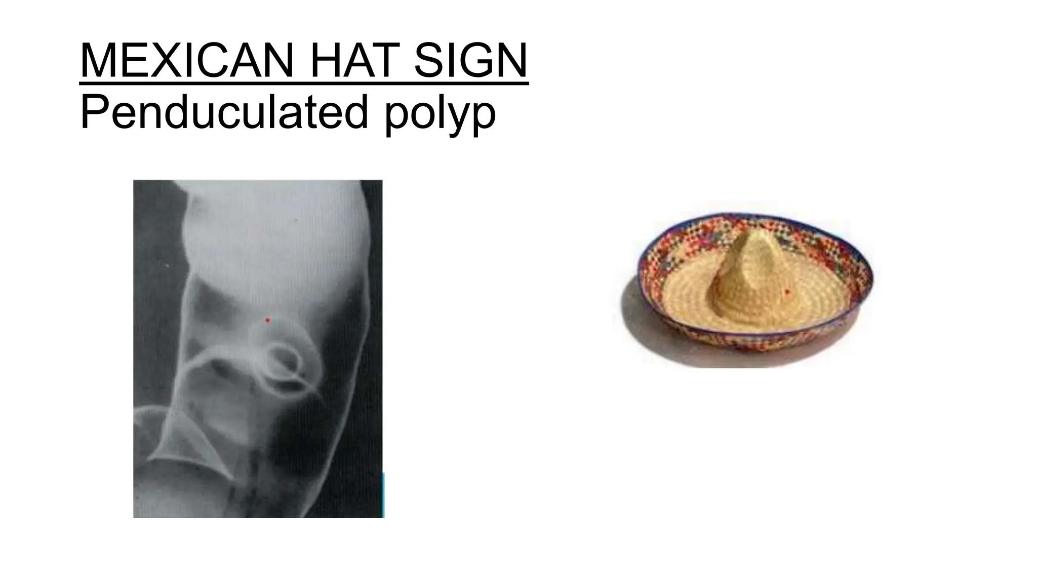
BARIUM ENEMA
PRESENTATION BY : DR MIRZA SANAULLA
MODERATOR : DR SHANKAR PATIL
BARIUM ENEMA
• It is the radiographic study of the large bowel by
administration of the contrast medium through the
rectum.
BARIUM
• Contrast medium of choice for GI studies is Barium sulphate.
• Barium has a high atomic number 56.
• Non absorbable, non-toxic.
• Insoluble in water/lipid.
• Inert to tissues.
• Can be used for double contrast studies.
Advantages
• 1. The main advantage of barium over water-soluble contrast
agents is that it results in better coating and thus better mucosal
detail.
• 2. Low cost.
Disadvantages
• 1. Subsequent abdominal CT is rendered difficult to interpret.
Patients may need to wait for up to 2 weeks to allow satisfactory
clearance of the barium.
• 2. High morbidity associated with barium in the peritoneal cavity.
IDEAL PREPARATION
• Stable suspension – carboxy methyl cellulose.
• Should not flocculate – sodium citrate.
• High density and low viscosity
• Antifoaming agent –dimethyl polysilicone , simethecone.
Adverse effects of barium
• Chemical perotinitis -50% mortaliy
• Barium inspissation
• Barium embolism
• Barium encephalopathy
• Barium granuloma
• Barium allergy (anaphylaxis)
BOWEL PREPARATION
DIET:
• Patient should be given a low residue (low fibre) diet for 2 days
prior to the examination.
• Patient should drink plenty of clear fluids on the day preceding
the examination.
• Iron containing medication should be stopped 2 days before the
examination
LAXATIVES
• Castor oil / Bisacodyl / magnesium citrate
• Bisacodyl (DULCOLAX ) 2 HS (15-20 mg ) for 2 days
Bowel wash
• Previous night.
• In the morning, 2 hours prior to the procedure.
INDICATIONS
• Rectal bleeding
• Suspected ca colon
• Polyposis
• Familial history of ca / polyposis
• Sinuses/fistulas
• Diverticulosis
• Ulcerative colitis
• Crhons disease
• Strictures
Contraindications
Absolute
• Toxic megacolon
• Pseudomembranous colitis
• Acute or fulminating inflammatory colon disease.
• Recent biopsy via:
a) rigid endoscope within previous 5 days (the biopsy forceps
used tend to be larger)
b) flexible endoscope within previous 24 h (the smaller biopsy
forceps only allows superficial mucosal biopsies).
Relative
• Incomplete bowel preparation
• Recent barium meal – it is advised to wait for 7–10 days
• Patient frailty.
METHODS
1. Double contrast – the method of choice to demonstrate
mucosal pattern.
2. Single contrast –
(a) localization of an obstructing colonic lesion
(b) gross pathology
(c) patient immobile
(c) reduction of an intussusception
SINGLE CONTRAST BARIUM ENEMA
Indications
• 1. Uncooperative, very debilitated or immobile patient.
• 2. Evaluation of acute obstruction or volvulus.
• 3. Reduction of intussusception.
• 4. Where only gross pathology is to be excluded.
TECHNIQUE
• Barium suspension : Low density (to promote see through effect with
a high kV ) 15% to 20% w /v.
• SCOUT FILM TAKEN
• Rectum and presacral space - Left lateral Frontal-prone
• Rectosigmoid - Prone right side down oblique
• Splenic flexure - Prone left side down oblique
• Hepatic flexure - Prone right side down oblique
• Entire colon - Supine and decubitus views
• After evacuation, mucosal relief film is exposed. Polyposis
and diverticulosis can be better visualized on post-evacuation films.
Millers tube and barium enema bag
DOUBLE CONTRAST BARIUM ENEMA
• Barium suspension : High density (slower flowing, better
coating) 75% to 95% w/v.
TECHNIQUE
• The patient lies on their left side, and the catheter is inserted
gently into the rectum. It is taped firmly in position. Connections
are made to the barium reservoir and the hand pump for
injecting air.
• An i.v. injection of Buscopan (20 mg) or glucagon (1 mg) is
given.
• 3. The infusion of barium is commenced. Intermittent screening is required to
check the progress of the barium. The barium is run to splenic flexure in the
left lateral position and then the patient is turned prone.
• Contrast is then run to the hepatic flexure and is stopped when it tips into the
right colon. Gentle puffs of air maybe needed to encourage the barium to flow.
The patient rolls onto their right and quickly onto their back. An adequate
amount of barium in the right colon is confirmed with fluoroscopy. The
column of barium within the distal colon is run back out by either lowering the
infusion bag to the floor or tilting the table to the erect position.
• 4. The catheter tube is occluded and air is gently pumped into the bowel to
produce the double-contrast effect. CO2 gas has been shown to reduce the
incidence of severe, post enema pain.
RECTOSIGMOID
Single contrast vs double contrast
AFTERCARE
• The patient must not drive until any blurring of vision produced
by Buscopan has resolved; usually within 30 minutes.
• Patients should be warned that their bowel motions will
be white for a few days after the examination. They may eat
normally and should drink extra fluids to avoid
barium impaction.
COMPLICATIONS
1. Cardiac arrhythmias induced by Buscopan or the procedure itself.
This is the most frequent cause of death after barium enema.
2. Perforation of the bowel. The second most common cause of
death after barium enema. Often associated with the rectal
catheter balloon
3. Transient bacteraemia..
4. Intramural barium.
5. Venous intravasation. This may result in a barium pulmonary
embolus, which carries an 80% mortality risk.
Normal mucosa in barium study
-smooth and featureless
MEXICAN HAT SIGN
Penduculated polyp
DIVERTICULUM
DIVERTICULOSIS
Apple core sign- colorectal carcinoma
Ulcerative colitis
Collar
button
abscess
Pseudopolyps
Lead pipe colon
Claw sign -INTUSSUSCEPTION
Bird beak sign- SIGMOID VOLVULUS
THANK YOU

barium enema121 study and its applications .pptx

  • 1.
    BARIUM ENEMA PRESENTATION BY: DR MIRZA SANAULLA MODERATOR : DR SHANKAR PATIL
  • 2.
    BARIUM ENEMA • Itis the radiographic study of the large bowel by administration of the contrast medium through the rectum.
  • 3.
    BARIUM • Contrast mediumof choice for GI studies is Barium sulphate. • Barium has a high atomic number 56. • Non absorbable, non-toxic. • Insoluble in water/lipid. • Inert to tissues. • Can be used for double contrast studies.
  • 4.
    Advantages • 1. Themain advantage of barium over water-soluble contrast agents is that it results in better coating and thus better mucosal detail. • 2. Low cost. Disadvantages • 1. Subsequent abdominal CT is rendered difficult to interpret. Patients may need to wait for up to 2 weeks to allow satisfactory clearance of the barium. • 2. High morbidity associated with barium in the peritoneal cavity.
  • 5.
    IDEAL PREPARATION • Stablesuspension – carboxy methyl cellulose. • Should not flocculate – sodium citrate. • High density and low viscosity • Antifoaming agent –dimethyl polysilicone , simethecone.
  • 6.
    Adverse effects ofbarium • Chemical perotinitis -50% mortaliy • Barium inspissation • Barium embolism • Barium encephalopathy • Barium granuloma • Barium allergy (anaphylaxis)
  • 7.
    BOWEL PREPARATION DIET: • Patientshould be given a low residue (low fibre) diet for 2 days prior to the examination. • Patient should drink plenty of clear fluids on the day preceding the examination. • Iron containing medication should be stopped 2 days before the examination LAXATIVES • Castor oil / Bisacodyl / magnesium citrate • Bisacodyl (DULCOLAX ) 2 HS (15-20 mg ) for 2 days
  • 8.
    Bowel wash • Previousnight. • In the morning, 2 hours prior to the procedure.
  • 9.
    INDICATIONS • Rectal bleeding •Suspected ca colon • Polyposis • Familial history of ca / polyposis • Sinuses/fistulas • Diverticulosis • Ulcerative colitis • Crhons disease • Strictures
  • 10.
    Contraindications Absolute • Toxic megacolon •Pseudomembranous colitis • Acute or fulminating inflammatory colon disease. • Recent biopsy via: a) rigid endoscope within previous 5 days (the biopsy forceps used tend to be larger) b) flexible endoscope within previous 24 h (the smaller biopsy forceps only allows superficial mucosal biopsies). Relative • Incomplete bowel preparation • Recent barium meal – it is advised to wait for 7–10 days • Patient frailty.
  • 11.
    METHODS 1. Double contrast– the method of choice to demonstrate mucosal pattern. 2. Single contrast – (a) localization of an obstructing colonic lesion (b) gross pathology (c) patient immobile (c) reduction of an intussusception
  • 12.
    SINGLE CONTRAST BARIUMENEMA Indications • 1. Uncooperative, very debilitated or immobile patient. • 2. Evaluation of acute obstruction or volvulus. • 3. Reduction of intussusception. • 4. Where only gross pathology is to be excluded.
  • 13.
    TECHNIQUE • Barium suspension: Low density (to promote see through effect with a high kV ) 15% to 20% w /v. • SCOUT FILM TAKEN • Rectum and presacral space - Left lateral Frontal-prone • Rectosigmoid - Prone right side down oblique • Splenic flexure - Prone left side down oblique • Hepatic flexure - Prone right side down oblique • Entire colon - Supine and decubitus views • After evacuation, mucosal relief film is exposed. Polyposis and diverticulosis can be better visualized on post-evacuation films.
  • 14.
    Millers tube andbarium enema bag
  • 18.
    DOUBLE CONTRAST BARIUMENEMA • Barium suspension : High density (slower flowing, better coating) 75% to 95% w/v.
  • 19.
    TECHNIQUE • The patientlies on their left side, and the catheter is inserted gently into the rectum. It is taped firmly in position. Connections are made to the barium reservoir and the hand pump for injecting air. • An i.v. injection of Buscopan (20 mg) or glucagon (1 mg) is given.
  • 20.
    • 3. Theinfusion of barium is commenced. Intermittent screening is required to check the progress of the barium. The barium is run to splenic flexure in the left lateral position and then the patient is turned prone. • Contrast is then run to the hepatic flexure and is stopped when it tips into the right colon. Gentle puffs of air maybe needed to encourage the barium to flow. The patient rolls onto their right and quickly onto their back. An adequate amount of barium in the right colon is confirmed with fluoroscopy. The column of barium within the distal colon is run back out by either lowering the infusion bag to the floor or tilting the table to the erect position. • 4. The catheter tube is occluded and air is gently pumped into the bowel to produce the double-contrast effect. CO2 gas has been shown to reduce the incidence of severe, post enema pain.
  • 21.
  • 24.
    Single contrast vsdouble contrast
  • 25.
    AFTERCARE • The patientmust not drive until any blurring of vision produced by Buscopan has resolved; usually within 30 minutes. • Patients should be warned that their bowel motions will be white for a few days after the examination. They may eat normally and should drink extra fluids to avoid barium impaction.
  • 26.
    COMPLICATIONS 1. Cardiac arrhythmiasinduced by Buscopan or the procedure itself. This is the most frequent cause of death after barium enema. 2. Perforation of the bowel. The second most common cause of death after barium enema. Often associated with the rectal catheter balloon 3. Transient bacteraemia.. 4. Intramural barium. 5. Venous intravasation. This may result in a barium pulmonary embolus, which carries an 80% mortality risk.
  • 27.
    Normal mucosa inbarium study -smooth and featureless
  • 29.
  • 30.
  • 31.
  • 32.
    Apple core sign-colorectal carcinoma
  • 33.
  • 34.
  • 35.
  • 36.
    Bird beak sign-SIGMOID VOLVULUS
  • 37.