Download to read offline
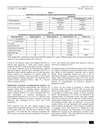
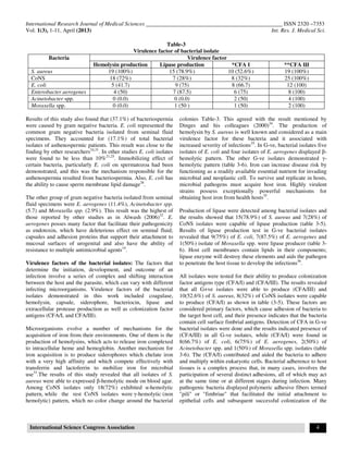

This study examined bacterial isolates associated with leukocytospermia in asthenospermic patients in Hilla City, Iraq. Semen samples were collected from 100 infertile men and divided into two groups based on the presence of leukocytes. Bacterial cultures were positive in 87.1% of samples with leukocytospermia compared to 0% without. Gram-positive bacteria like coagulase-negative staphylococci and Staphylococcus aureus were the most common isolates. Virulence factors including hemolysins, colonization factors, lipases and proteases were detected in many of the isolates. The isolates showed resistance to many antibiotics but were susceptible to imipenem, meropenem and


