Download to read offline
![2 Hussein Oleiwi Muttaleb Al-Dahmoshi et al, 2014
Advances in Natural and Applied Sciences, 8(15) December 2014, Pages: 1-8
to cell apoptosis or gene upregulation (Waterston, A. and M. Bower, 2004). TNF-α is attractive therapeutic
protein with a broad range of potent anti-cancer effects, without inhibiting normal cell growth (Christel, L., et
al., 2007). The apoptosis in tumor cells induced by TNF-α via activation of downstream effector Caspases and
disruption of mitochondrial integrity (Bocangel, D.B., et al., 2005). Also there are an evidence for involvement
of endothelial cell integrin avβ3 in disruption of tumor vasculature induced by combination of TNF-α and IFN-γ
(Christel, L., et al., 2007). TNF-α level determined by ELISA using culture medium increase between 5-10 fold
, 48hr. following exposure to radiation with observed in cell in which TNF-α was constitutively expressed under
Cytomegaloviruspromoter (pCMV- TNF-α) (Jung, M., et al., 2011)
Hallahan et al. (1989)[8]
show that TNF-α and TNF-α mRNA increase significantly after exposure to 500
cGy X-ray in tumor sarcoma cell lines and not elevated in supernatant from normal human fibroblast cell lines.
TNF-α increased in cell that exposed to linear energy transfer (LED) radiation (0.1-2 Gy). The TNF-α as low as
0.1 ng/ml initiated increased DNA damage in cell treated with LED cell as compare with control (Natarajan, M.,
et al., 2007). The nuclear factor, κβ (NF-κβ) activation by TNF-α, the specific processes involved in the
activation of transcription factor by ionizing radiation (IR) not completely defined (Russell, J.S. and P.J.
Tofilon, 2002). There is evidence that implicated IR-induced NF-κβ mediated initiation of TNF-α dependent a
positive feedback mechanism. TNF-α can activate NF-κβ through TNF receptor associated factor. The blocking
of NF-κβ has been demonstrated sensitize cancer cells to TNF-α induced killing (Veeraraghavan, J., et al.,
2001).
Interferons (IFNs) are family of cytokines that can induce diverse biological functions, such as antiviral,
antitumor and immunomodulatory activities. IFNs can be divided into type I, II and III. Type I consist of seven
classes, mainly IFN-α and IFN-β, whereas type II has only one, IFN-γ. IFN-γ has important immunoregulatory
functions including activation of macrophage, antiproleferative effects on transformed cell as well as
antitumorigenic effects (Kim, K.S., et al., 2012).
IFN-γ treatment can activate STAT (Signal Transducer and Activator of Transcription), which essential
for cell differential, cell cycle control and development. IFN-α induced apoptosis in both A431 and Hela cells
(Chin, Y.E., et al., 1997). Many of inhibitory of effect of IFN-α appear to be mediated by IFN regulatory factor-
1 (IRF-1). IRF-1 regulates expression of several genes involved in apoptosis, such as the pro apoptotic protein
Bax, the tumor suppressor p53, Caspase-1 and PKR (Kim, K.S., et al., 2012). Macrophage become cytotoxic
and destroy tumor cell through production of reactive oxygen intermediates and TNF-α after activation with
IFN-γ. The ionizing radiation can mimic cytokine signals and lead to enhanced states of activation (McKinney,
L.C., et al., 1998).
Han et.al (2002) shows the gamma irradiation can reduce IFN-γ mRNA expression and reduce both of
STAT1 and IF-1. It has been shown that the phagocytic function of macrophage is quite resistant to irradiation.
Macrophages have been observed phagocytizing debris of dead cells in irradiated lymphoid tissues and also
noted in alveolar macrophages isolated from mice after radon exposure (Trollip, A.P., 2007).
MATERIALS AND METHODS
Samples:
Venous blood samples (5-10ml) were withdrawn from inadvertent radium irradiated person and putted in
Gel tube to separate the serum. The serum then divided and reserved in 1.5 ml sterile eppendrof tube and frozen
at -20˚C.
Subject and Control:
Fourty two inadvertent radium irradiated person were taken from Nader 3 District, Hilla City- Iraq who
have been inadvertently exposed to radium-226(226
Ra) radiation, that enrolled in Iraqi ministry of science and
technology, and ministry of health and ministry of environment. The control group was comprised of healthy
with no history of irradiation.
Age Groups:
The inadvertent radium irradiated person were divided into three age groups as follow: group1 (< 27 years),
group2 (27-37 years) and group3 (>37 years). The same groups non irradiated consider as control.
Ethics:
The all inadvertent radium irradiated person were told about the importance and details of research and
fully explain aims of this study and the protocol of ethics approved by ethical guide. It is important to know that
many of the subjects were refuse and don’t agree to give the serum samples.](https://image.slidesharecdn.com/30c3f0dd-748d-4d86-bbc9-7a9980764cc8-161102182220/85/Radium-2-320.jpg)
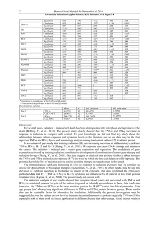

This study investigated hematological changes, serum TNF-α and IFN-γ levels in 42 people inadvertently exposed to radium-226 in Hilla City, Iraq. The study found significantly higher levels of the pro-inflammatory cytokines TNF-α and IFN-γ in exposed individuals compared to unexposed controls. There was a negative correlation between cytokine levels and complete blood count results. The results suggest that tiny doses of radium could induce TNF-α and IFN-γ, and that these cytokines may serve as biomarkers for radium exposure. Radium exposure was found to cause changes in pro-inflammatory cytokine levels and hematological parameters.

![[Interdisciplinary Toxicology] Evaluation of miR-9 and miR-143 expression in ...](https://cdn.slidesharecdn.com/ss_thumbnails/aaa8c98e-b367-4a4e-8eda-52087a22aee0-160921073900-thumbnail.jpg?width=640&height=640&fit=bounds)


![20120112111947555[1]](https://cdn.slidesharecdn.com/ss_thumbnails/201201121119475551-120113075518-phpapp02-thumbnail.jpg?width=640&height=640&fit=bounds)


