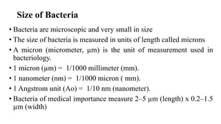
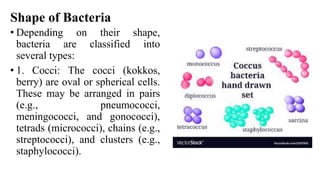
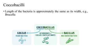
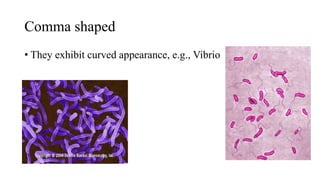
2. MICROORGANSIMS
• All living beings can be classified into three kingdoms:
• Plant, Animal, and Protista
• Microorganisms are a heterogeneous group of several distinct living
structures of microscopic size, classified under the kingdom Protista.
The kingdom Protista includes unicellular organisms, such as
bacteria, fungi, protozoa, and algae.
• Based on the differences in cellular organization and biochemistry, the
kingdom Protista has been divided into three groups:
• Prokaryotes, eukaryotes, and the most recently described
archaebacteria.
3. Prokaryotes
• Bacteria and blue green algae are prokaryotes
• Bacteria are unicellular free-living organisms having both DNA and
RNA
• They are capable of performing all essential processes of life, e.g.,
growth, reproduction, and metabolism
• They do not show any true branching except Actinomycetales, the
higher bacteria
• Bacteria lack chlorophyll unlike blue green algae, which contain
chlorophyll
4. Eukaryotes
• Fungi, algae other than blue green, protozoa, and slime
moulds are eukaryotes
• Archaebacteria
• These are more closely related to eukaryotes than prokaryotes
• They however do not include any human pathogens
5. Size of Bacteria
• Bacteria are microscopic and very small in size
• The size of bacteria is measured in units of length called microns
• A micron (micrometer, µm) is the unit of measurement used in
bacteriology.
• 1 micron (µm) = 1/1000 millimeter (mm).
• 1 nanometer (nm) = 1/1000 micron ( mm).
• 1 Angstrom unit (Ao) = 1/10 nm (nanometer).
• Bacteria of medical importance measure 2–5 µm (length) x 0.2–1.5
µm (width)
6. Microscopy
• Microscopy is an important component of diagnostic microbiology
• Bacteria being very small cannot be visualized by the naked eye,
because the limit of resolution with the unaided eye is about 200
microns
• So, the study of bacteria requires the use of microscopes
• A microscope is an instrument that uses one or more lenses to
produce a magnified image of an object that is invisible to the
unaided eye
7.
8. Shape of Bacteria
• Depending on their shape,
bacteria are classified into
several types:
• 1. Cocci: The cocci (kokkos,
berry) are oval or spherical cells.
These may be arranged in pairs
(e.g., pneumococci,
meningococci, and gonococci),
tetrads (micrococci), chains (e.g.,
streptococci), and clusters (e.g.,
staphylococci).
9. Bacilli
• The bacilli (bacillus, rod) are rod
shaped. These bacilli may show
either of the following
arrangement
16. Structure and Functions of Bacterial Cell Envelope
• The outer layer or cell envelope provides a structural and physiological barrier
between the protoplasm (inside) of the cell and the external environment
• The cell envelope protects bacteria against osmotic lysis and gives bacteria
rigidity and shape
• The cell envelope primarily consists of two components: a cell wall and
cytoplasmic or plasma membrane
• It encloses the protoplasm, which consists of (i) cytoplasm, (ii) cytoplasmic
inclusions (mesosomes, ribosomes, inclusion granules, vacuoles), and (iii) a single
circular DNA
17.
18. Cell Wall
• Prokaryotic cells almost always are bounded by a rigid and
chemically complex structure present between the cell membrane
and capsule/slime layer called the cell wall
• Peptidoglycan is the main component of the cell wall and is
responsible for the shape and strength of the cell
• It is a disaccharide and contains two sugar derivatives—N-
acetylglucosamine and N-acetylmuramic acid—joined together by
short peptide chains
• N-acetylmuramic acid carries a tetrapeptide side chain consisting of
D- and L-amino acids (D-glutamic acid and L-alanine) with
mesodiaminopimelic acid (Gram-negative bacteria) or L-lysine
(Gram-positive bacteria)