Download to read offline
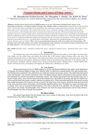


1) During a routine anatomy dissection, researchers observed an unusual branching pattern of the brachial artery in a male cadaver. 2) Specifically, they found that the ulnar artery originated from the lateral side of the brachial artery, rather than in the cubital fossa as is typical. 3) The variant ulnar artery descended on the lateral forearm and crossed to the medial side, following a superficial path along the arm.


