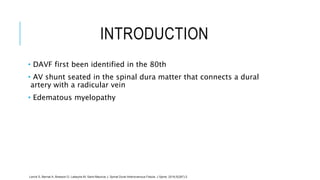
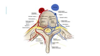
SPINAL DURAL
ARTERIOVENOUS FISTULA
Ade Wijaya – June 2018
OUTLINE:
• Introduction
• Anatomy
• Pathophysiology
• Clinical Presentation
• MRI
• Treatment
• Summary
INTRODUCTION
• DAVF first been identified in the 80th
• AV shunt seated in the spinal dura matter that connects a dural
artery with a radicular vein
• Edematous myelopathy
Lenck S, Bernat A, Bresson D, Labeyrie M, Saint-Maurice J. Spinal Dural Arteriovenous Fistula. J Spine. 2016;5(287):2.
PATHOPHYSIOLOGY
• The venous hyper pressure induced by arterialization of the anterior
and/or posterior spinal veins hinders venous return to the spinal
cord, thus resulting in a congestive myelopathy.
• Thrombosis of a radicular vein can aggravate the myelopathy by
restricting venous drainage of the fistula.
• If there are enough functional radicular veins, the fistula may be
asymptomatic
Jeng Y, Chen DY, Hsu HL, Huang YL, Chen CJ, et al. (2015) Spinal Dural Arteriovenous Fistula: Imaging Features and Its Mimics. Korean J
CLINICAL PRESENTATION
• Middle aged men.
• Progressive myelopathy.
• The symptoms can rapidly become worse, and this can sometimes
take place following a strenuous effort; a change in position; or occur
spontaneously.
• Average diagnostic limbo of 10 to 15 months.
• The neurological prognosis depends on the initial clinical condition
and also on an early therapeutic treatment
• Spontaneous course of this pathology can result in irreversible para-
or tetraplegia
Lenck S, Bernat A, Bresson D, Labeyrie M, Saint-Maurice J. Spinal Dural Arteriovenous Fistula. J Spine. 2016;5(287):2.
MRI
• The hyper signal most often extends over more than 2 vertebrae and
involves the terminal cone for approximately 80% of cases.
• The presence of flow voids and vascular dilation in T2 sequence is
highly suggestive of DAVF.
• Enhanced contrast of the perimedullary spaces.
Luetmer PH, Lane JI, Gilbertson JR, Bernstein MA, Huston J, et al. (2005) Preangiographic evaluation of spinal dural arteriovenous fistulas with elliptic centric contrast-enhanced MR Angiography and effect on
radiation dose and volume of iodinated contrast material. AJNR Am J Neuroradiol. 26: 711-718.
Hurst RW, Grossman RI (2000) Peripheral spinal cord hypointensity on J Spine ISSN: 2165-7939 JSP, an open access journal T2-weighted MR images: a reliable imaging sign of venous hypertensive myelopathy.
TREATMENT
• Endovascular
• Surgical
• Post endovascular / surgical
Lenck S, Bernat A, Bresson D, Labeyrie M, Saint-Maurice J. Spinal Dural Arteriovenous Fistula. J Spine. 2016;5(287):2.
SUMMARY
• Rare; often undiagnosed
• Early diagnosis and treatment is a major prognostic factor
• Diagnosis: MRI & Medullar angiography
• Embolization failure or contraindication; or reccurent after
embolization  surgery
•In case of recurrence of the symptoms, medullar angiography should
be performed to rule out a recurrence of the DAVF, a new fistula, or a
medullary venous insufficiency.
Spinal Dural Arteriovenous Fistula

Spinal Dural Arteriovenous Fistula

  • 1.
  • 2.
    OUTLINE: • Introduction • Anatomy •Pathophysiology • Clinical Presentation • MRI • Treatment • Summary
  • 3.
    INTRODUCTION • DAVF firstbeen identified in the 80th • AV shunt seated in the spinal dura matter that connects a dural artery with a radicular vein • Edematous myelopathy Lenck S, Bernat A, Bresson D, Labeyrie M, Saint-Maurice J. Spinal Dural Arteriovenous Fistula. J Spine. 2016;5(287):2.
  • 5.
    PATHOPHYSIOLOGY • The venoushyper pressure induced by arterialization of the anterior and/or posterior spinal veins hinders venous return to the spinal cord, thus resulting in a congestive myelopathy. • Thrombosis of a radicular vein can aggravate the myelopathy by restricting venous drainage of the fistula. • If there are enough functional radicular veins, the fistula may be asymptomatic Jeng Y, Chen DY, Hsu HL, Huang YL, Chen CJ, et al. (2015) Spinal Dural Arteriovenous Fistula: Imaging Features and Its Mimics. Korean J
  • 6.
    CLINICAL PRESENTATION • Middleaged men. • Progressive myelopathy. • The symptoms can rapidly become worse, and this can sometimes take place following a strenuous effort; a change in position; or occur spontaneously. • Average diagnostic limbo of 10 to 15 months. • The neurological prognosis depends on the initial clinical condition and also on an early therapeutic treatment • Spontaneous course of this pathology can result in irreversible para- or tetraplegia Lenck S, Bernat A, Bresson D, Labeyrie M, Saint-Maurice J. Spinal Dural Arteriovenous Fistula. J Spine. 2016;5(287):2.
  • 7.
    MRI • The hypersignal most often extends over more than 2 vertebrae and involves the terminal cone for approximately 80% of cases. • The presence of flow voids and vascular dilation in T2 sequence is highly suggestive of DAVF. • Enhanced contrast of the perimedullary spaces. Luetmer PH, Lane JI, Gilbertson JR, Bernstein MA, Huston J, et al. (2005) Preangiographic evaluation of spinal dural arteriovenous fistulas with elliptic centric contrast-enhanced MR Angiography and effect on radiation dose and volume of iodinated contrast material. AJNR Am J Neuroradiol. 26: 711-718. Hurst RW, Grossman RI (2000) Peripheral spinal cord hypointensity on J Spine ISSN: 2165-7939 JSP, an open access journal T2-weighted MR images: a reliable imaging sign of venous hypertensive myelopathy.
  • 8.
    TREATMENT • Endovascular • Surgical •Post endovascular / surgical Lenck S, Bernat A, Bresson D, Labeyrie M, Saint-Maurice J. Spinal Dural Arteriovenous Fistula. J Spine. 2016;5(287):2.
  • 9.
    SUMMARY • Rare; oftenundiagnosed • Early diagnosis and treatment is a major prognostic factor • Diagnosis: MRI & Medullar angiography • Embolization failure or contraindication; or reccurent after embolization  surgery •In case of recurrence of the symptoms, medullar angiography should be performed to rule out a recurrence of the DAVF, a new fistula, or a medullary venous insufficiency.