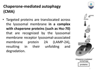
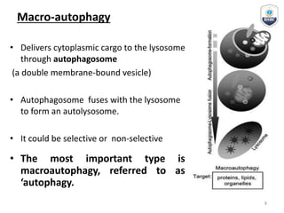
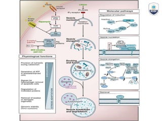
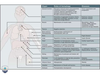
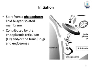
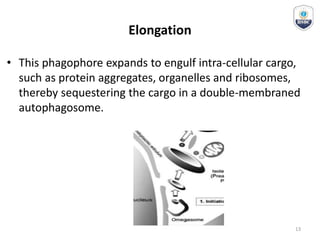
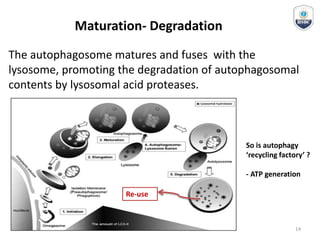
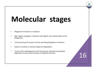
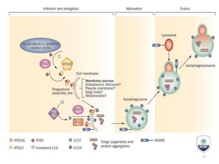
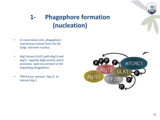
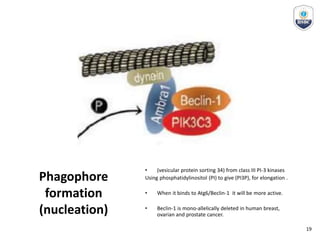
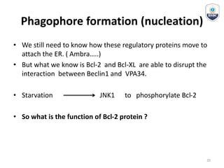
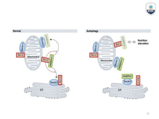
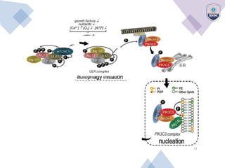
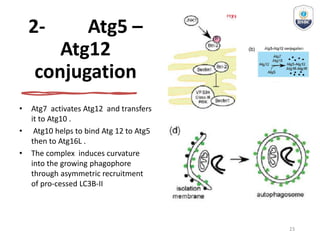
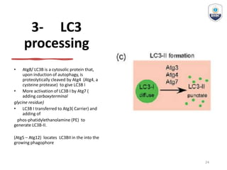
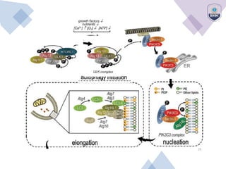
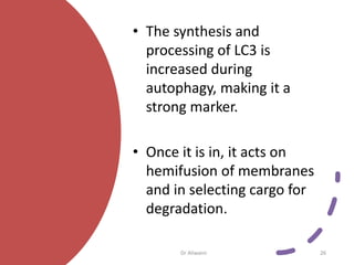
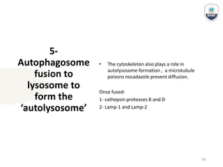
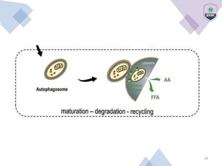
Autophagy
1
Autophagy
• Introduction
• Types of autophagy
• Cellular and Molecular mechanism
• Signaling pathways
• Estimation of Autophagic Activity
2
Introduction
• Auto-phagy = Self- eating
• Autophagy is a lysosomal degradation pathway that is essential for
survival, differentiation, development, and homeostasis.
• Autophagy principally to protect organisms against diverse pathologies,
including infections, cancer, neurodegeneration, aging, and heart
disease.
• First autophagy in rat liver cells
• But it was not clear why? and how it works?
• Starvation, Hypoxia, Stress…
3
Introduction
• How autophagy is regulated and executed at the
molecular level have been made in yeast.
• 32 different autophagy-related genes (Atg)
• many of these genes are conserved in slime
mould, plants, worms, flies and mammals.
4
Types of autophagy
General feature for all of them : proteolytic degradation of
cytosolic components at the lysosome.
5
Micro-autophagy
Chaperone-mediated autophagy
(CMA)
• Targeted proteins are translocated across
the lysosomal membrane in a complex
with chaperone proteins (such as Hsc-70)
that are recognized by the lysosomal
membrane receptor lysosomal-associated
membrane protein 2A (LAMP-2A),
resulting in their unfolding and
degradation.
7
Macro-autophagy
• Delivers cytoplasmic cargo to the lysosome
through autophagosome
(a double membrane-bound vesicle)
• Autophagosome fuses with the lysosome
to form an autolysosome.
• It could be selective or non-selective
• The most important type is
macroautophagy, referred to as
‘autophagy.
8
The basic
machinery of
autophagy
11
Initiation
• Start from a phagophore:
lipid bilayer isolated
membrane
• Contributed by the
endoplasmic reticulum
(ER) and/or the trans-Golgi
and endosomes
12
Elongation
• This phagophore expands to engulf intra-cellular cargo,
such as protein aggregates, organelles and ribosomes,
thereby sequestering the cargo in a double-membraned
autophagosome.
13
Maturation- Degradation
The autophagosome matures and fuses with the
lysosome, promoting the degradation of autophagosomal
contents by lysosomal acid proteases.
Re-use
So is autophagy
‘recycling factory’ ?
- ATP generation
14
Molecular
Mechanism
15
Molecular stages
• Phagophore formation or nucleation
• Atg5 –Atg12 conjugation, interaction with Atg16L and multimerization at the
phagophore.
• LC3 processing and insertion into the extending phagophore membrane.
• Capture of random or selective targets for degradation.
• Fusion of the autophagosome with the lysosome, followed by proteolytic
degradation by lyso-somal proteases of engulfed molecules.
16
17
1- Phagophore formation
(nucleation)
• In mammalian cells, phagophore
membranes initiate from the ER,
Golgi and even nucleus.
• Atg1 kinase (ULK1) with Atg13 and
Atg17 regulate Atg9 activity which
promotes lipid recruitment to the
expanding phagophore .
• TOR Kinase prevent Atg 13 to
interact Atg 1
18
Phagophore
formation
(nucleation)
• (vesicular protein sorting 34) from class III PI-3 kinases
Using phosphatidylinositol (PI) to give (PI3P), for elongation .
• When it binds to Atg6/Beclin-1 it will be more active.
• Beclin-1 is mono-allelically deleted in human breast,
ovarian and prostate cancer.
19
Phagophore formation (nucleation)
• We still need to know how these regulatory proteins move to
attach the ER. ( Ambra…..)
• But what we know is Bcl-2 and Bcl-XL are able to disrupt the
interaction between Beclin1 and VPA34.
• Starvation JNK1 to phosphorylate Bcl-2
• So what is the function of Bcl-2 protein ?
20
21
22
2- Atg5 –
Atg12
conjugation
• Atg7 activates Atg12 and transfers
it to Atg10 .
• Atg10 helps to bind Atg 12 to Atg5
then to Atg16L .
• The complex induces curvature
into the growing phagophore
through asymmetric recruitment
of pro-cessed LC3B-II
23
3- LC3
processing
• Atg8/ LC3B is a cytosolic protein that,
upon induction of autophagy, is
proteolytically cleaved by Atg4 (Atg4, a
cysteine protease) to give LC3B I
• More activation of LC3B I by Atg7 (
adding carboxyterminal
glycine residue)
• LC3B I transferred to Atg3( Carrier) and
adding of
phos-phatidylethanolamine (PE) to
generate LC3B-II.
(Atg5 – Atg12) locates LC3BII in the into the
growing phagophore
24
25
• The synthesis and
processing of LC3 is
increased during
autophagy, making it a
strong marker.
• Once it is in, it acts on
hemifusion of membranes
and in selecting cargo for
degradation.
Dr Aliwaini 26
4- Capture of
random or
selective
targets for
degradation.
• LC3B-II, acting as a ‘receptor’ to interacts
with ‘adaptor’ molecules as p62/SQSTM1
and NBR1on the target to promote their
selective uptake and degradation.
27
5-
Autophagosome
fusion to
lysosome to
form the
‘autolysosome’
• The cytoskeleton also plays a role in
autolysosome formation , a microtubule
poisons nocadazole prevent diffusion.
Once fused:
1- cathepsin proteases B and D
2- Lamp-1 and Lamp-2
28
29

Autophagy Lecture from HNO Skill Development Centre

  • 1.
  • 2.
    Autophagy • Introduction • Typesof autophagy • Cellular and Molecular mechanism • Signaling pathways • Estimation of Autophagic Activity 2
  • 3.
    Introduction • Auto-phagy =Self- eating • Autophagy is a lysosomal degradation pathway that is essential for survival, differentiation, development, and homeostasis. • Autophagy principally to protect organisms against diverse pathologies, including infections, cancer, neurodegeneration, aging, and heart disease. • First autophagy in rat liver cells • But it was not clear why? and how it works? • Starvation, Hypoxia, Stress… 3
  • 4.
    Introduction • How autophagyis regulated and executed at the molecular level have been made in yeast. • 32 different autophagy-related genes (Atg) • many of these genes are conserved in slime mould, plants, worms, flies and mammals. 4
  • 5.
    Types of autophagy Generalfeature for all of them : proteolytic degradation of cytosolic components at the lysosome. 5
  • 6.
  • 7.
    Chaperone-mediated autophagy (CMA) • Targetedproteins are translocated across the lysosomal membrane in a complex with chaperone proteins (such as Hsc-70) that are recognized by the lysosomal membrane receptor lysosomal-associated membrane protein 2A (LAMP-2A), resulting in their unfolding and degradation. 7
  • 8.
    Macro-autophagy • Delivers cytoplasmiccargo to the lysosome through autophagosome (a double membrane-bound vesicle) • Autophagosome fuses with the lysosome to form an autolysosome. • It could be selective or non-selective • The most important type is macroautophagy, referred to as ‘autophagy. 8
  • 11.
  • 12.
    Initiation • Start froma phagophore: lipid bilayer isolated membrane • Contributed by the endoplasmic reticulum (ER) and/or the trans-Golgi and endosomes 12
  • 13.
    Elongation • This phagophoreexpands to engulf intra-cellular cargo, such as protein aggregates, organelles and ribosomes, thereby sequestering the cargo in a double-membraned autophagosome. 13
  • 14.
    Maturation- Degradation The autophagosomematures and fuses with the lysosome, promoting the degradation of autophagosomal contents by lysosomal acid proteases. Re-use So is autophagy ‘recycling factory’ ? - ATP generation 14
  • 15.
  • 16.
    Molecular stages • Phagophoreformation or nucleation • Atg5 –Atg12 conjugation, interaction with Atg16L and multimerization at the phagophore. • LC3 processing and insertion into the extending phagophore membrane. • Capture of random or selective targets for degradation. • Fusion of the autophagosome with the lysosome, followed by proteolytic degradation by lyso-somal proteases of engulfed molecules. 16
  • 17.
  • 18.
    1- Phagophore formation (nucleation) •In mammalian cells, phagophore membranes initiate from the ER, Golgi and even nucleus. • Atg1 kinase (ULK1) with Atg13 and Atg17 regulate Atg9 activity which promotes lipid recruitment to the expanding phagophore . • TOR Kinase prevent Atg 13 to interact Atg 1 18
  • 19.
    Phagophore formation (nucleation) • (vesicular proteinsorting 34) from class III PI-3 kinases Using phosphatidylinositol (PI) to give (PI3P), for elongation . • When it binds to Atg6/Beclin-1 it will be more active. • Beclin-1 is mono-allelically deleted in human breast, ovarian and prostate cancer. 19
  • 20.
    Phagophore formation (nucleation) •We still need to know how these regulatory proteins move to attach the ER. ( Ambra…..) • But what we know is Bcl-2 and Bcl-XL are able to disrupt the interaction between Beclin1 and VPA34. • Starvation JNK1 to phosphorylate Bcl-2 • So what is the function of Bcl-2 protein ? 20
  • 21.
  • 22.
  • 23.
    2- Atg5 – Atg12 conjugation •Atg7 activates Atg12 and transfers it to Atg10 . • Atg10 helps to bind Atg 12 to Atg5 then to Atg16L . • The complex induces curvature into the growing phagophore through asymmetric recruitment of pro-cessed LC3B-II 23
  • 24.
    3- LC3 processing • Atg8/LC3B is a cytosolic protein that, upon induction of autophagy, is proteolytically cleaved by Atg4 (Atg4, a cysteine protease) to give LC3B I • More activation of LC3B I by Atg7 ( adding carboxyterminal glycine residue) • LC3B I transferred to Atg3( Carrier) and adding of phos-phatidylethanolamine (PE) to generate LC3B-II. (Atg5 – Atg12) locates LC3BII in the into the growing phagophore 24
  • 25.
  • 26.
    • The synthesisand processing of LC3 is increased during autophagy, making it a strong marker. • Once it is in, it acts on hemifusion of membranes and in selecting cargo for degradation. Dr Aliwaini 26
  • 27.
    4- Capture of randomor selective targets for degradation. • LC3B-II, acting as a ‘receptor’ to interacts with ‘adaptor’ molecules as p62/SQSTM1 and NBR1on the target to promote their selective uptake and degradation. 27
  • 28.
    5- Autophagosome fusion to lysosome to formthe ‘autolysosome’ • The cytoskeleton also plays a role in autolysosome formation , a microtubule poisons nocadazole prevent diffusion. Once fused: 1- cathepsin proteases B and D 2- Lamp-1 and Lamp-2 28
  • 29.