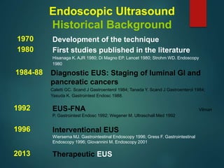
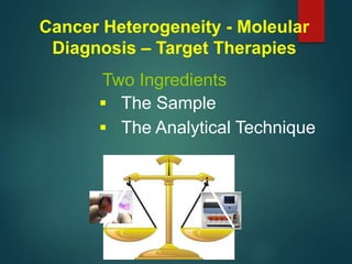
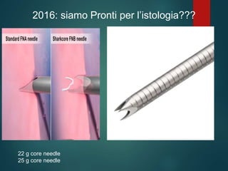
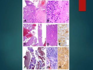
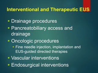
Fabia Attili MD, PhD
Digestive Endoscopy Unit
European Endoscopy Training Centre
Catholic University, Rome
INNOVAZIONI in ECOENDOSCOPIA
COS’E’
L’ECOENDOSCOPIA
E’ la combinazione di endoscopia ed ecografia
COS’E’L’ECOENDOSCOPIA
Strumento radiale
Strumento lineare
EUS radiale (imaging) EUS lineare (operativa)
1980 First studies published in the literature
Hisanaga K. AJR 1980; Di Magno EP. Lancet 1980; Strohm WD. Endoscopy
1980
1984-88 Diagnostic EUS: Staging of luminal GI and
pancreatic cancers
Caletti GC. Scand J Gastroenterol 1984; Tanada Y. Scand J Gastroenterol 1984;
Yasuda K. Gastrointest Endosc 1988.
1992 EUS-FNA Vilman
P. Gastrointest Endosc 1992; Wegener M. Ultraschall Med 1992
1970 Development of the technique
1996 Interventional EUS
Wiersema MJ. Gastrointestinal Endoscopy 1996; Gress F. Gastrointestinal
Endoscopy 1996; Giovannini M. Endoscopy 2001
2013 Therapeutic EUS
Endoscopic Ultrasound
Historical Background
ECOENDOSCOPIA E ONCOLOGIA
CANCRO
Tipo di neoplasia
Marcatori Prognostitici predittivi
Diagnosi molecolare
Terapie Mirate
 The Sample
 The Analytical Technique
Two Ingredients
Cancer Heterogeneity - Moleular
Diagnosis – Target Therapies
CITOLOGIA o ISTOLOGIA ???
Histology advantages over Cytology
 Future tissue profiling to guide individualized
therapies highly dependent on tissue
availability
 Easier to interpret and to perform
immunohistochemicals studies than cytology
 Cytopathology expertise is not available in
many centers
 The need for cytopathology has limited the
widespread use of EUS throughout the world
Why Histology?
Gastrointest Endosc 2005;62:768-74.
Gastrointest Endosc 2004;59:185-90.
19 g
22 g
25 g
2015 : Siamo Pronti per L’istologia??
 19 G/Procore/Flex: very good results but
difficult to use and fear of complications
 22 Procore: too few results to draw
conclusions
 25 Procore: too small to get enough tissue
What we need to search for:
A needle that can be used by the large majority
of endosonographers able to provide the
adequate amount of tissue to perform all the
studies you need
2015 : Siamo pronti per L’istologia??
NO
Contrast-enhanced EUS
 Signals from microbubbles appear approximately 10 seconds
after infusion and peak 20-30 seconds after infusion
Pancreas head image before
infusion of SonoVue
CH-EUS image 13 sec. after
infusion of contrast agent. Spotty
signals from microbubbles and
fine branching vessels
CH-EUS image 21 sec. After
infusion of contrast agent. Diffuse
parenchymal perfusion of
microbubbles. Bile Duct and MPD
are non-enhanced structures
Kitano, 2015
Contrast-enhanced EUS
Hypo-enhancement
Ductal carcinoma, hypoechoic area of
23 mm
Iso-enhancement
Tumor-forming chroning pancreatitis,
hypoechoic area of 22 mm
Hyper-enhancement
Neuroendocrine tumor, hypoechoic area
of 14 mm
Kitano, 2015
Contrast-enhanced EUS
CH-EUS for FNA
80-90% of false negative cases in EUS-FNA were correctly
classified by CH-EUS
CH-EUS could help to decide between surgery and follow-
up when then results of FNA are inconclusive
CH-EUS is also useful for identification of the target of
FNA, improving depiction of very small lesions and
identification of avascular sites (necrotic areas and
hemorragic risks)
Contrast-enhanced EUS
Kitano, 2015
Elastography
The science of creating
noninvasive images of
mechanical characteristics of
tissues
Real-time elastography during
the endoscopic ultrasound is
the study of tissue stiffness
and orients the diagnosis
towards different pathological
entities
EUS Elastography
2016: siamo Pronti per l’istologia???
22 g core needle
25 g core needle
2016: siamo pronti per l’istologia???
SI !!!!!!
 Drainage procedures
 Pancreatobiliary access and
drainage
 Oncologic procedures
 Fine needle injection, implantation and
EUS-guided directed therapies
 Vascular interventions
 Endosurgical interventions
Interventional and Therapeutic EUS
Attili F. L'Ecoendoscopia. ASMaD 2016

Attili F. L'Ecoendoscopia. ASMaD 2016

  • 1.
    Fabia Attili MD,PhD Digestive Endoscopy Unit European Endoscopy Training Centre Catholic University, Rome INNOVAZIONI in ECOENDOSCOPIA
  • 2.
  • 3.
  • 4.
  • 5.
    EUS radiale (imaging)EUS lineare (operativa)
  • 6.
    1980 First studiespublished in the literature Hisanaga K. AJR 1980; Di Magno EP. Lancet 1980; Strohm WD. Endoscopy 1980 1984-88 Diagnostic EUS: Staging of luminal GI and pancreatic cancers Caletti GC. Scand J Gastroenterol 1984; Tanada Y. Scand J Gastroenterol 1984; Yasuda K. Gastrointest Endosc 1988. 1992 EUS-FNA Vilman P. Gastrointest Endosc 1992; Wegener M. Ultraschall Med 1992 1970 Development of the technique 1996 Interventional EUS Wiersema MJ. Gastrointestinal Endoscopy 1996; Gress F. Gastrointestinal Endoscopy 1996; Giovannini M. Endoscopy 2001 2013 Therapeutic EUS Endoscopic Ultrasound Historical Background
  • 7.
    ECOENDOSCOPIA E ONCOLOGIA CANCRO Tipodi neoplasia Marcatori Prognostitici predittivi Diagnosi molecolare Terapie Mirate
  • 8.
     The Sample The Analytical Technique Two Ingredients Cancer Heterogeneity - Moleular Diagnosis – Target Therapies
  • 9.
  • 10.
    Histology advantages overCytology  Future tissue profiling to guide individualized therapies highly dependent on tissue availability  Easier to interpret and to perform immunohistochemicals studies than cytology  Cytopathology expertise is not available in many centers  The need for cytopathology has limited the widespread use of EUS throughout the world Why Histology?
  • 11.
  • 12.
  • 14.
    2015 : SiamoPronti per L’istologia??  19 G/Procore/Flex: very good results but difficult to use and fear of complications  22 Procore: too few results to draw conclusions  25 Procore: too small to get enough tissue What we need to search for: A needle that can be used by the large majority of endosonographers able to provide the adequate amount of tissue to perform all the studies you need
  • 15.
    2015 : Siamopronti per L’istologia?? NO
  • 16.
  • 17.
     Signals frommicrobubbles appear approximately 10 seconds after infusion and peak 20-30 seconds after infusion Pancreas head image before infusion of SonoVue CH-EUS image 13 sec. after infusion of contrast agent. Spotty signals from microbubbles and fine branching vessels CH-EUS image 21 sec. After infusion of contrast agent. Diffuse parenchymal perfusion of microbubbles. Bile Duct and MPD are non-enhanced structures Kitano, 2015 Contrast-enhanced EUS
  • 18.
    Hypo-enhancement Ductal carcinoma, hypoechoicarea of 23 mm Iso-enhancement Tumor-forming chroning pancreatitis, hypoechoic area of 22 mm Hyper-enhancement Neuroendocrine tumor, hypoechoic area of 14 mm Kitano, 2015 Contrast-enhanced EUS
  • 19.
    CH-EUS for FNA 80-90%of false negative cases in EUS-FNA were correctly classified by CH-EUS CH-EUS could help to decide between surgery and follow- up when then results of FNA are inconclusive CH-EUS is also useful for identification of the target of FNA, improving depiction of very small lesions and identification of avascular sites (necrotic areas and hemorragic risks) Contrast-enhanced EUS Kitano, 2015
  • 20.
    Elastography The science ofcreating noninvasive images of mechanical characteristics of tissues Real-time elastography during the endoscopic ultrasound is the study of tissue stiffness and orients the diagnosis towards different pathological entities EUS Elastography
  • 21.
    2016: siamo Prontiper l’istologia??? 22 g core needle 25 g core needle
  • 23.
    2016: siamo prontiper l’istologia??? SI !!!!!!
  • 24.
     Drainage procedures Pancreatobiliary access and drainage  Oncologic procedures  Fine needle injection, implantation and EUS-guided directed therapies  Vascular interventions  Endosurgical interventions Interventional and Therapeutic EUS