NEW INSTANT DIGITAL PATHOLOGY: THE REVOLUTION IN REAL-TIME MICROSCOPIC ASSESSMENT DIRECTLY FROM BIOLOGICAL SPECIMENS. A STUDY FOR USE IN PANCREATIC CANCER DIAGNOSTICS.
NEW INSTANT DIGITAL PATHOLOGY: THE REVOLUTION IN REAL-TIME MICROSCOPIC ASSESSMENT DIRECTLY FROM BIOLOGICAL SPECIMENS. A STUDY FOR USE IN PANCREATIC CANCER DIAGNOSTICS.
Similar to NEW INSTANT DIGITAL PATHOLOGY: THE REVOLUTION IN REAL-TIME MICROSCOPIC ASSESSMENT DIRECTLY FROM BIOLOGICAL SPECIMENS. A STUDY FOR USE IN PANCREATIC CANCER DIAGNOSTICS.
Confocal Laser Endomicroscopy and In Vivo Optical Coherence TomographyDr Felipe Templo Jr
Similar to NEW INSTANT DIGITAL PATHOLOGY: THE REVOLUTION IN REAL-TIME MICROSCOPIC ASSESSMENT DIRECTLY FROM BIOLOGICAL SPECIMENS. A STUDY FOR USE IN PANCREATIC CANCER DIAGNOSTICS. (20)
Personnel and Equipment - Code and Rapid Response Workshop
NEW INSTANT DIGITAL PATHOLOGY: THE REVOLUTION IN REAL-TIME MICROSCOPIC ASSESSMENT DIRECTLY FROM BIOLOGICAL SPECIMENS. A STUDY FOR USE IN PANCREATIC CANCER DIAGNOSTICS.
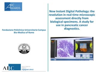
1. Fondazione Policlinico Universitario Campus
Bio-Medico of Rome
New Instant Digital Pathology: the
revolution in real-time microscopic
assessment directly from
biological specimens. A study for
use in pancreatic cancer
diagnostics.
2. Il gruppo di lavoro
This collaborative project brings together a multi-professional team
with synergistic and complementary skills.
Pathologists:
Anna Crescenzi
Chiara Taffon
Endoscopists:
Francesco Maria Di Matteo
Serena Stigliano
Biotechnologist:
Martina Verri
Company:
Roberto Banchi by Vivascope GmbH, Munich, Germany.
3. Descrizione
Ex-vivo fluorescence confocal microscopes (FCMs) allow for fast microscopic
assessment in the fresh state of the tissue with accuracy in the visualization of
both cellular and architectural details, without any slides preparation. This
technology is known as Instant Digital Pathology and uses different lasers
sources and fusion images to generate optical plans directly from native
tissues, similar to hematoxylin/eosin stained sections. FCM requires minimal
tissue preparation, and do not cause any damage in the native tissue, even in
very small specimens. Pancreatic cancer is the third leading cause of cancer
death worldwide. Early detection of pancreatic tumors has relevant impact on
clinical behavior.
Our team developed a consisted and reproducible protocol for instant digital
imaging of cells and micro-histological core of tissue from endoscopic
ultrasound guided fine needle aspiration/biopsy (EUS-FNB) of pancreatic solid
lesions.
4. Obiettivi e destinatari del lavoro
First aim:
Develop a consistent and reproducible protocol for submitting cellular
material and micro-fragments of tissue from pancreatic EUS-FNB to instant
digital pathology assessment.
Second aim:
Evaluate the performance of this approach by comparing sample adequacy
assessment and diagnostic evaluation on these digital biopsies with their
paired conventional histology.
Patients with pancreatic lesions will benefit from immediate evidence of
biopsy quality to avoid the occurrence of inadequate sampling. Clinicians will
be able to start patients’ management at the time of the EUS-FNB without
diagnostic delay. A collaboration between Physicians and Engineers is
expected for extensive development of instant digital pathology for both
artificial intelligence algorithms and characterization of cell and tissue by
ancillary techniques to improve rapidity in targeted treatments.
5. Risultati
90 cases of EUS-FNB samples from solid pancreatic lesions were observed from
April 2020 to May 2021 at the Fondazione Policlinico Universitario Campus Bio-
Medico of Rome. All patients signed the informed consent. The protocol for sample
preparation was developed using a polymeric matrix as specimens holder and by
modulating the laser power and/or the incubation period of the specimen in the
fluorescent solution (acridine orange), to obtain the best possible visualization of
microscopic details. From the performance point of view, we have found an
accuracy of 97% and a positive predictive value of 99%.
Our study paves the way to an innovative real time bedside approach, available for
remote sharing, and with immediate effects on fast diagnostic accuracy.