WHAT IS
PERIPHERY
• theoutermost , boundary, parts,
or surface of an area
• the outer parts of a town, city or area
• The outer part or uppermost layer of
something
• A place for meeting ort socializing
with others
5.
LEARNING OBJECTIVES
At theend of this module, the students will be able to:
• Identify the functions of the peripheral vascular system
• Discuss the different peripheral vascular diseases and reduction of
risk factors.
• Demonstrate the proper assessment of the peripheral vascular
system
• Discuss the nursing considerations in peripheral vascular
assessment
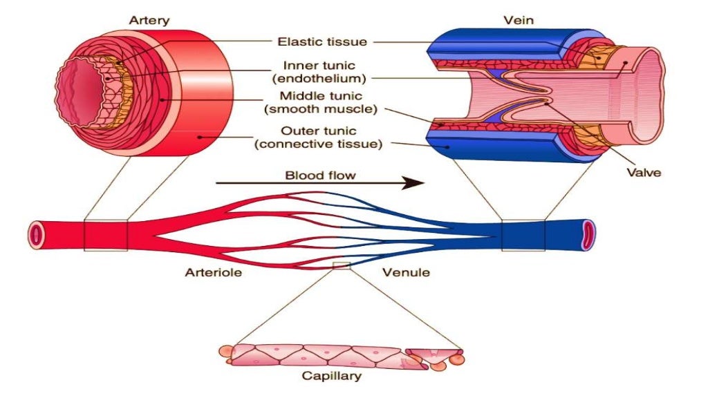
ARTERIES
•A vessel thatcarries blood high in oxygen
content away from the heart to the farthest
reaches of the body
•Since blood in arteries is usually full of oxygen,
the hemoglobin in the RBC is oxygenated.
•The resultant form of hemoglobin
(oxyhemoglobin) is what makes arterial blood
look bright red.
Reference:
Definition of Artery. (n.d.). MedicineNet. https://www.medicinenet.com/artery/definition.htm
11.
ARTERIES
• Theyare partof the efferent wing of the circulatory system
• ("Efferent"from theLatin:
"ex“ - out
"ferre“ - to bear
=to bearout or carry away
Whatthearteries arecarrying awayisblood from the heart.
Reference:
Definition of Artery. (n.d.). MedicineNet. https://www.medicinenet.com/artery/definition.htm
12.
VEINS
• They areblood vessels thatcarry blood lowin oxygen contentfrom the body back
to the heart
• The deoxygenated form of hemoglobin(deoxyhemoglobin) in venous blood makes
it appear dark.
• Veins are part of the afferentwing of thecirculatory system which returns bloodto
theheart
Reference:
Definition of Artery. (n.d.). MedicineNet. https://www.medicinenet.com/artery/definition.htm
13.
VEINS
•Unlike the arteries,venous network is a low -
pressure system since there is no force to propel
the blood,
- thus, its walls are thinner and larger in
diameter than the arteries to help reduce
workload on the heart.
•70% of the blood volume is all contained in the
veins
14.
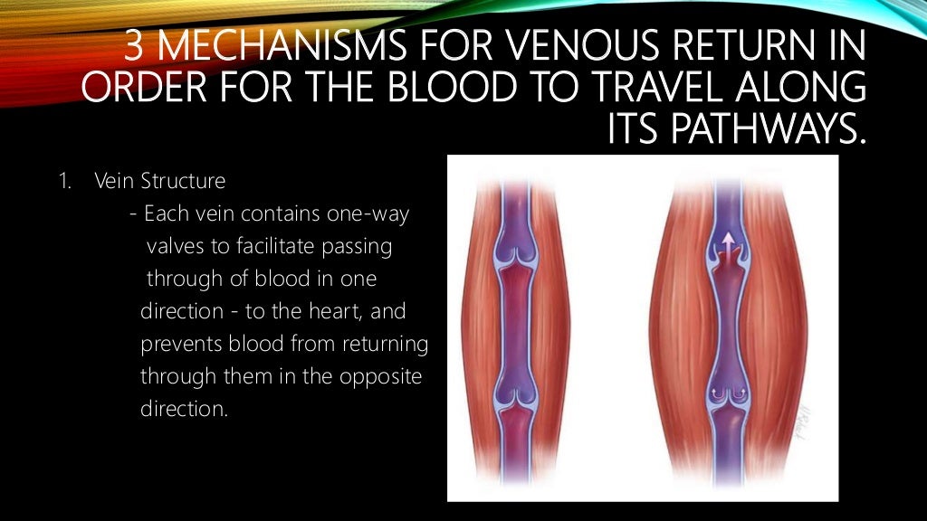
3 MECHANISMS FORVENOUS RETURN IN
ORDER FOR THE BLOOD TO TRAVEL ALONG
ITS PATHWAYS.
1. Vein Structure
- Each vein contains one-way
valves to facilitate passing
through of blood in one
direction - to the heart, and
prevents blood from returning
through them in the opposite
direction.
15.
3 MECHANISMS FORVENOUS RETURN IN
ORDER FOR THE BLOOD TO TRAVEL ALONG
ITS PATHWAYS.
2. Muscle contraction
- As the skeletal muscle contracts
during movement, it squeezes the
blood toward the heart through
the one-way valves.
- This is why movement, such as
walking and exercise are all
important factors to improve
one’s blood circulation.
16.
3 MECHANISMS FORVENOUS RETURN IN
ORDER FOR THE BLOOD TO TRAVEL ALONG
ITS PATHWAYS.
3. Respiratory pump
- During inspiration, the
intrathoracic pressure
decreases as the lungs
expands, thereby decreasing
pressure in the right atrium of
the heart as well
-the abdominal pressure increases
and the diaphragm contracts
creating a pressure gradient,
squeezing the inferior vena cava
and pushing the blood towards the
right atrium of the heart.
17.
CAPILLARIES
• They arethe smallest blood vessels in the body, connecting the
smallest arteries to the smallest veins.
• These vessels are often referred to as the "microcirculation.“
• Only two layers of cells thick (endothelial cells-inner layer, and
epithelial cells-outer layer),
• approximately 5 micrometers in diameter
• the purpose/central role of capillaries:
- deliver oxygen in the blood to the tissues
- pick up carbon dioxide to be eliminated
- they are also the place where nutrients are
delivered to feed all of the cells of the body.
Reference:
Eldridge, L. (2009). Capillary Structure and Function in the Body. [online] Verywell Health. Available at: https://www.verywellhealth.com/what-are-capillaries-2249069.
18.
CAPILLARIES
•If all thecapillaries in the human
body were lined up in single file, the
line would stretch over 100,000 miles
•From the capillaries, blood flows into
the smaller venules and then into
veins, flowing back to the heart
Reference:
Eldridge, L. (2009). Capillary Structure and Function in the Body. [online] Verywell Health. Available at: https://www.verywellhealth.com/what-are-capillaries-2249069.
19.
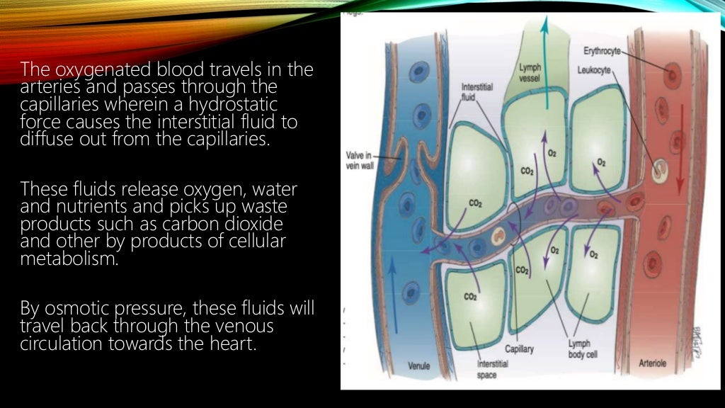
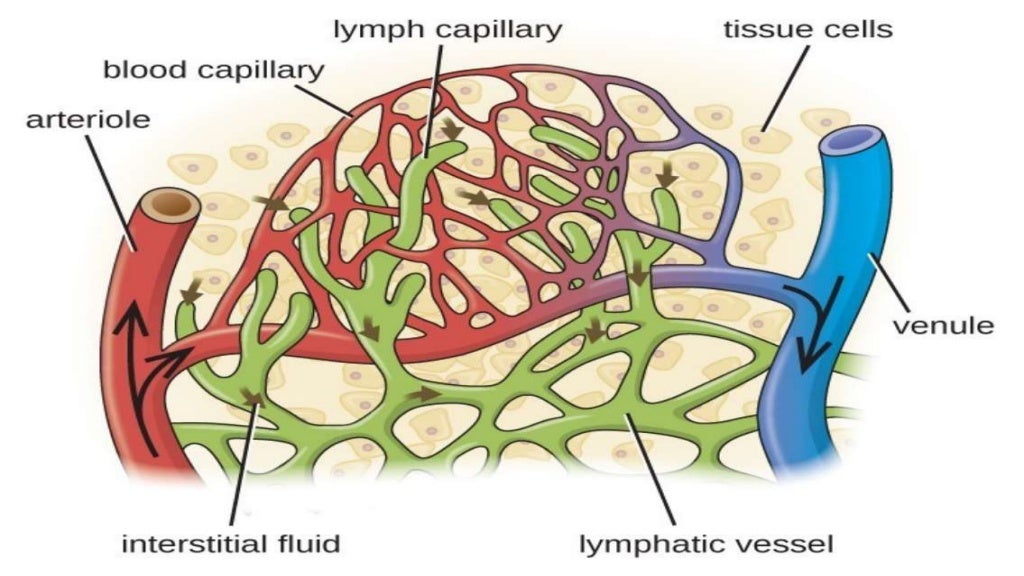
CAPILLARIES
The oxygenated bloodtravels in the
arteries and passes through the
capillaries wherein a hydrostatic
force causes the interstitial fluid to
diffuse out from the capillaries.
These fluids release oxygen, water
and nutrients and picks up waste
products such as carbon dioxide
and other by products of cellular
metabolism.
By osmotic pressure, these fluids will
travel back through the venous
circulation towards the heart.
MAJOR ARTERIES OFTHE ARM
• Axillary
- the subclavian artery, it exits the torso and enters the
arm
• Brachial
- delivers blood to the upper region of the arm
• Radial and ulnar
- run alongside the two bones of the forearm where they
eventually divide to deliver blood to the wrist and hand
Reference:
major arteries of the arm - Search. (n.d.). Www.bing.com. Retrieved June 28, 2022, from https://www.bing.com/search?q=major+arteries+of+the+arm&cvid=e8308701370540419477e1f49e7a7575&aqs=edge.0.0l9.6977j0j1&pglt=771&FORM=ANNTA1&PC=NMTS
23.
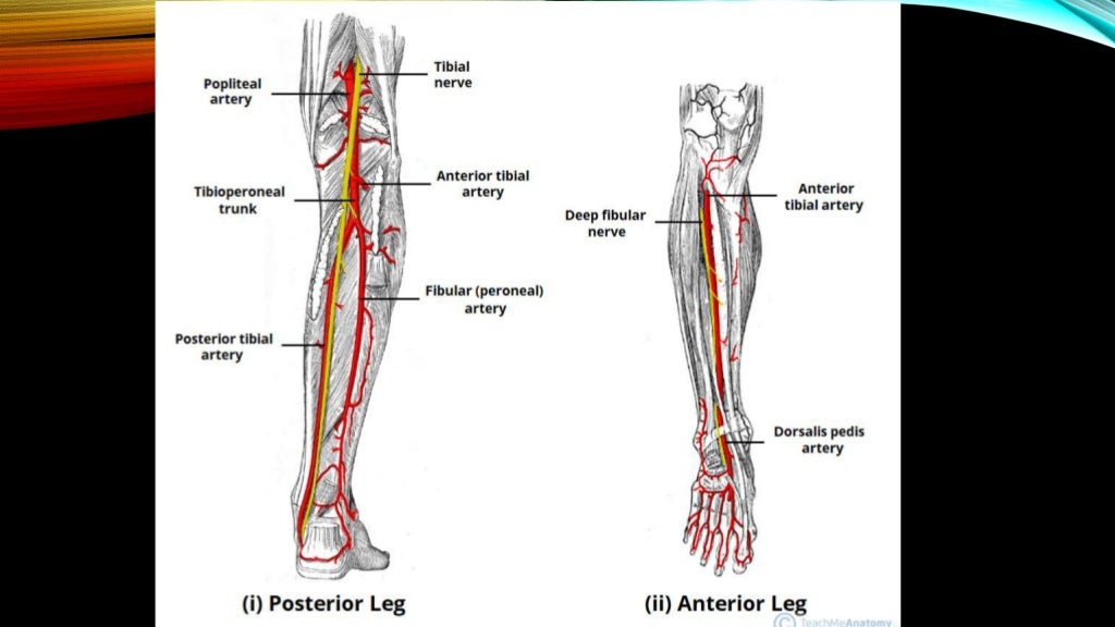
MAJOR ARTERIES OFTHE LEGS
• Femoral
- derived from the external iliac artery
- this artery supplies blood to the thigh and divides into the
various smaller arteries that supply the legs.
• Genicular
- supplies blood to the knee region.
• Popliteal
- This is the name given to the femoral artery
- it passes below the knee.
• Anterior and posterior tibial
- derived from the popliteal artery
- supply blood to the lower portion of the leg
- when they reach the ankle , they divide further to supply the ankle and foot
region
Reference:
Healthline. (2019). Arteries of the Body: Picture, Anatomy, Definition & More. [online] Available at:
https://www.healthline.com/health/arteries-of-the-body#leg-arteries.
25.
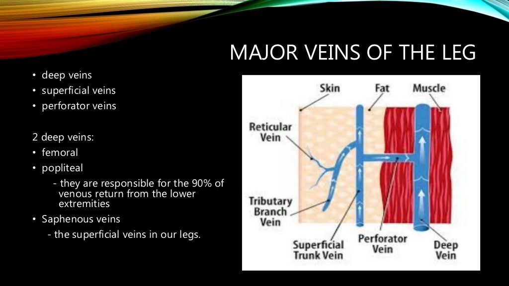
MAJOR VEINS OFTHE LEG
• deep veins
• superficial veins
• perforator veins
2 deep veins:
• femoral
• popliteal
- they are responsible for the 90% of
venous return from the lower
extremities
• Saphenous veins
- the superficial veins in our legs.
26.
The photo showsthe
Different location of
the major veins in our
Legs
• Femoral
• popliteal
• saphenous veins
29.
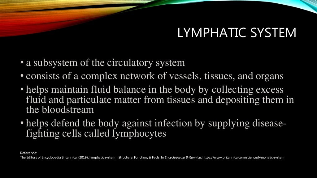
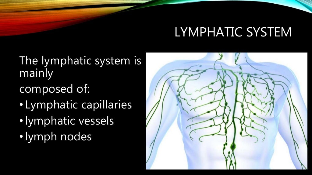
LYMPHATIC SYSTEM
• asubsystem of the circulatory system
• consists of a complex network of vessels, tissues, and organs
• helps maintain fluid balance in the body by collecting excess
fluid and particulate matter from tissues and depositing them in
the bloodstream
• helps defend the body against infection by supplying disease-
fighting cells called lymphocytes
Reference:
The Editors of Encyclopedia Britannica. (2019). lymphatic system | Structure, Function, & Facts. In Encyclopædia Britannica. https://www.britannica.com/science/lymphatic-system
LYMPHATIC CIRCULATION
•The lymphaticsystem can be thought of as a
drainage system, as blood circulates through the
body, blood plasma leaks into tissues through the
thin walls of the capillaries
• The portion of blood plasma that escapes is called
interstitial or extracellular fluid, and it contains
oxygen, glucose, amino acids and other nutrients
needed by tissue cells.
Reference:
The Editors of Encyclopedia Britannica. (2019). lymphatic system | Structure, Function, & Facts. In Encyclopædia Britannica. https://www.britannica.com/science/lymphatic-system
33.
LYMPHATIC CIRCULATION
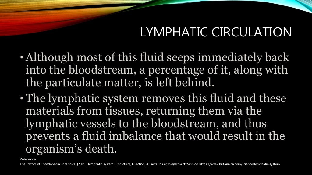
•Although mostof this fluid seeps immediately back
into the bloodstream, a percentage of it, along with
the particulate matter, is left behind.
•The lymphatic system removes this fluid and these
materials from tissues, returning them via the
lymphatic vessels to the bloodstream, and thus
prevents a fluid imbalance that would result in the
organism’s death.
Reference:
The Editors of Encyclopedia Britannica. (2019). lymphatic system | Structure, Function, & Facts. In Encyclopædia Britannica. https://www.britannica.com/science/lymphatic-system
34.
3 MAIN FUNCTIONSOF THE
LYMPHATIC SYSTEM
1. It drains excess body fluids and proteins back to the venous
system
- the excess fluids from the right part of the body will
travel through the right lymphatic duct and will be
drained in the right subclavian vein
- the excess fluid from the left part of the body will all be
reabsorbed and travel through the thoracic duct and
will drain in the left subclavian vein
36.
3 MAIN FUNCTIONSOF THE
LYMPHATIC SYSTEM
2. Filters out microorganisms, foreign materials, dead
blood cells, abnormal cells
- As the lymph travels in the lymphatics vessels,
they will pass through filters known as lymph
nodes to filter out microorganisms, foreign
materials, dead cells and abnormal cells where
they will be trapped and destroyed
- This is the reason why you will have swollen lymph
nodes that are nearest to a wound or to the
source of infection
38.
3 MAIN FUNCTIONSOF THE
LYMPHATIC SYSTEM
3. Absorb fats
(lipids) from small
intestine into
bloodstream by the
lymphoid tissues in
the intestines called
Peyer’s patches.
39.
PEYER’S PATCHES
• Thegut-associated
lymphoid tissue (GALT)
consists of isolated or
aggregated lymphoid
follicles forming Peyer's
patches.
• By their ability to
transport luminal
antigens and bacteria,
PPs can be considered
as the immune sensors
of the intestine.
40.
PEYER’S PATCHES
• anadult has 30 to 40 Peyer’s
patches on average in the region
of the small intestine
• The location of these patches is
in the mucosa of the intestinal
lining
• usually found in humans in the
lowest portion of the small
intestine, mainly in the distal
jejunum and the ileum, but also
could be detected in the
duodenum.
LYMPHATIC SYSTEM OFTHE HEAD
AND NECK
Reference:
The Editors of Encyclopedia Britannica. (2019). lymphatic system | Structure, Function, & Facts. In Encyclopædia Britannica. https://www.britannica.com/science/lymphatic-system
ABNORMAL FINDINGS
SKIN ANDHAIR ABNORMALITIES
• Warm Skin may indicate conditions causing
fever or increased cardiac output
• Absence of body hair on the arms or legs may
indicate diminished arterial blood flow to
these areas
• Cyanosis, pallor, or cool skin may indicate
poor cardiac output and tissue perfusion
AORTIC ANEURYSM
• abnormalbulge in the
aorta
• It can occur any where
in the aorta
• may be in a tube
form or round
shaped
Reference:
MSN. (n.d.). Www.msn.com. Retrieved June 28, 2022, from https://www.msn.com/en-
ph/health/condition/aortic+aneurysm?ocid=entnewsntp
•
71.
CEREBROVASCULAR DISEASE
• refersto a group of conditions,
diseases, and disorders that
affect the blood vessels and
blood supply to the brain
• Causes:
• Atherosclerosis- narrowing of the
arteries
• Thrombosis - where a blood clot creates
a blockage in a blood vessel
• Embolic arterial blood clot - which is a
blood clot in an artery of the brain
• cerebral venous thrombosis - which is a
blood clot in a vein of the brain
Reference:
Kraft, S. (2019). Cerebrovascular disease: Causes,
symptoms, and treatment. [online]
www.medicalnewstoday.com. Available at:
https://www.medicalnewstoday.com/articles/184601.
73.
ATHEROSCLEROSIS
• A plaquebuild-up
• made up of fat, cholesterol, calcium,
and other substances found in the
blood
• Over time, plaque hardens and
narrows your arteries
• This limits the flow of oxygen-rich
blood to your organs and other
parts of your body
Reference:
• MD, S. (n.d.). Atherosclerosis: Symptoms, Risk Factors, and Health Complications - Cardiology
Specialist Houston - Arsalan Shahzad, M.D. F.A.C.C. [online] https://www.care4heart.com/.
Available at: https://www.care4heart.com/education/heart-disease/atherosclerosis-
symptoms-risk-factors-and-health-complications [Accessed 28 Jun. 2022].
74.
PERIPHERAL ARTERIAL OCCLUSIVE
DISEASE(PAD)
• is chronic arterial occlusive
disease of the lower
extremities caused by
atherosclerosis.
Reference:
Aronow, W.S. (2012). State of the art paper Peripheral
arterial disease of the lower extremities. Archives of Medical
Science, [online] 2, pp.375–388.
doi:10.5114/aoms.2012.28568.
75.
PULMONARY EMBOLISM AND
VENOUSTHROMBOSIS
• Pulmonary embolism (PE) and
deep venous thrombosis (DVT)
exist on the spectrum of venous
thromboembolic disease (VTE)
• PE results when thrombus
migrates from the venous
circulation to the pulmonary
vasculature and lodges in the
pulmonary arterial system.
• Reference:
Turetz, M., Sideris, A., Friedman, O., Triphathi, N. and Horowitz, J. (2018). Epidemiology,
Pathophysiology, and Natural History of Pulmonary Embolism. Seminars in Interventional
Radiology, 35(02), pp.92–98. doi:10.1055/s-0038-1642036.
•


![CAPILLARIES
• They are the smallest blood vessels in the body, connecting the
smallest arteries to the smallest veins.
• These vessels are often referred to as the "microcirculation.“
• Only two layers of cells thick (endothelial cells-inner layer, and
epithelial cells-outer layer),
• approximately 5 micrometers in diameter
• the purpose/central role of capillaries:
- deliver oxygen in the blood to the tissues
- pick up carbon dioxide to be eliminated
- they are also the place where nutrients are
delivered to feed all of the cells of the body.
Reference:
Eldridge, L. (2009). Capillary Structure and Function in the Body. [online] Verywell Health. Available at: https://www.verywellhealth.com/what-are-capillaries-2249069.](https://image.slidesharecdn.com/peripheralvascularsystem-220708111633-d8a6dcc6/95/PERIPHERAL-VASCULAR-SYSTEM-pptx-17-1024.jpg)
![CAPILLARIES
•If all the capillaries in the human
body were lined up in single file, the
line would stretch over 100,000 miles
•From the capillaries, blood flows into
the smaller venules and then into
veins, flowing back to the heart
Reference:
Eldridge, L. (2009). Capillary Structure and Function in the Body. [online] Verywell Health. Available at: https://www.verywellhealth.com/what-are-capillaries-2249069.](https://image.slidesharecdn.com/peripheralvascularsystem-220708111633-d8a6dcc6/95/PERIPHERAL-VASCULAR-SYSTEM-pptx-18-1024.jpg)


![MAJOR ARTERIES OF THE LEGS
• Femoral
- derived from the external iliac artery
- this artery supplies blood to the thigh and divides into the
various smaller arteries that supply the legs.
• Genicular
- supplies blood to the knee region.
• Popliteal
- This is the name given to the femoral artery
- it passes below the knee.
• Anterior and posterior tibial
- derived from the popliteal artery
- supply blood to the lower portion of the leg
- when they reach the ankle , they divide further to supply the ankle and foot
region
Reference:
Healthline. (2019). Arteries of the Body: Picture, Anatomy, Definition & More. [online] Available at:
https://www.healthline.com/health/arteries-of-the-body#leg-arteries.](https://image.slidesharecdn.com/peripheralvascularsystem-220708111633-d8a6dcc6/95/PERIPHERAL-VASCULAR-SYSTEM-pptx-23-1024.jpg)


![CEREBROVASCULAR DISEASE
• refers to a group of conditions,
diseases, and disorders that
affect the blood vessels and
blood supply to the brain
• Causes:
• Atherosclerosis- narrowing of the
arteries
• Thrombosis - where a blood clot creates
a blockage in a blood vessel
• Embolic arterial blood clot - which is a
blood clot in an artery of the brain
• cerebral venous thrombosis - which is a
blood clot in a vein of the brain
Reference:
Kraft, S. (2019). Cerebrovascular disease: Causes,
symptoms, and treatment. [online]
www.medicalnewstoday.com. Available at:
https://www.medicalnewstoday.com/articles/184601.](https://image.slidesharecdn.com/peripheralvascularsystem-220708111633-d8a6dcc6/95/PERIPHERAL-VASCULAR-SYSTEM-pptx-71-1024.jpg)

![ATHEROSCLEROSIS
• A plaque build-up
• made up of fat, cholesterol, calcium,
and other substances found in the
blood
• Over time, plaque hardens and
narrows your arteries
• This limits the flow of oxygen-rich
blood to your organs and other
parts of your body
Reference:
• MD, S. (n.d.). Atherosclerosis: Symptoms, Risk Factors, and Health Complications - Cardiology
Specialist Houston - Arsalan Shahzad, M.D. F.A.C.C. [online] https://www.care4heart.com/.
Available at: https://www.care4heart.com/education/heart-disease/atherosclerosis-
symptoms-risk-factors-and-health-complications [Accessed 28 Jun. 2022].](https://image.slidesharecdn.com/peripheralvascularsystem-220708111633-d8a6dcc6/95/PERIPHERAL-VASCULAR-SYSTEM-pptx-73-1024.jpg)
![PERIPHERAL ARTERIAL OCCLUSIVE
DISEASE (PAD)
• is chronic arterial occlusive
disease of the lower
extremities caused by
atherosclerosis.
Reference:
Aronow, W.S. (2012). State of the art paper Peripheral
arterial disease of the lower extremities. Archives of Medical
Science, [online] 2, pp.375–388.
doi:10.5114/aoms.2012.28568.](https://image.slidesharecdn.com/peripheralvascularsystem-220708111633-d8a6dcc6/95/PERIPHERAL-VASCULAR-SYSTEM-pptx-74-1024.jpg)


