Ascitis -
•Download as PPTX, PDF•
15 likes•5,365 views

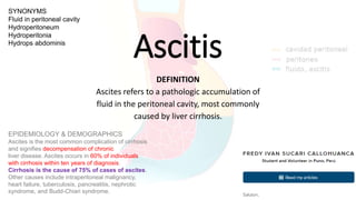
Ascitis es la acumulación patológica de fluido en la cavidad peritoneal, la causa más común es la cirrosis hepática.

More Related Content
What's hot
What's hot (20)
Similar to Ascitis -
Similar to Ascitis - (20)
More from Fredy Ivan SUCARI CALLOHUANCA
More from Fredy Ivan SUCARI CALLOHUANCA (15)
Recently uploaded
Recently uploaded (20)
Ascitis -
- 1. Ascitis DEFINITION Ascites refers to a pathologic accumulation of fluid in the peritoneal cavity, most commonly caused by liver cirrhosis. SYNONYMS Fluid in peritoneal cavity Hydroperitoneum Hydroperitonia Hydrops abdominis EPIDEMIOLOGY & DEMOGRAPHICS Ascites is the most common complication of cirrhosis and signifies decompensation of chronic liver disease. Ascites occurs in 60% of individuals with cirrhosis within ten years of diagnosis. Cirrhosis is the cause of 75% of cases of ascites. Other causes include intraperitoneal malignancy, heart failure, tuberculosis, pancreatitis, nephrotic syndrome, and Budd-Chiari syndrome.
- 2. CLINICAL PRESENTATION Important information to elicit within history: 1. History of viral hepatitis 2. Alcoholism 3. Intravenous drug use, intranasal cocaine use 4. Sexual history (i.e., men who have sex with men) 5. History of transfusions, tattoos, piercings, imprisonment 6. Symptoms suggestive of peritoneal malignancy (e.g., weight loss, pain, palpable masses, rectal/vaginal bleeding) 7. Other liver disease symptoms (e.g., increasing abdominal girth, jaundice, pruritis, confusion, pedal edema) 8. Cardiac symptoms (e.g., pedal edema, shortness of breath, orthopnea, chest pains)
- 3. Important physical exam findings: 1. Bulging flanks (can be present in obesity) 2. Flank dullness to percussion 3. Fluid wave on abdominal exam 4. Lower extremity edema 5. Shifting dullness on abdominal exam 6. Physical signs associated with liver cirrhosis: spider angiomas, jaundice, loss of body hair, skeletal muscle wasting (sarcopenia), Dupuytren’s contracture, bruising, palmar erythema, gynecomastia, testicular atrophy, rectal varices, and caput medusae
- 5. Diagnóstico: LABORATORY TESTS • Initial evaluation should always include: 1. Diagnostic paracentesis. Laboratory tests on this fluid should include a cell count, cytology, albumin, total protein, culture, and Gram stain. A serum-ascites albumin gradient (SAAG) should be calculated in all patients. a. If the SAAG is greater than 1.1, the cause of ascites can be attributed to portal hypertension. b. If SAAG is less than 1.1, a non-portal hypertension etiology of ascites must be sought. Optional tests on paracentesis fluid include amylase, LDH, acid-fast bacilli, and glucose levels. c. Causes of ascites in the normal or diseased peritoneum by SAAG are summarized in Table 1. d. Fig. 2 illustrates an algorithm for the approach to the differential diagnosis of ascites. 2. AST, ALT, total and direct bilirubin, albumin, alkaline phosphatase, GGTP 3. CBC, coagulation studies 4. Electrolytes, BUN, creatinine IMAGING STUDIES • Abdominal ultrasound (Fig. 3) is the most sensitive measure for detecting ascitic fluid; a CT or MRI scan is a viable alternative. Doppler studies of portal and hepatic veins should be added to rule out vascular etiology. • Endoscopy of the upper GI tract to evaluate for esophageal varices if ascites is secondary to portal hypertension.
- 7. Diagnóstico: LABORATORY TESTS • Initial evaluation should always include: 1. Diagnostic paracentesis. Laboratory tests on this fluid should include a cell count, cytology, albumin, total protein, culture, and Gram stain. A serum-ascites albumin gradient (SAAG) should be calculated in all patients. a. If the SAAG is greater than 1.1, the cause of ascites can be attributed to portal hypertension. b. If SAAG is less than 1.1, a non-portal hypertension etiology of ascites must be sought. Optional tests on paracentesis fluid include amylase, LDH, acid-fast bacilli, and glucose levels. c. Causes of ascites in the normal or diseased peritoneum by SAAG are summarized in Table 1. d. Fig. 2 illustrates an algorithm for the approach to the differential diagnosis of ascites. 2. AST, ALT, total and direct bilirubin, albumin, alkaline phosphatase, GGTP 3. CBC, coagulation studies 4. Electrolytes, BUN, creatinine IMAGING STUDIES • Abdominal ultrasound (Fig. 3) is the most sensitive measure for detecting ascitic fluid; a CT or MRI scan is a viable alternative. Doppler studies of portal and hepatic veins should be added to rule out vascular etiology. • Endoscopy of the upper GI tract to evaluate for esophageal varices if ascites is secondary to portal hypertension.
- 9. Ascites, ultrasound. Ultrasound is useful for detection of ascites. Simple fluids such as ascites are excellent sound transmission media, reflecting almost no sound waves. As a consequence, they appear quite hypoechoic (black) on ultrasound. This view of the right lower quadrant shows loops of bowel surrounded by fluid. During the ultrasound, the bowel loops would be seen to undergo peristalsis and drift back and forth in the ascitic fluid with patient movement. Ultrasound cannot distinguish the composition of the fluid; ascites, liquid blood, liquid bile, urine, and infectious fluids have a similar appearance, with a few exceptions. Blood may coagulate and form septations within the fluid collection. Infectious fluids also frequently form loculated fluid collections that may be recognized on ultrasound, although the exact composition cannot be determined.
- 15. Gastrointestinal malignancies Unfortunately, clinical symptoms appear most often at the late stages of disease and therefore are associated with poor patient survival. Some tumors induce diffuse dissemination of the tumor cells into the peritoneal cavity. The appearance of peritoneal carcinomatosis (PCA) is associated with tumor progression and an unfavorable outcome (survival ranging from weeks to months), with ascites and bowel obstruction as the main symptoms. Expression of MicroRNAs in the Ascites of Patients With Peritoneal Carcinomatosis and Peritonitis Diagnostic is based on imaging of PCA and cytological evaluation of ascites followed by an invasive approach such as laparoscopy or laparotomy. Computed tomography and magnetic resonance imaging deliver the most reliable results for the nodal type of PCA and pre-dominantly in advanced stages of disease. cytology remains the gold standard that allows for the definitive diagnosis with high specificity, although the sensitivity is only approximately 40% to 60%
- 16. Expression of MicroRNAs in the Ascites of Patients With Peritoneal Carcinomatosis and Peritonitis patients with cirrosis and ascites may develop spontaneous bacterial peritonitis (SBP), which occurs in 10% to 30% of cases. limited progress in the development of alternative molecular-based methods for the diagnosis of PCA and SBP. MicroRNAs (miRNAs) small, endogenous, non-coding RNA molecules miRNA is a consequence of the complex transcription and RNA processing. During this process, miRNA binds to Argonaute proteins and controls the gene expression as the RNA-induced silencing complex (RISC). various body fluids such as blood, urine, feces, cerebrospinal fluid, etc.
- 17. Department of Gastroenterology, Hepatology and Infectious Diseases at Otto-von-Guericke-University in Magdeburg, Germany. 45 patients The ascites samples (approximately 25 mL) were collected from patients with various conditions during diagnostic or therapeutic paracentesis. 1. 15 patients with PCA (detection of neoplastic cells in cytology); 2. 15 patients with sponatenous and secondary bacterial peritonitis (SBP) (polymorphonuclear leukocytes count [PMN] 250 cells/mm3 and/or positive bacterial culture); and 3. 15 patients with ascites due to portal hypertension without complications (no SBP/PCA) (negative cytology, PMNs 250 cells/mm3 , and negative bacterial culture).
- 19. miRNAs en ascítis Análisis por reacción de cadena de transcriptasa reversa y polimerasa en forma cuantitativa The results of the current study demonstrate that miRNAs can be easily isolated from ascites samples. Hipertensión portal Sobre expresado miR-21 miR-186 miR-222 miR-483 infraexpresado miR-266 Peritonitis bact. Espont. “Sobre expresado” miR-223 Carcinomatosis peritoneal ^^ Sobre expresado miR-21 miR-186
- 20. Gracias Dankon