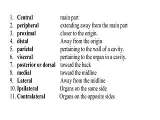
2. Definitions
• No medicine without physiology
• No surgery without Anatomy
• Physiology: This is the study of function of organ or
body or cell.
• Anatomy: The study of structure (shape, size, location)
of an organ or a cell is called anatomy.
• Pathology: The study of abnormalities or diseases is
called pathology.
• Etiology: The study of causes of diseases is known as
etiology.
3. • Cell: The basic structural and functional
unit of life.
• Organelles (i.e. cell membrane, nucleus,
ribosomes). A small organ of a cell,
which performs a particular function.
4. • Tissues (i.e. epithelial, connective, muscle, nervous).
A tissue is defined as a group of similar cells that
performs a specialized function. Two or more tissues
combine to form organ.
• Organ (i.e. skin, heart, brain). An organ is defined as
a structure consisting of a group of tissues that
performs a specialized function. Two or more organs
combine to form system.
• Organ systems (i.e. integumentary, cardiovascular).
An organ system is defined as a group of organs that
act together to carry on a specialized function. The
eleven organ systems collectively form the organism.
5. • Diffusion: The movement of molecules of a
substance from the area of its higher
concentration to the area of its lower
concentration is called diffusion.
For example, Exchange of gases in the lungs or
body tissues.
• Osmosis: The movement of water molecules
from its higher concentration to its lower
concentration through a semi permeable
membrane.
e.g. Absorption of water by small intestine.
6. • Facilitated Diffusion: The movement of molecules
from the area of its greater (higher) concentration to
the area of its lesser (lower) concentration with the
help of channels or carriers. Eg, Intake of glucose by
cells.
• Active Transport: The movement of molecules from
its lower concentration to the higher concentration
with the help of consumption of cellular energy (in
the form of ATP). e.g. Sodium Potassium Pump
(Na-K pump).
• Filtration: The movement of water and dissolved
substances from the area of its higher pressure to
the area of its lower pressure is called filtration.
e.g. formation of urine in the renal tubules.
7. • Phagocytosis: The engulfment (swallowing) of a
solid substance (e.g. bacteria etc.) by phagocytic
cells is called phagocytosis.
Example: White Blood Cells engulf bacteria.
• Pinocytosis: The uptake of fluid and dissolved
substance.
Example: Cells of the kidney tubules reabsorb
dissolved proteins
Endocytosis
Exocytosis
8. Organization level of a body
• Atoms molecules & compounds
cells tissues organs
systems body
9. Directional and Non-Directional Terms
1. Superficial towards the surface
2. perineal pelvic floor
3. Plantar sole of foot
4. Popliteal back of knee
5. Femoral thigh
6. Frontal front
7. Gastric stomach
8. gluteal buttock
9. Brachial upper arm
10. Buccal/oral mouth
11. Cardiac heart
12. survical neck
13. Cranial of head
14. Cranium head
15. Deltoid shoulder
16. volar palm
17. Superior above
18. Temporal side of head
19. Umbilical navel
Patellar knee cap
20. Hepatic liver
21. Iliac hip
22. Inguinal groin
23. Lumber small back (loin)
24. Mammary breast
25. Nasal nose
26. Inferior below/lower
27. Anterior or ventral toward the front
28. Axillary Arm pit
29. Internal within interior to
30. External outside
31. Occipital back of head
32. Orbital eye related
33. Parietal crown of head
34. Pulmonary lungs
35. Renal kidney
36. Sacral base of spine
37. Mental Chin
10. 1. Central main part
2. peripheral extending away from the main part
3. proximal closer to the origin.
4. distal Away from the origin
5. parietal pertaining to the wall of a cavity.
6. visceral pertaining to the organ in a cavity.
7. posterior or dorsal toward the back
8. medial toward the midline
9. Lateral Away from the midline
10. Ipsilateral Organs on the same side
11. Contralateral Organs on the opposite sides
12. Relationship between Anatomy and Physiology
• Anatomy explains the structure while
physiology explains the function
• Anatomical Position:
• standing erect, face forward, upper limbs
at sides, palms forward.
13. • Anatomy and physiology are the sciences
that most closely relate to the human body.
Anatomy explains the structure while
physiology explains the function of the body
of a human being. Thereby it is clear that
both are interrelated to one another intently
and both need to be learned and understood
parallel to each other.
14. SOLUTIONS
• When a solute is dissolved in a solvent it forms
a solution. Eg. Sugar dissolved in water.
• Strength of a solution: The amount of solute
particles dissolved per unit volume of a
• solution is called strength or tonicity of the
solution.
• When we compare the tonicity of one solution
to another solution we use the terms as:
--- hypotonic, isotonic, and hypertonic.
15. SOLUTIONS Cont…
• Hypotonic Solution
• A solution having strength (concentration) lesser
than the other one is said to be
• hypotonic solution.
• Isotonic Solution
• A solution having strength equal to the other
one is considered to be isotonic solution.
• Hypertonic Solution
• A solution having strength greater than the
other one is called hypertonic solution.
16. BODY CAVITIES
Dorsal and Ventral Cavities
• Dorsal Cavities
Cranial cavity-----brain
Vertebral canal-----spinal cord
20. Body Planes and Sections
• A plane is an imaginary line that separates two portions
of the body or an organ.
• Sections and planes are:
• Frontal (coronal) Section
• Divides a body into front and back portions.
• Sagittal Section
• cuts a body into right and left portion.
• A mid sagittal plane gives equal right and left halves.
• A parasagittal plane cuts a body or an organ into two
unequal right and left parts.
• Transverse Section
• Divides a body into upper and lower portions.
• Longitudinal Section
• Cut along the long axis of a body or an organ.
22. Abdominopelvic Regions and
Quadrants
• To describe the location of abdominal and pelvic
organs easily, the abdomenopelvic region is
divided into two ways—
• 1) Either into Nine regions or 2) Four quadrants.
• The nine regions are made by drawing two
transverse and two vertical lines
• The top horizontal, the subcostal line is drawn
just inferior to the rib cage, across the inferior
portion of the stomach. The bottom horizontal
one is transtubercular line.
• The two vertical lines are midclavicular lines.
27. Abdominopelvic Regions and Quadrants
• Right Hypochondriac Region Contains
• Right lobe of Liver, gall bladder, a part of right
kidney
• Left Hypochondriac Region Contains
• A Part of stomach, spleen, a small part of
pancreas, a part of left kidney
28. Abdominopelvic Regions and Quadrants
• Right Lumber Region
• Part of small intestine,
• Superior part of cecum
• Ascending colon,
• Inferior part of right kidney
• Left Lumber Region
• Part of small intestine,
• Descending colon,
• An inferior part of left kidney
29. Abdominopelvic Regions and Quadrants
• Right Iliac Region
• Lower end of cecum, appendix
• Left Iliac Region
• Part of small intestine,
• Part of sigmoid colon
30. Abdominopelvic Regions and Quadrants
• Epigastric Region
• Greater part of liver, greater part of stomach,
• Greater part of pancreas
• Umbilical Region
• Part of small intestine,
• Transverse colon of large intestine
• Hypogastric Region
• Part of sigmoid colon
• Urinary bladder when full
Editor's Notes
* Note that the kidneys, adrenal glands, pancreas, and ureters are behind the abdominopelvic cavity. This is referred to as RETROPERITONEAL.