Antitubercular agents in TB patients with Chronic Liver disease (CLD)
•Download as PPTX, PDF•
31 likes•10,648 views

Antitubercular agents in TB patients with Chronic Liver disease (CLD). Brief review on how to initiate and maintan ATT IN CLD patients.
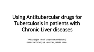
![Introduction
• It is quite evident that more than 90 percent of tuberculosis are reactivation of
their latent form that usually present in patients with some level of
immunodeficient or immunosuppressed state.
• So, Cirrhosis being itself an immunodeficient state[1], it would be aggreable to
state that prevalence of tuberculosis is much higher in cirrhotic population when
compared to normal population.
• A study conducted in Western India showed that the prevalence rate was 15
times higher than in the general population[2].
• Another study from India showed that there is nearly five times higher
prevalence of TB in cirrhosis patients (8.1%) compared to the general population
(1.6%), with pulmonary TB being the commonest form[3].
1. Cho YJ, Lee SM, Yoo CG, Kim YW, Han SK, Shim YS, Yim JJ. Clinical characteristics of tuberculosis in patients with liver cirrhosis. Respirology. 2007;12:401–405.
2. Saigal S, Nandeesh HP, Agarwal SR, Misra A, Jain SK, Sarin SK. High prevalence and profile of tuberculosis in chronic liver disease patients. Gastroenterology. 1998;114:A38.
3. Baijal R, Praveenkumar HR, Amarapurkar DN, Nagaraj K, Jain M. Prevalence of tuberculosis in patients with cirrhosis of liver in western India. Trop Doct. 2010;40:163–164.](data:image/gif;base64,R0lGODlhAQABAIAAAAAAAP///yH5BAEAAAAALAAAAAABAAEAAAIBRAA7)
Recommended
More Related Content
What's hot
What's hot (20)
Similar to Antitubercular agents in TB patients with Chronic Liver disease (CLD)
Similar to Antitubercular agents in TB patients with Chronic Liver disease (CLD) (18)
More from Pratap Tiwari
More from Pratap Tiwari (20)
Recently uploaded
Recently uploaded (20)
Antitubercular agents in TB patients with Chronic Liver disease (CLD)
- 1. Using Antitubercular drugs for Tuberculosis in patients with Chronic Liver diseases Pratap Sagar Tiwari, MD (Internal Medicine) DM HEPATOLOGY, BIR HOSPITAL, NAMS, NEPAL
- 2. Introduction • It is quite evident that more than 90 percent of tuberculosis are reactivation of their latent form that usually present in patients with some level of immunodeficient or immunosuppressed state. • So, Cirrhosis being itself an immunodeficient state[1], it would be aggreable to state that prevalence of tuberculosis is much higher in cirrhotic population when compared to normal population. • A study conducted in Western India showed that the prevalence rate was 15 times higher than in the general population[2]. • Another study from India showed that there is nearly five times higher prevalence of TB in cirrhosis patients (8.1%) compared to the general population (1.6%), with pulmonary TB being the commonest form[3]. 1. Cho YJ, Lee SM, Yoo CG, Kim YW, Han SK, Shim YS, Yim JJ. Clinical characteristics of tuberculosis in patients with liver cirrhosis. Respirology. 2007;12:401–405. 2. Saigal S, Nandeesh HP, Agarwal SR, Misra A, Jain SK, Sarin SK. High prevalence and profile of tuberculosis in chronic liver disease patients. Gastroenterology. 1998;114:A38. 3. Baijal R, Praveenkumar HR, Amarapurkar DN, Nagaraj K, Jain M. Prevalence of tuberculosis in patients with cirrhosis of liver in western India. Trop Doct. 2010;40:163–164.
- 3. Drug induced hepatotoxicity: ATT • Anti-TB chemotherapy containing isoniazid (H), rifampicin (R) and pyrazinamide (Z) has proved to be highly effective but hepatotoxic and the risk further increases when these drugs are combined. • The development of DIH during chemotherapy for TB is one of the most common reason leading to interruption of therapy. • Wide variations have been found in the incidence of hepatotoxic reactions from different countries, with the reported incidence being 3 % in the USA[1], 4 % in the UK[1], 11 % in Germany[2], 13 % in HK[3], 36 % in Japan[4], 26 % in Taiwan[4] and 8-36 % in India[5,6]. 1. Ormerod LP, Skinner C, Wales J. Hepatotoxicity of antituberculosis drugs. Thorax 1996; 51 : 111-3. 2. Garg PK, Tandon RK. Antituberculosis treatment induced hepatotoxicity. In: Sharma SK, Mohan A, editors. Tuberculosis 2nd ed. New Delhi: Jaypee Brothers Medical Publishers; 2009.p. 783-95. 3. Lauterburg BH, Smith CV, Todd EL, Mitchell JR. Pharmacokinetics of the toxic hydrazino metabolites formed from isoniazid in humans. J Pharmacol Exp Ther 1985; 235 : 566-70. 4. Ellard GA, Mitchison DA, Girling DJ, Nunn AJ, Fox W. The hepatic toxicity of isoniazid among rapid and slow acetylators of the drug. Am Rev Respir Dis 1978; 118 : 628-9. 5. Parthasarthy R, Sarma GR, Janardhanam B, Ramachandran P, Santha T, Sivasubramania S, et al. Hepatic toxicity in south Indian patients during treatment of tuberculosis with short- course regimens containing isoniazid, rifampicin and pyrazinamide. Tubercle 1986; 67 : 99-108. 6. Mehta S. Malnutrition and drugs: clinical implications. Dev Pharmacol Ther 1990; 15 : 159-65.
- 4. Risk Factors • Why only some pts who receive anti-TB drugs develop hepatitis is not clear. Even from the incidence datas, it can be derived that hepatotoxicity occurs more in patients of Asian origin, so ethnicity and environment seems to play a role aswell. • Several studies have searched for host factors, or genetic factors such as HLA typing[1], cytochrome P450 2E1[2] or acetylator status[3]. RISK FACTORS Advance age[4-10] Women [4,6,12,13] Extrapulmonary TB [10,11] Greater disease severity on chest radiograph [10,14]. Poor nutritional status [9,10,15,16]. PEM[17], MAC < 20 cm and hypoalbuminaemia [10] References are at the end of the slides Now it is noteworthy to state that patients with cirrhosis already have some of these risk factors like advanced age, poor nutritional status and hypoalbuminemia.
- 5. Studies on hepatotoxicity of ATT drugs in combination therapy References are at the end of the slides
- 6. Studies on hepatotoxicity of ATT drugs in combination therapy References are at the end of the slides
- 7. Risk Factors • Treating a tuberculosis with drugs among which many are hepatotoxic, in CLD patients who are already in immunodeficient state and moreover have these risk factors poses a great challenge. • Treatment of TB in decompensated cirrhosis is challenging because treatment is a double-edged sword. Treatment may lead to hepatotoxicity and progressive TB may lead to liver decompensation. • However the real picture is not scary as I have just explained. Anti- tubercular drugs can be safely used in these patients with CLD when some appropriate measures are applied.
- 8. Principle of Safe ATT Therapy in CLD 1. Monitoring 2. Modification
- 9. Monitoring • LFT should be carried out before initiating ATT, for baseline levels[1] as it has been seen that pts with abnormal baseline ATs levels are at ↑ risk of developing hepatic injury eventually.[2] • These should be repeated twice weekly for the first 2 wks followed by wkly monitoring till the end of 2 months and then monthly till the end of the RX.[3] • If the serum AT is >3 X normal before the initiation of treatment,[4] the modified regimens should be considered. 1. American Thoracic Society and Centers for Disease Control and Prevention. Targeted tuberculin testing and treatment of latent tuberculosis infection. Am J Respir Crit Care Med. 2000;161:S221–47. 2. Teleman MD, Chee CB, Earnest A , Wang YT . Hepatotoxicity of tuberculosis chemotherapy under general programme conditions in Singapore. Int J Tuberc Lung Dis.2002;6:699–705. 3. Saukkonen JJ, Cohn DL, Jasmer RM, Schenker S, Jereb JA, Nolan CM, et al. An official ATS statement: hepatotoxicity of antituberculosis therapy. Am J Respir Crit Care Med. 2006;174:935–52. 1 Note that TB itself may involve the liver and cause abnormal liver function.
- 10. Modification: • Two hepatotoxic drugs REGIME: — 9 HRE — 2 HRSE + 6 HR — 6–9 ZRE • One hepatotoxic drug REGIME: — 2 SHE + 10 HE • No hepatotoxic drugs REGIME: — 18–24 SEO. WHO 4th ed: TB treatment Guideline Regimes without pyrazinamide (Z) Regime without isoniazid (H) 2 HRE + 7 HR 1. American thoracic society, CDC, infectious disease society of America. Treatment of tuberculosis. Morbidity and mortality weekly report: recommendations and reports.2003;52(RR-11):1–77. [1] Most preferred regime Most preferred regime
- 11. Modification: 1. Sonika U. Tuberculosis and liver disease: management issues. Tropical Gastroenterology 2012;33(2):102–106 However, in some situations like early CLD, the patient will obviously fall under CLASS A despite relatively preserved function, so in such situations,it isn’t necessary to stick to CPA OR B, First line drugs HRZE can be initiated with close monitoring. Moreover at times, it is better to deal case by case basis. For eg Though Pyrazinamide is a weak bactericidal but its stong sterilizing properties may prove to be beneficial while treating tb.
- 12. A study on hepatotoxic drug regime containing Z • The regime containing H+Z+E+O for 2 months followed by H+E+O for 10 months was found to be less hepatotoxic and better tolerated than rifampicin containing regimes.[1] • 26.6% pts on regimen (2 HRE + 7 HR ) developed hepatotoxicity as compared to none on regimen B (2 HZEO + 10 HEO )(P = 0.043).[1] [N=31] • Hepatotoxicity was diagnosed if ALT increased > 5X from the baseline or to > 400 IU/L, or if bilirubin increased by > 2.5 mg/dL from the baseline. 1. Saigal S, Agarwal SR, Nandeesh HP, Sarin SK. Safety of an ofloxacin-based antitubercular regimen for the treatment of tubrculosis in patients with underlying chronic liver disease: a preliminary report. J Gastroenterol Hepatol. 2001;16:1028–32. 2 HZEO + 10 HEO
- 13. • The American Thoracic Society (ATS) guidelines advise the use of E+O+ cycloserine + capreomycin or aminoglycoside for 18-24 mo if the patient has liver cirrhosis with encephalopathy[1]. Proposed regimens are: [2] (1) REO ± Aminoglycoside for 9-12 mo (2) HEO ± Aminoglycoside for 9-12 mo (3) EO ± Aminoglycoside for 12-24 mo. Note: Role of aminoglycosides may be limited due to ↓ renal reserve in these pts. The role of streptomycin is slowly being eliminated and it is also not included in proposed regime for future (see next slide). 1. American Thoracic Society. CDC; Infectious Diseases Society of America. Treatment of tuberculosis. MMWR Recomm Rep. 2003;52:1–77.
- 14. Summary of the existing classifications of anti-tuberculosis drugs (1 and 2) and possible future evolutions based on recent evidence (3)
- 15. Summary of the existing classifications of anti-tuberculosis drugs (1 and 2) and possible future evolutions based on recent evidence (3) Jose A. Caminero, Anna Scardigli. Classification of antituberculosis drugs: a new proposal based on the most recent evidence.European Respiratory Journal 2015 46: 887-893
- 16. Summary of the existing classifications of anti-tuberculosis drugs (1 and 2) and possible future evolutions based on recent evidence (3)
- 17. Definition of hepatotoxicity • The definition of hepatotoxicity in pts with liver diseases is controversial, because of difficulty in defining the influence of the natural evolution of the underlying liver disease. • There is a need to define better the level of AST/ALT and serum bilirubin at which to consider hepatotoxicity to avoid unnecessary treatment withdrawal and to avoid dangerous continuation of antitubercular therapy when hepatotoxicity has set in. • Although it is generally recommended that therapy be interrupted when AT levels ↑ to 3-5 X ULN, this limit has not been defined in pts with AT values already elevated before starting therapy[1]. 1. Lew W, Pai M, Oxlade O, Martin D, Menzies D. Initial drug resistance and tuberculosis treatment outcomes: systematic review and meta-analysis. Ann Intern Med 2008; 149: 123- 134
- 18. Definition of hepatotoxicity • Schenker et al[1] reported that elevations in the ALT and/or AST levels to 50-100 IU/L more than the baseline levels might define toxicity. • In a study by Saigal et al[2], hepatotoxicity was diagnosed if ALT/AST levels ↑ to >5X of the baseline level, or >400 IU/L, or if the bilirubin ↑ by 2.5 mg/dL after exclusion of superimposed acute hepatitis. 1. Schenker S, Martin RR, Hoyumpa AM. Antecedent liver disease and drug toxicity. J Hepatol 1999; 31: 1098-1105 2. Saigal S, Agarwal SR, Nandeesh HP, Sarin SK. Safety of an ofloxacin-based antitubercular regimen for the treatment of tuberculosis in patients with underlying chronic liver disease: a preliminary report. J Gastroenterol Hepatol 2001; 16: 1028-1032
- 19. • All confounding factors like superimposed acute viral hepatitis and recidivism towards alcohol should be investigated. • Usually, transaminase elevation falls back to baseline after stopping the drugs. • When the initial antitubercular regimen has been interrupted due to hepatotoxicity, it is reasonable to go either of the 2 ways a: modified regime or b: reintroduction one at a time (BTS/ATS) 1. Schenker S, Martin RR, Hoyumpa AM. Antecedent liver disease and drug toxicity. J Hepatol 1999; 31: 1098-1105 2. Saigal S, Agarwal SR, Nandeesh HP, Sarin SK. Safety of an ofloxacin-based antitubercular regimen for the treatment of tuberculosis in patients with underlying chronic liver disease: a preliminary report. J Gastroenterol Hepatol 2001; 16: 1028-1032
- 20. ATDT ESTABLISHED Stoppage of all hepatotoxic drugs Continue 2-3 non hepatotoxic drug Wait for AST/ALT and Bilirubin to return to baseline or < 2ULN Complete full ATT Stop temporary drugs Similarly introduce Isoniazid if required Start/Continue 2-3 non hepatotoxic drug Increase by 150 mg every 7-14 d till full dose (450 mg/d) Restart with 150 mg/d rifampicin Development of nausea, vomiting, abdominal pain, jaundice LFT every 3-7 d Rifampicin tolerated
- 21. • If a second episode of ATDT occurs after full institution of antitubercular therapy, all hepatotoxic drugs should be stopped and extended duration antitubercular therapy with no potentially hepatotoxic drugs should be provided OR the last drug added should be stopped. • One advise is to start with R because it is less likely than H or Z to cause hepatotoxicity and is the most effective agent [1,2]. • In pts who have experienced jaundice but tolerate the reintroduction of R and H, it is advisable to avoid Z. 1. American Thoracic Society, CDC, Infectious Diseases Society of America. Treatment 7. of tuberculosis. Morbidity and Mortality Weekly Report: Recommendations and Reports, 2003,52(RR-11):1–77. 2. Saukkonen JJ et al. An official ATS statement: hepatotoxicity of antituberculosis 8. therapy. American Journal of Respiratory and Critical Care Medicine, 2006, 174:935–952.
- 22. Prevention of ATDH • N-Acetyl cysteine (NAC) has been shown in one study to prevent ATT-induced hepatotoxicity[1]. • In that RCT, 60 new TB pts aged ≥ 60 years were randomized into two groups. In Group Ⅰ (n = 32), the drug regimen included daily doses of HRZE. Pts in Group Ⅱ (n = 28) were treated with the same regimen and NAC. • The mean values of AT were significantly higher in group Ⅰ than in group Ⅱ (with NAC) after 1 and 2 wk of treatment[1]. Hepatotoxicity occurred in 12 pts with (37.5%) group I and none in group II. • This study proved that NAC protects against antitubercular-drug-induced hepatotoxicity. • A hepatoprotective effect of silymarin on ATDH has been shown in rats[2]. 1. Baniasadi S, Eftekhari P, Tabarsi P, Fahimi F, Raoufy MR, Masjedi MR, Velayati AA. Protective effect of N-acetylcysteine on antituberculosis drug-induced hepatotoxicity. Eur J Gastroenterol Hepatol 2010; 22: 1235-1238 2. Tasduq SA, Peerzada K, Koul S, Bhat R, Johri RK. Biochemical manifestations of anti-tuberculosis drugs induced hepatotoxicity and the effect of silymarin. Hepatol Res 2005; 31:132- 135
- 23. END OF SLIDES
- 24. References to slide: Studies on hepatotoxicity 61. Døssing M, Wilcke JT, Askgaard DS, Nybo B. Liver injury during antituberculosis treatment: an 11-year study. Tuber Lung Dis. 1996;77:335–340. 62. Ormerod LP, Horsfield N. Frequency and type of reactions to antituberculosis drugs: observations in routine treatment. Tuber Lung Dis. 1996;77:37–42. 63. Tost JR, Vidal R, Caylà J, Díaz-Cabanela D, Jiménez A, Broquetas JM. Severe hepatotoxicity due to anti-tuberculosis drugs in Spain. Int J Tuberc Lung Dis. 2005;9:534–540. 64. van Hest R, Baars H, Kik S, van Gerven P, Trompenaars MC, Kalisvaart N, Keizer S, Borgdorff M, Mensen M, Cobelens F. Hepatotoxicity of rifampin-pyrazinamide and isoniazid preventive therapy and tuberculosis treatment. Clin Infect Dis. 2004;39:488–496. 65. Teleman MD, Chee CB, Earnest A, Wang YT. Hepatotoxicity of tuberculosis chemotherapy under general programme conditions in Singapore. Int J Tuberc Lung Dis. 2002;6:699–705. 66. Fernández-Villar A, Sopeña B, Fernández-Villar J, Vázquez-Gallardo R, Ulloa F, Leiro V, Mosteiro M, Piñeiro L. The influence of risk factors on the severity of anti-tuberculosis drug-induced hepatotoxicity. Int J Tuberc Lung Dis. 2004;8:1499–1505. 67. Pukenyte E, Lescure FX, Rey D, Rabaud C, Hoen B, Chavanet P, Laiskonis AP, Schmit JL, May T, Mouton Y, et al. Incidence of and risk factors for severe liver toxicity in HIV-infected patients on anti-tuberculosis treatment. Int J Tuberc Lung Dis. 2007;11:78–84. 68. Schaberg T, Rebhan K, Lode H. Risk factors for side-effects of isoniazid, rifampin and pyrazinamide in patients hospitalized for pulmonary tuberculosis. Eur Respir J. 1996;9:2026–2030. 69. Saigal S, Agarwal SR, Nandeesh HP, Sarin SK. Safety of an ofloxacin-based antitubercular regimen for the treatment of tuberculosis in patients with underlying chronic liver disease: a preliminary report. J Gastroenterol Hepatol. 2001;16:1028– 1032. 70. Breen RA, Miller RF, Gorsuch T, Smith CJ, Schwenk A, Holmes W, Ballinger J, Swaden L, Johnson MA, Cropley I, et al. Adverse events and treatment interruption in tuberculosis patients with and without HIV co-infection. Thorax. 2006;61:791–794. 71. Huang YS, Chern HD, Su WJ, Wu JC, Chang SC, Chiang CH, Chang FY, Lee SD. Cytochrome P450 2E1 genotype and the susceptibility to antituberculosis drug-induced hepatitis. Hepatology. 2003;37:924–930. 72. Sharma SK, Balamurugan A, Saha PK, Pandey RM, Mehra NK. Evaluation of clinical and immunogenetic risk factors for the development of hepatotoxicity during antituberculosis treatment. Am J Respir Crit Care Med. 2002;166:916–919. 73. Ungo JR, Jones D, Ashkin D, Hollender ES, Bernstein D, Albanese AP, Pitchenik AE. Antituberculosis drug-induced hepatotoxicity. The role of hepatitis C virus and the human immunodeficiency virus. Am J Respir Crit Care Med. 1998;157:1871– 1876. 74. Sharifzadeh M, Rasoulinejad M, Valipour F, Nouraie M, Vaziri S. Evaluation of patient-related factors associated with causality, preventability, predictability and severity of hepatotoxicity during antituberculosis [correction of antituberclosis] treatment. Pharmacol Res. 2005;51:353–358. 75. Pande JN, Singh SP, Khilnani GC, Khilnani S, Tandon RK. Risk factors for hepatotoxicity from antituberculosis drugs: a case-control study. Thorax. 1996;51:132–136. 48. Yee D, Valiquette C, Pelletier M, Parisien I, Rocher I, Menzies D. Incidence of serious side effects from first-line antituberculosis drugs among patients treated for active tuberculosis. Am J Respir Crit Care Med. 2003;167:1472–1477. 38. Park WB, Kim W, Lee KL, Yim JJ, Kim M, Jung YJ, Kim NJ, Kim DH, Kim YJ, Yoon JH, et al. Antituberculosis drug-induced liver injury in chronic hepatitis and cirrhosis. J Infect. 2010;61:323–329.
- 25. References to slide: RISK FACTORS 1. Sharma SK, Balamurugan A, Saha PK, Pandey RM, Mehra NK. Evaluation of clinical and immunogenetic risk factors for the development of hepatotoxicity during antituberculosis treatment. Am J Respir Crit Care Med 2002; 166 : 916-9. 2. Singh J, Arora A, Garg PK, Thakur VS, Pande JN, Tandon RK. Antituberculosis treatment-induced hepatotoxicity: role of predictive factors. Postgrad Med J 1995; 71 : 359-62 3. Garg PK, Tandon RK. Antituberculosis treatment induced hepatotoxicity. In: Sharma SK, Mohan A, editors. Tuberculosis 2nd ed. New Delhi: Jaypee Brothers Medical Publishers; 2009.p. 783-95. 4. Ormerod LP, Horsfield N. Frequency and type of reactions to antituberculosis drugs: observations in routine treatment. Tuber Lung Dis 1996; 77 : 37-42. 5. Yee D, Valiquette C, Pelletier M, Parisien I, Rocher I, Menzies D. Incidence of serious side effects from first-line antituberculosis drugs among patients treated for active tuberculosis. Am J Respir Crit Care Med 2003; 167 : 1472-7. 6. Dossing M, Wilcke JT, Askgaard DS, Nybo B. Liver injury during antituberculosis treatment: an 11-year study. Tuber Lung Dis 1996; 77 : 335-40. 7. Teleman MD, Chee CB, Earnest A, Wang YT. Hepatotoxicity of tuberculosis chemotherapy under general programme conditions in Singapore. Int J Tuberc Lung Dis 2002; 6 : 699-705. 8. Hwang SJ, Wu JC, Lee CN, Yen FS, Lu CL, Lin TP, et al. A prospective clinical study of isoniazid-rifampicinpyrazinamide-induced liver injury in an area endemic for hepatitis B. J Gastroenterol Hepatol 1997; 12 : 87-91. 9. Ohkawa K, Hashiguchi M, Ohno K, Kiuchi C, Takahashi S, Kondo S, et al. Risk factors for antituberculous chemotherapyinduced hepatotoxicity in Japanese pediatric patients. Clin Pharmacol Ther 2002; 72 : 220-6. 10. Singla R. Evaluation of risk factors for antituberculosis treatment induced hepatotoxicity. Indian J Med Res 132, July 2010, pp 81-86 11. Anand AC, Seth AK, Paul M, Puri P. Risk factors of hepatotoxicity during anti-tuberculosis treatment. MJAFI 2006; 62 : 45-9. 12. [No authors listed]. Controlled clinical trial comparing a 6-month and a 12-month regimen in the treatment of pulmonary tuberculosis in the Algerian Sahara Algerian Working Group/British Medical Research Council cooperative study. Am Rev Respir Dis 1984; 129 : 921-8. 13. Shakya R, Rao BS, Shrestha B. Incidence of hepatotoxicity due to antitubercular medicines and assessment of risk factors. Ann Pharmacother 2004; 38 : 1074-9. 14. Pande JN, Singh SP, Khilnani GC, Khilnani S, Tandon RK. Risk factors for hepatotoxicity from antituberculosis drugs: a case-control study. Thorax 1996; 51 : 132-6. 15. Singh J, Garg PK, Tandon RK. Hepatotoxicity due to antituberculosis therapy: clinical profile and reintroduction of therapy. J Clin Gastroenterol 1996; 22 : 211-4. 16. Krishnaswamy K, Prasad CE, Murthy KJ. Hepatic dysfunction in undernourished patients receiving isoniazid and rifampicin. Trop Geogr Med 1991; 43 : 156-60. 17. Buchanan N, Eyberg C, David MD. Isoniazid pharmacokinetics in kwashiorkor. S Afr Med J 1979; 56 : 299-300.