1. Tumours of the parapharyngeal space
The parapharyngeal space, as the name implies ,lie laterally on either side of the
pharynx(nasopharynx& oropharynx). They are spaces, filled with fat &areolar tissue & bounded&
subdivided by various condensations of fascia.
Medial boundary as the pharyngobasilar fascia & the superior constrictor muscle.
Laterally by the ramus of mandible.
Posteriorly boundary consists of prevertibral fascia& muscles.
The styloid process & with its muscles (staloglossus & stalopharyngeus) & condensations of fascia
divided the PPS into a prestyloid & post-styloid compartments.
The main structure of anterior compartments(pre-styloid compartment) are pterygoid & tensor
palate muscles, fat & deep lobe of parotid gland. Nearly all tumours of this prestyloid space are
deep lobe parotid tumours.
Post-styloid compartment contains the carotid sheath, with carotid artery, internal jugular vein &
sympathetic trunk, the IX, X, XI, XII cranial nerve & major part of internal maxillary artery. Most
tumour of the post-styloid compartment are either paragangliomas or schwanoma. In addition,
the great majority ot tumour are benign.
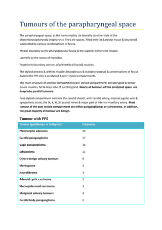
Tumour with PPS
Tumour type(Benign or malignant) Frequency
Pleomorphic adenoma
Carotid paraganglioma
Vagal paraganglioma
Schwanoma
Others benign salivary tumours
Meningioma
Neurofibroma
34
17
16
11
8
2
3
Adenoid cystic carcinoma
Mucoepidermoid carcinoma
Malignant salivary tumours
Carotid body paraganglioma
5
3
4
2
2. Vagal paraganglioma
Lymphoma
1
2
Salivary gland tumours
These are the common neoplasms to involve the PPS & invariably located in the prestyloid
compartment & almost arise from the deep lobe of the parotid gland. A minority , however, arise
from extraparotid salivary gland tissue in PPS.
Histologically these lesion are almost always pleomorphic adenoma. Rarely warthin’s tumour,
oncocytoma, benign lymphepithelial lesions. Very rarely malignant salivary cancer include
mucoepidermoid carcinoma, malignant mixed tumour, adenoid cystic carcinoma, adenocarcinoma.
Neurogegic tumours
As opposed to salivary neoplasm, these tumours involve the poststyloid compartment. Three
neurogenic neoplasm are involved in this area are1) paraganglioma, 2)schwannoma &
3)neurofibroma. Others tumour are extremely rare.
Carotid body Paragangliomas
arising from carotid body were previously known as carotid body tumours or chemodectomas.
The paragangliomas are sensitive to change particularly of carbon dioxide but also of oxygen & pH.
Tumour arise at high altitude, female to male 8:1, unilateral, family history absent( 1%), majority are
benign(3% malignant).
Tumour arise at low altitude,female to male 2: 1, bilaterally common, family history usually present,
Paragangliomas is a encapsulated tumour.
Histologically : typical appearance of epithelial cell clusters in extremely vascular & fibrous tissue.
The clusters have been given the name zellballen. The tumours are histologically similarly to the
adrenal medullary phaeochromocytoma.
Vagal paragangliomas
These are the rare tumour of neural crest. They are slow growing & usually occur in the PPS involving
the nodosa ganglion, but can occur anywhere along the course of vagus nerve& ite branches
including the middle ear.
A large jugular bulb paraganglioma, may be impossible to differentiate from a large nodosa ganglion
paraganglioma.
Carotid body paraganglioma very rarely secrecte catecholamine. It is common is vagal
paragangliomas.
3. Clinical feature first, history of hypertension, labile hypertension, facial fiushing or tachycardia,
serum catechecolamine & a 24hours uninary vanillylmandelic acid(VMA).
Schwannoma & neurofibromas
The histological appearance of a schwannoma fall into two groups antoni type A & B. The type A
pattern is of elongated spindle cells, forming a palisade of nuclei around a central mass of
cytoplasm(verocay body). Type B is not really apattern but consist of cells in loose myxoid stroma.
Occasionally malignant phenotype polymorphism, increased mitotic figures.
Although , the schwannoma is the commonest neurogenic tumour to be found in the PPS. They arise
from the sympathetic chain or vagus nerve. Very occasionally malignant. All schwannoma are in the
post-styloid compartment.
Neurofibromas probably arise from Schwann cells & usually seen in subcutaneous site.multiple,
often associated with von Recklinghausen’s disease. Histologically neurofibroma do not have
capsule but nerve fibre are within the tumour.
Metastases to the parapharyngeal space
A more common metastasis to the PPS is from nasopharyngeal carcinoma, maxillary carcinoma,
occult thyroid carcinoma.
Symptoms & signs
A lump in the neck is the most common finding approximatly 50%.
The great majority of patients are benign 80%, the most common neoplasm are tumours of the deep
lobe of the parotid. Large tumour of superficial lobe of the parotid may pass into area of deep lobe
via the stylomandibular tunnel, giving a typical dumb-bell shaped tumour, result in medial
displacement of the lateral pharyngeal wall & tonsil .
More superficial placed tumour may expand laterally to produce a lump in the neck, behind the
angle of the mandible.
In case of the jugular foramen itself, an expansion within it may produce jugular foramen syndrome.
Palsy of IX, X &XI. Hypoglossal nerve may also involve.
In the anterior compartment(masticator space), due to pressure on masticator muscles or invasion
of muscles will cause trismus.
High post-styloid lesions may encompress the cartilaginous part of the Eustachian tube, causing
middle ear effusion & hearing impairment. Obstructive sleep apnoea may occur when medial
displacement of the lateral pharyngeal wall is pronounced.
Diagnosis & investigations
The majority of these patients present with a mass normally in the neck but sometimes as a bulge in
the orapharynx.
Cranial nerve palsies should be sought.
4. A Bruit may be detectable over a carotid body paraganglion. Pulse & Bp should be measured.
FNAC if as a lump is the usual presenting feature.
Imaging : MRI & CT scaning
Carotid angiography : The main indication of carotid angiography is in planning surgical treatment.
Without angiography vagal paragangliomas can be difficult to distinguish radiologically from a
carotid body paragangliomas. More recent developments using digital subtraction technique in
association with MRI will show accurate diagnosis in most cases. There is no other method of
imaging that accurately demonstrates the feeding vessels.
Carotid body paragangliomas are characteristically located between the internal & external carotid
arteries at the bifurcation. Tend to displace carotid artery posteriorly(tumour anteriorly)
Most vagal paragangliomas tend to displace the internal carotid artery anteriorly , tumour
posteriorly(in addition, they are high in postion).
Glomus jugulare tumours are in the jugular bulb & can be seen to be expansion the jugular foramen.
Surgical treatment
There are two standard approach to the PPS.
Tansparotid approach primarily used to access for larger deep lobe tumours in the prestyloid
compartment. This approach by a routine superficial parotidectomy procedure, preferable healthy
parotid gland & with full identification & preservation of the facial nerve. The division of
stylomandibular ligament(diffuse condensation of fascia seen at operation) allows anterior
displacement of the mandible with improve access.
Transcervical approach: for most tumours of the poststyloid approach is adequate. A reasonably
extensive excision is made from a point 2cm below the mandibular ramus, starting some 2cm in
front of the mandibular angle & exended backwards & upwards. The size & accessibility of the
tumour will dictate how large the incision needs to be.
Facial artery is ligated & divided. Submandibular gland retract anteriorly or removed. Division of
diagastric tendon then allows excellent exposure with direct visualtion & proper removal of the
tumour.
Extended transmandibular approach for malignant PPS tumour particularly oropharyngeal
involvement.( transpharyngeal approach see in the chapter benign salivary tumours).