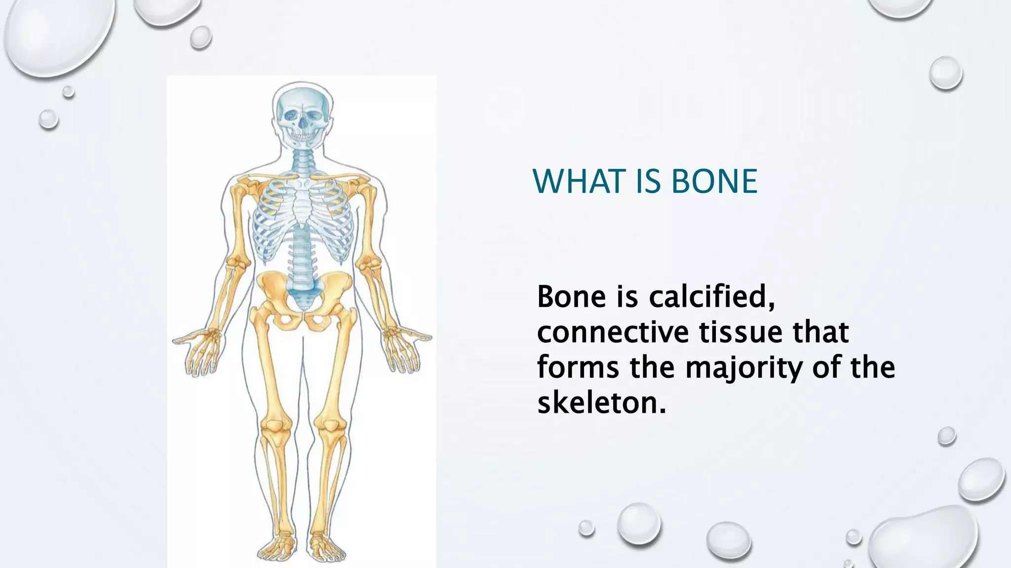
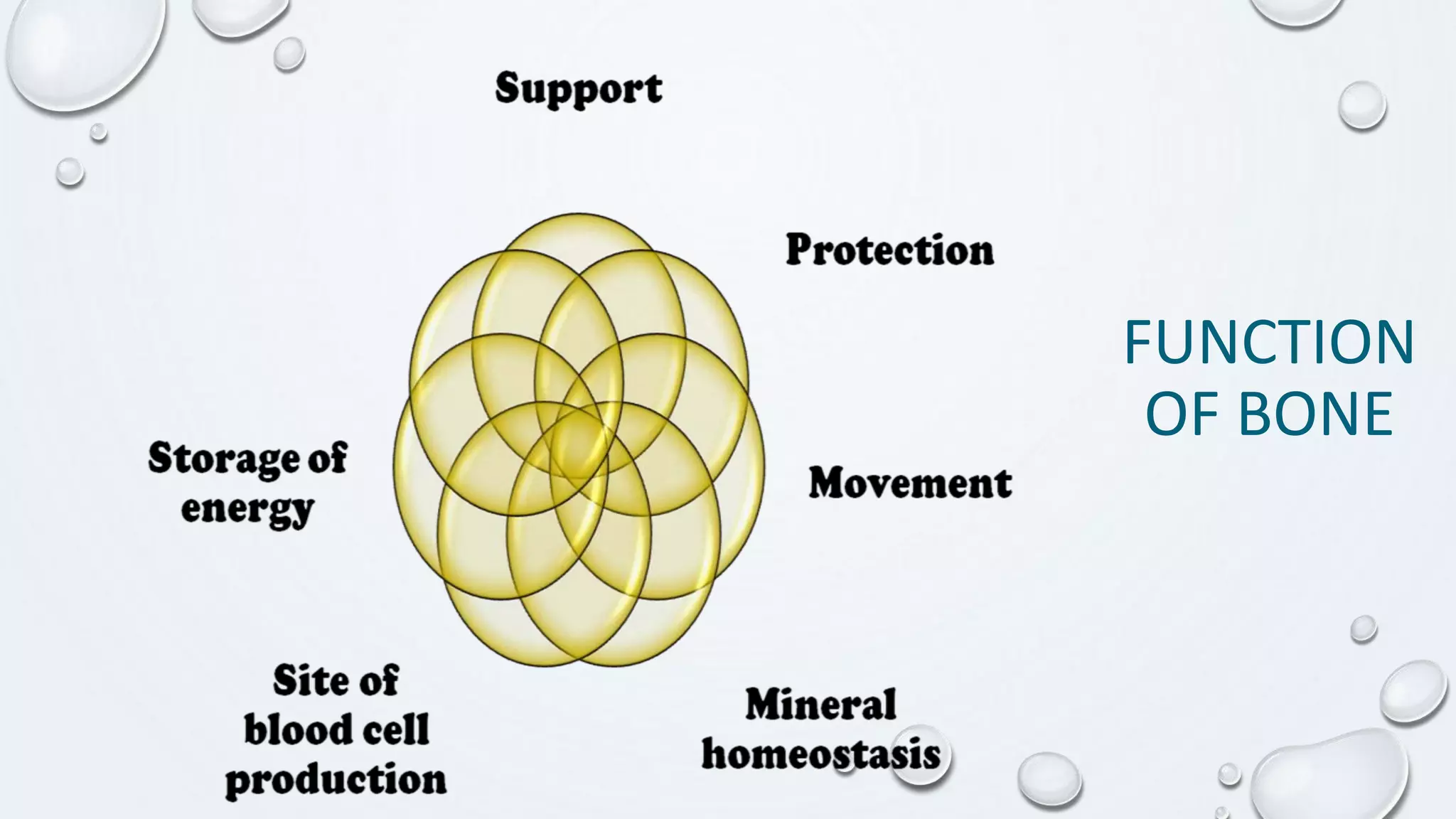
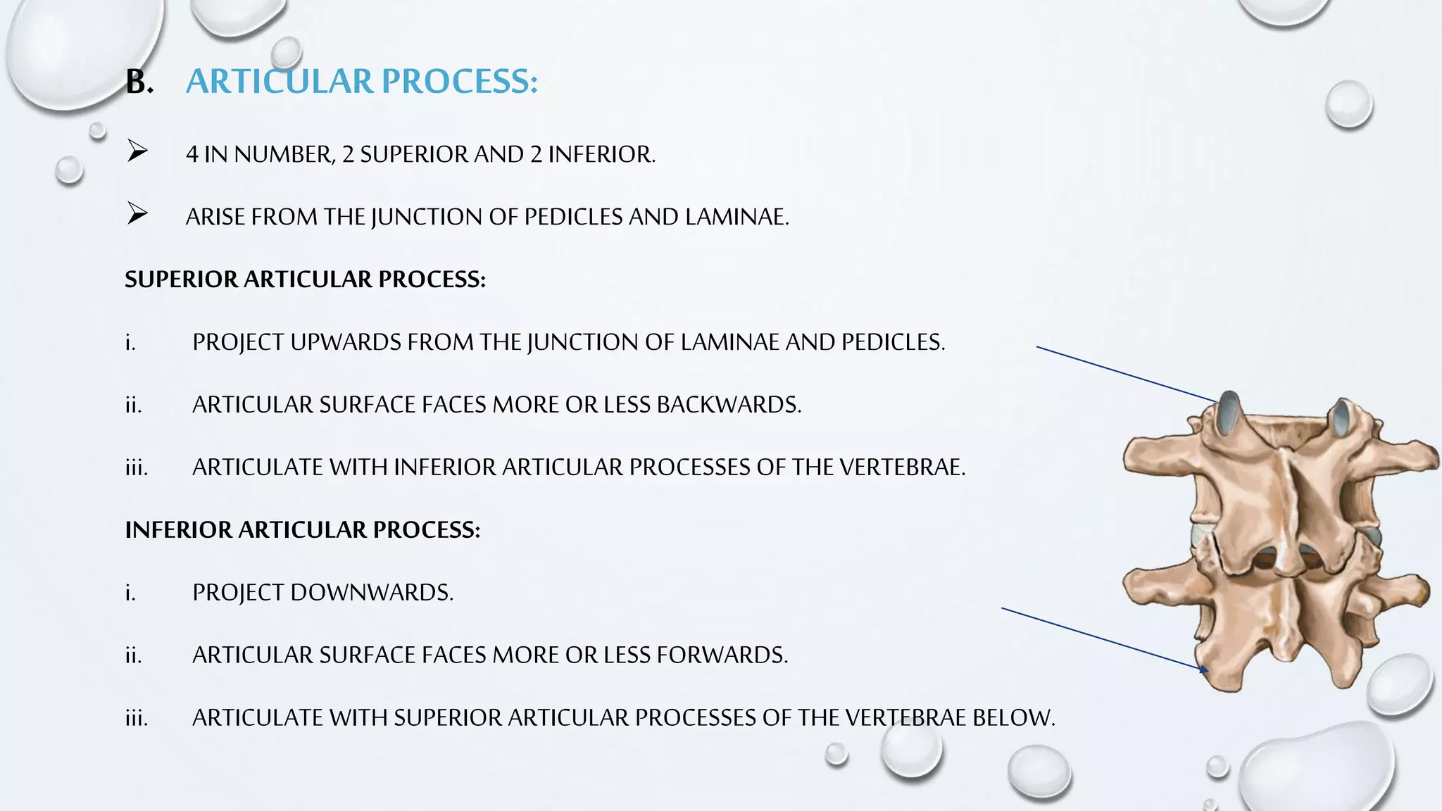
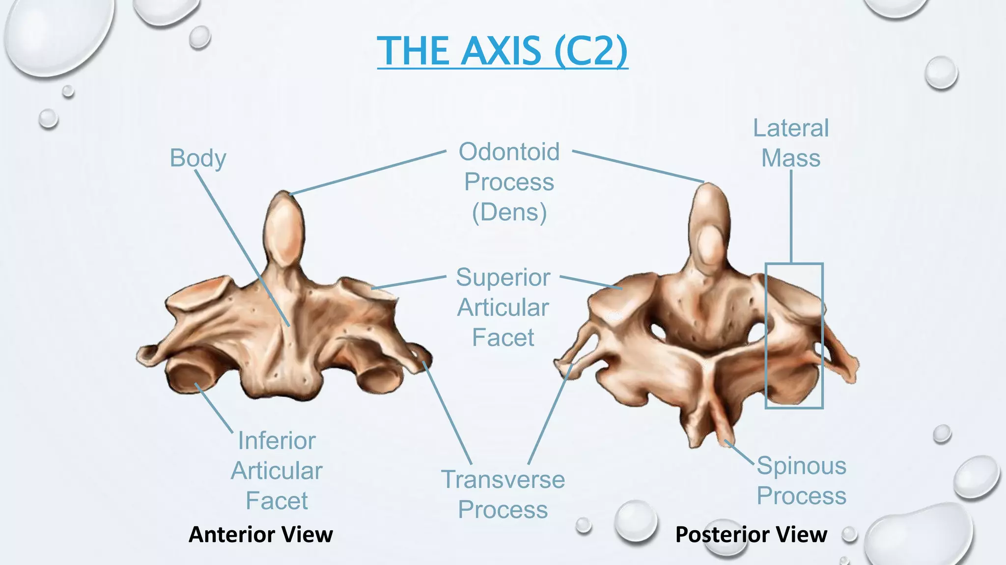
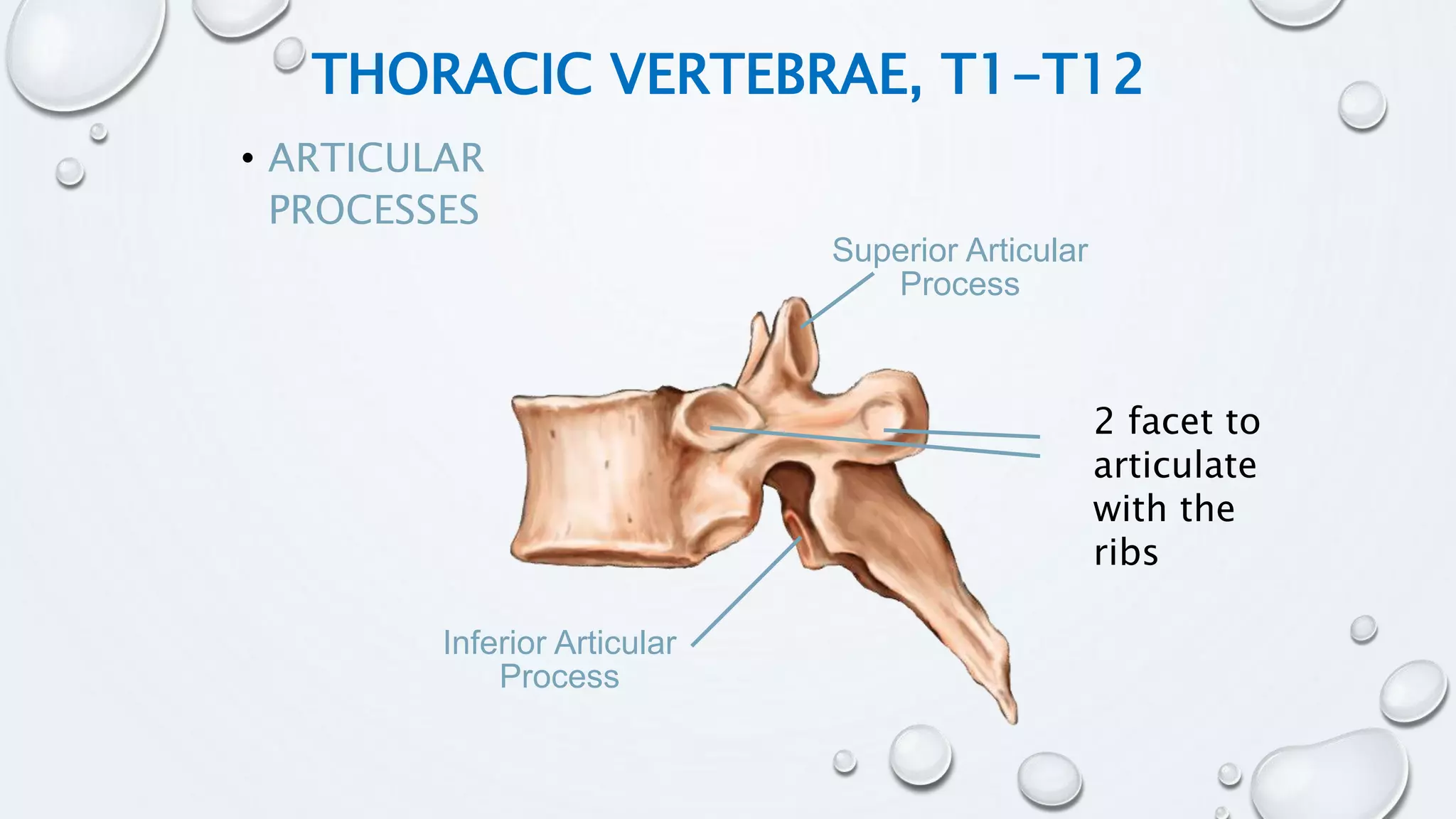
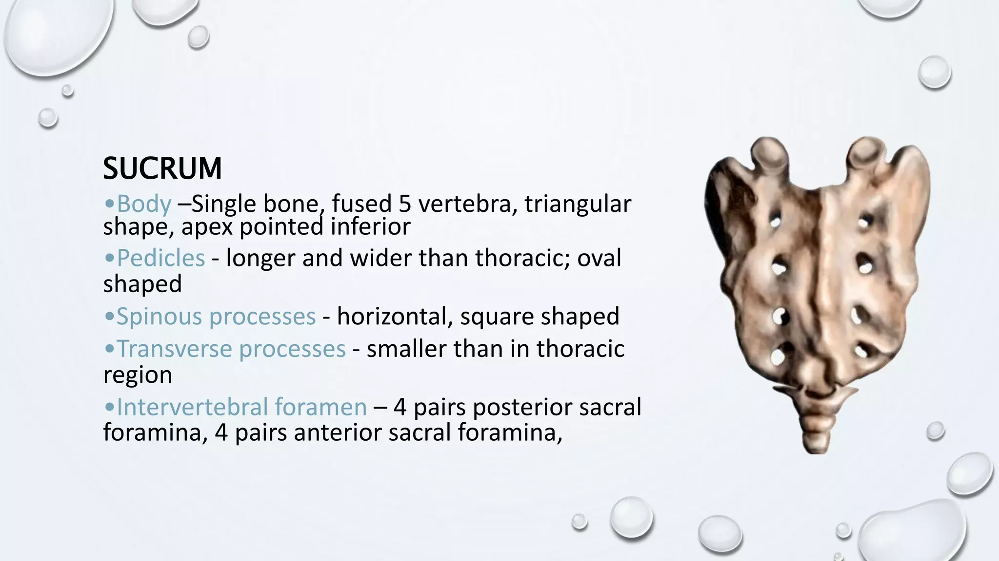
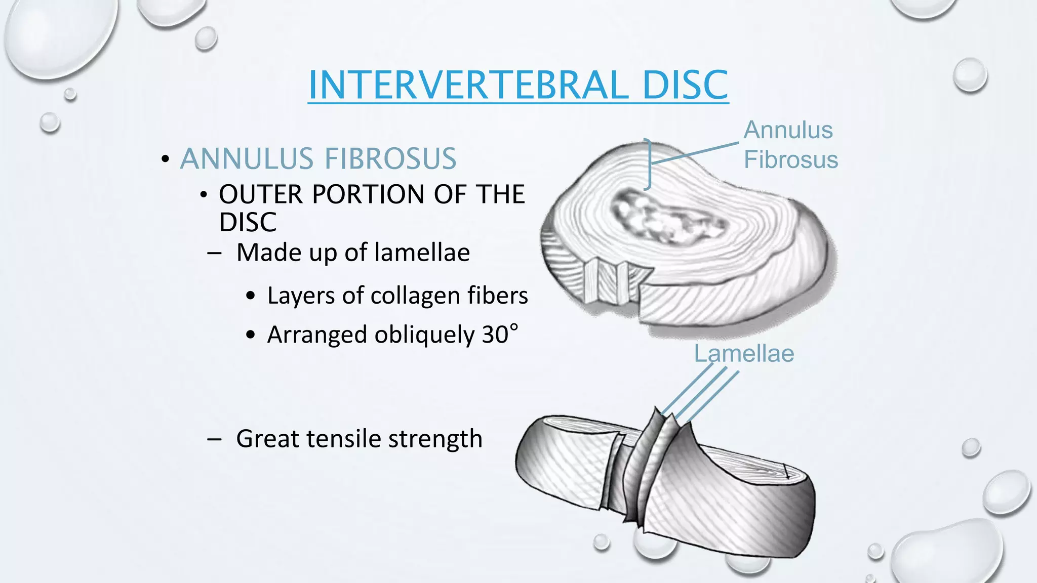
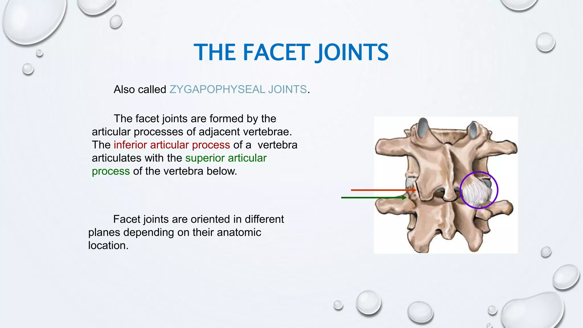
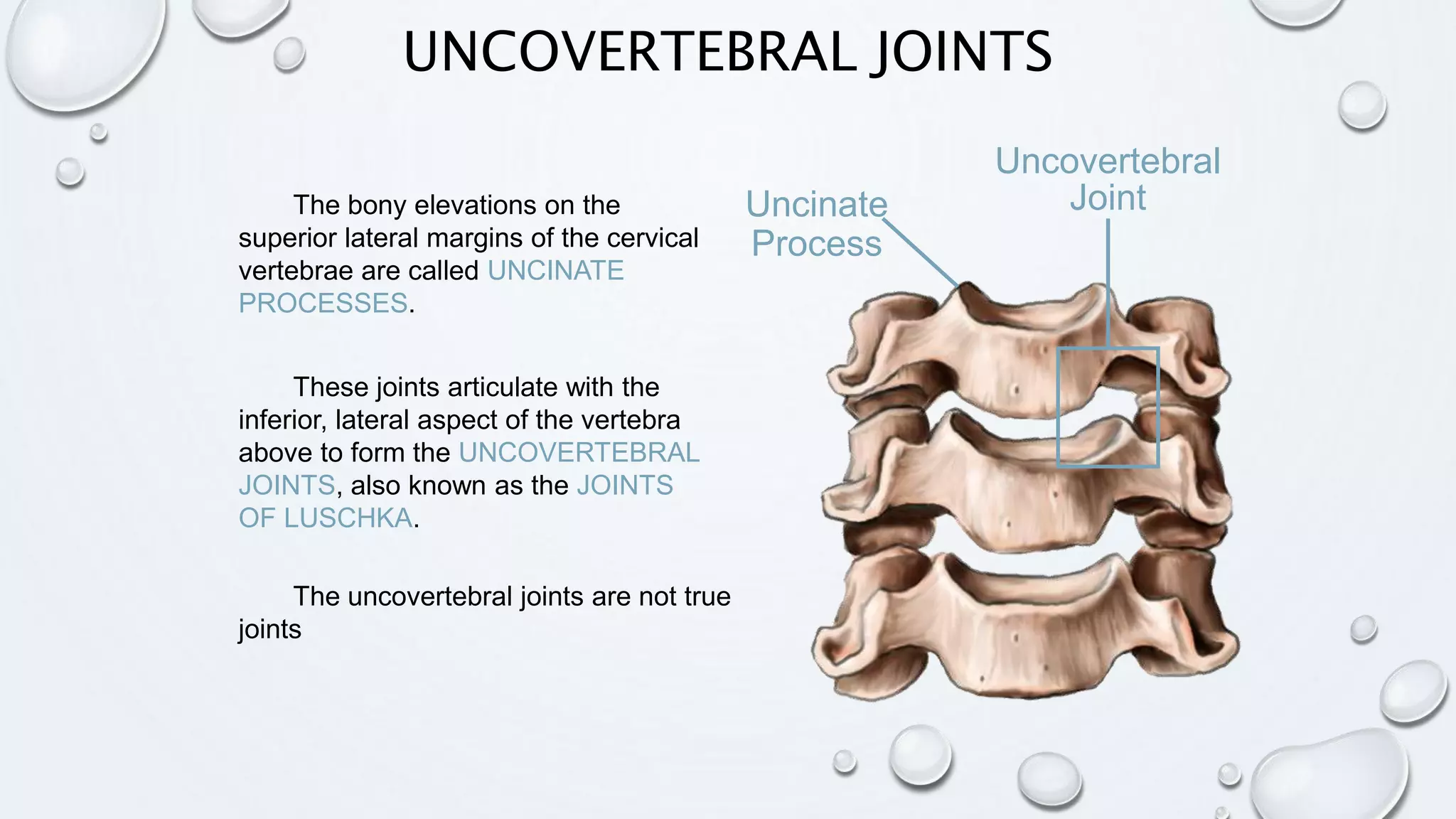
ANATOMY OF
BONES
ALIO MOHAMED HERSI
WHAT IS BONE
Bone is calcified,
connective tissue that
forms the majority of the
skeleton.
FUNCTION
OF BONE
SKELETAL ORGANIZATION
• The actual number of bones in the human
skeleton varies from person to person
• Typically there are about 206 bones
• For convenience the skeleton is divided into the:
• Axial skeleton
• Appendicular skeleton
SKELETAL
ORGANIZATION
CLASSIFICATION OF BONES BY
SHAPE
 Long bones
 Short bones
 Flat bones
 Irregular bones
 Pneumatized bones
 Sesamoid bones
TYPES OF
BONE
ANATOMY OF
VERTEBRAL COLUMN
VERTEBRAL COLUMN
•Vertebralcolumn consists ofa numberof
separateirregularbonescalledVERTEBRAE
•Forms the centralaxis of the body.
FUNCTION
Structuralsupportand balancefor upright
posture
 Protects spinalcord.
Supports andtransmitsbodyweight.
Provides attachmentto axial muscles.
Provides movement of the trunk.
FLEXIBILITY OF MOTION IN SIX DEGREES OF
FREEDOM
Flexion and Extension
Left and Right
Side Bending
Left and Right Rotation
SAGITTAL PLANE CURVES
•Primary Curves
•Secondary Curves
REGIONS OF THE SPINE
• Cervical
– Upper cervical: C1-C2
– Lower cervical: C3-C7
• Thoracic: T1-T12
•Lumbar: L1- L5
•Sacrococcygeal: 9 fused
vertebrae in the sacrum and
coccyx.
BASIC VERTEBRAL STRUCTURES
Cervical Thoracic Lumbar
CHARACTERISTICS OF A VERTEBRAE:
• A TYPICAL VERTEBRAE HAS TWO PARTS:
1. BODY-ANTERIOR PART.
2. ARCH-POSTERIOR PART.
• VERTEBRALFORAMEN:LIES BETWEEN THE BODY AND ARCH.
• VERTEBRAE ARE PLACED ONE ABOVE THE OTHER WITH INTERVERTEBRAL DISCS
BETWEEN THEM AND FORMS A VERTEBRAL CANAL(SPINAL CANAL) WHICH LODGES
THE SPINAL CORD WITH ITS MENINGES AND BLOOD VESSELS.
1. BODY OF VERTEBRAE:
• IT POSSESSES6 SURFACES: UPPER, LOWER, ANTERIOR, POSTERIOR AND TWO
LATERAL.
• BODIES OFVERTEBRAE LIE ONE ABOVE ANOTHER SEPARATED BY INTERVERTEBRAL
DISC AND FORM THE CENTRAL AXIS.
2. VERTEBRAL (NEURAL) ARCH:
• THE VERTEBRAL ARCH CONSISTS OF:
I. A PAIR OFPEDICLES.
II. A PAIR OFLAMINA.
III. SEVEN PROCESSES:
o SPINOUS ----ONE
o ARTICULAR ---- FOUR
o TRANSVERSE --- TWO
pedicles
LAMINA
PROCESSES:
 The arch isconnected tothe bodyby pedicles.
Pedicles:
Short, thickprocesses pass backwardfrom the junction
of lateraland posterior surfaces of the body.
Possess2 surfacesand4 borders.
LAMINA:
• BROAD PLATES OF BONE LYING BEHIND AND MEDIAL TO PEDICLES.
• THEY FUSE BEHIND IN THE MEDIAN PLANE INTO THE SPINOUS PROCESS.
• THEY FORMPOSTERIOR BOUNDARY OFVERTEBRAL FORAMEN.
PROCESSES:
A. SPINOUSPROCESS----
 PASSESBACKWARDSANDDOWNWARDSFROMTHEJUNCTION OFTHE TWOLAMINAE.
 SHAPEANDSIZE VARIESIN VERTEBRAEOFDIFFERENTREGIONS.
 GIVESATTACHMENTTOMUSCLESANDLIGAMENTS.
B. ARTICULARPROCESS:
 4 IN NUMBER, 2 SUPERIOR AND 2 INFERIOR.
 ARISE FROM THE JUNCTION OF PEDICLES AND LAMINAE.
SUPERIOR ARTICULAR PROCESS:
i. PROJECT UPWARDS FROM THE JUNCTION OF LAMINAE AND PEDICLES.
ii. ARTICULAR SURFACE FACES MORE OR LESS BACKWARDS.
iii. ARTICULATE WITH INFERIOR ARTICULAR PROCESSES OF THE VERTEBRAE.
INFERIOR ARTICULAR PROCESS:
i. PROJECT DOWNWARDS.
ii. ARTICULAR SURFACE FACES MORE OR LESS FORWARDS.
iii. ARTICULATE WITH SUPERIOR ARTICULAR PROCESSES OF THE VERTEBRAE BELOW.
C. TRANSVERSE PROCESSES:
• 2 IN NUMBER.
• PROJECT LATERALLY FROMTHE JUNCTION OFPEDICLE AND LAMINA.
• IN THORACIC REGION THEY ARTICULATE WITH RIBS.
Vertebral
Body
Pedicle
Lamina
Superior
Articular
Process
Spinous
Process
Transverse
Process
Vertebral
Foramen
VERTEBRAL STRUCTURES
Superior
Articular
Process
Inferior
Articular
Process
Zygapophyseal
Joint
(Facet Joint)
Pars
VERTEBRAL STRUCTURES
• PEDICLE NOTCHES
Slight
Notch
Deep
Notch
Intervertebral
Foramen
• INTERVERTEBRAL FORAMEN
through which the spinal nerve roots
leave the spinal cord
VERTEBRAL STRUCTURES
• ANTERIOR ARCH
COMPRISED OF:
• VERTEBRAL BODY
• ANTERIOR 1/3 OF THE
PEDICLES
• Posterior Arch
Comprised of:
– Posterior 2/3 of the pedicles
– Lamina
– Processes
VERTEBRAL STRUCTURES
THE ATLAS (C1)
Transverse
Process
Transverse
Foramen
Anterior
Tubercle
Articular Facet
for Dens
Lateral
Mass
LaminaPosterior
Tubercle
Superior
Articular
Facet
Superior View
THE AXIS (C2)
Odontoid
Process
(Dens)
Body
Transverse
Process
Inferior
Articular
Facet
Superior
Articular
Facet
Anterior View Posterior View
Lateral
Mass
Spinous
Process
LOWER CERVICAL VERTEBRAE
• C3 TO C7
• MAY BE REFERRED
TO AS THE
SUBAXIAL
REGION
• DISC AT EVERY
LEVEL
• VERTEBRAL
STRUCTURES ARE
SIMILAR
LOWER CERVICAL VERTEBRAE
C3 - C7
Transverse
ProcessBody
Sulcus for
Spinal Nerve
Lateral
Mass
Lamina
Pedicle
Superior
Articular Facet
Vertebral
Foramen
Bifid Spinous Process
Transverse
Foramen
Axial View
THORACIC VERTEBRAE
• BODY - PROGRESSIVE INCREASE
IN MASS FROM T1 TO T12
• Pedicles - small diameter
• Laminae - vertical, with “roof tile”
arrangement
• Spinous processes - long,
overlapping, projected downward
• Intervertebral foramen - larger
THORACIC VERTEBRAE, T1-T12
• BODY - HEART SHAPED
WHEN VIEWED SUPERIORLY.
• Vertebral foramen - round
• Pedicles - small in diameter
• Spinous processes - long and
projected downwards
• ARTICULAR
PROCESSES
Superior Articular
Process
Inferior Articular
Process
THORACIC VERTEBRAE, T1-T12
2 facet to
articulate
with the
ribs
• Body - L1 to L5 progressive increase
in mass, cylindrical
• Pedicles - longer and wider than
thoracic; oval shaped
• Spinous processes - horizontal,
square shaped
• Transverse processes - smaller than
in thoracic region
• Intervertebral foramen – large,
triangular shape
LUMBAR VERTEBRAE, L1-L5
The most important distinguishing feature of
vertebrae of different regions is-----
Cervical: Presence of a foramen in the transverse process,
calledForamen Transversarium.
Thoracic:Presence of costal facets on the body
Lumbar: No foramen transversarium and nocostal facets
onthe body.
SUCRUM
•Body –Single bone, fused 5 vertebra, triangular
shape, apex pointed inferior
•Pedicles - longer and wider than thoracic; oval
shaped
•Spinous processes - horizontal, square shaped
•Transverse processes - smaller than in thoracic
region
•Intervertebral foramen – 4 pairs posterior sacral
foramina, 4 pairs anterior sacral foramina,
THE SACRUM
Sacral Horns
Sacral Ala
Pedicles
Dorsal
Foramina
Sacral
Hiatus
Coccyx
Posterior View
Inverted triangle shape
Coccyx
Lateral View
Sacral
Promontory
Sacral Tilt
30°-60°
Sacral Canal
1
2
3
4
5
Sacral Hiatus
THE SACRUM
NO ARCH OR
CANAL
• INTERVERTEBRAL DISC
• End plate
• Apophyseal ring
– Cartilaginous layer
– Bony layer
INTERVERTEBRAL DISC
• FIBROCARTILAGINOUS
JOINT OF THE MOTION
SEGMENT
• MAKE UP ¼ THE LENGTH
OF THE SPINAL COLUMN
• PRESENT AT LEVELS C2-
C3 TO L5-S1
• ALLOWS COMPRESSIVE,
TENSILE, AND
ROTATIONAL MOTION
• LARGEST AVASCULAR
STRUCTURES IN THE
BODY
INTERVERTEBRAL DISC
INTERVERTEBRAL DISC
• ANNULUS FIBROSUS
• OUTER PORTION OF THE
DISC
Lamellae
– Great tensile strength
– Made up of lamellae
Annulus
Fibrosus
• Layers of collagen fibers
• Arranged obliquely 30°
INTERVERTEBRAL DISC
• NUCLEUS PULPOSUS
Nucleus
Pulposus
– Inner structure
– Gelatinous
– High water content
– Resists axial forces
ATLANTOAXIAL JOINT
C1
C2
Dens
Zygapophyseal
joints
JOINT between the atlas (C1) and the
axis (C2); has a range of motion in the
transverse plane for rotation.
The DENS of C2 acts as a
pivot point for the rotation of
C1.
The articulating surfaces of the two
vertebrae form ZYGAPOPHYSEAL
(FACET) JOINTS that allow flexion-
extension, side bending, and
rotational movements.
Also called ZYGAPOPHYSEAL JOINTS.
The facet joints are formed by the
articular processes of adjacent vertebrae.
The inferior articular process of a vertebra
articulates with the superior articular
process of the vertebra below.
Facet joints are oriented in different
planes depending on their anatomic
location.
THE FACET JOINTS
REGIONS OF THE SPINE
• Cervical
Slope inferiorly from anterior to posterior.
Facilitates flexion and extension
• Thoracic: T1-T12
Oriented vertically and limit flexion and extension
Facilitate rotation
•Lumbar: L1- L5
The joint surface are curved and adjacent processes interlock
limiting range of movement
Major movements flexion and extension.
UNCOVERTEBRAL JOINTS
Uncovertebral
JointThe bony elevations on the
superior lateral margins of the cervical
vertebrae are called UNCINATE
PROCESSES.
The uncovertebral joints are not true
joints
These joints articulate with the
inferior, lateral aspect of the vertebra
above to form the UNCOVERTEBRAL
JOINTS, also known as the JOINTS
OF LUSCHKA.
Uncinate
Process
THANK YOU

Anatomy of bones

  • 1.
  • 2.
    WHAT IS BONE Boneis calcified, connective tissue that forms the majority of the skeleton.
  • 3.
  • 4.
    SKELETAL ORGANIZATION • Theactual number of bones in the human skeleton varies from person to person • Typically there are about 206 bones • For convenience the skeleton is divided into the: • Axial skeleton • Appendicular skeleton
  • 5.
  • 6.
    CLASSIFICATION OF BONESBY SHAPE  Long bones  Short bones  Flat bones  Irregular bones  Pneumatized bones  Sesamoid bones
  • 8.
  • 9.
  • 10.
    VERTEBRAL COLUMN •Vertebralcolumn consistsofa numberof separateirregularbonescalledVERTEBRAE •Forms the centralaxis of the body.
  • 11.
    FUNCTION Structuralsupportand balancefor upright posture Protects spinalcord. Supports andtransmitsbodyweight. Provides attachmentto axial muscles. Provides movement of the trunk.
  • 12.
    FLEXIBILITY OF MOTIONIN SIX DEGREES OF FREEDOM Flexion and Extension Left and Right Side Bending Left and Right Rotation
  • 13.
    SAGITTAL PLANE CURVES •PrimaryCurves •Secondary Curves
  • 14.
    REGIONS OF THESPINE • Cervical – Upper cervical: C1-C2 – Lower cervical: C3-C7 • Thoracic: T1-T12 •Lumbar: L1- L5 •Sacrococcygeal: 9 fused vertebrae in the sacrum and coccyx.
  • 15.
  • 16.
    CHARACTERISTICS OF AVERTEBRAE: • A TYPICAL VERTEBRAE HAS TWO PARTS: 1. BODY-ANTERIOR PART. 2. ARCH-POSTERIOR PART. • VERTEBRALFORAMEN:LIES BETWEEN THE BODY AND ARCH. • VERTEBRAE ARE PLACED ONE ABOVE THE OTHER WITH INTERVERTEBRAL DISCS BETWEEN THEM AND FORMS A VERTEBRAL CANAL(SPINAL CANAL) WHICH LODGES THE SPINAL CORD WITH ITS MENINGES AND BLOOD VESSELS.
  • 17.
    1. BODY OFVERTEBRAE: • IT POSSESSES6 SURFACES: UPPER, LOWER, ANTERIOR, POSTERIOR AND TWO LATERAL. • BODIES OFVERTEBRAE LIE ONE ABOVE ANOTHER SEPARATED BY INTERVERTEBRAL DISC AND FORM THE CENTRAL AXIS.
  • 18.
    2. VERTEBRAL (NEURAL)ARCH: • THE VERTEBRAL ARCH CONSISTS OF: I. A PAIR OFPEDICLES. II. A PAIR OFLAMINA. III. SEVEN PROCESSES: o SPINOUS ----ONE o ARTICULAR ---- FOUR o TRANSVERSE --- TWO pedicles LAMINA PROCESSES:
  • 19.
     The archisconnected tothe bodyby pedicles. Pedicles: Short, thickprocesses pass backwardfrom the junction of lateraland posterior surfaces of the body. Possess2 surfacesand4 borders.
  • 20.
    LAMINA: • BROAD PLATESOF BONE LYING BEHIND AND MEDIAL TO PEDICLES. • THEY FUSE BEHIND IN THE MEDIAN PLANE INTO THE SPINOUS PROCESS. • THEY FORMPOSTERIOR BOUNDARY OFVERTEBRAL FORAMEN. PROCESSES: A. SPINOUSPROCESS----  PASSESBACKWARDSANDDOWNWARDSFROMTHEJUNCTION OFTHE TWOLAMINAE.  SHAPEANDSIZE VARIESIN VERTEBRAEOFDIFFERENTREGIONS.  GIVESATTACHMENTTOMUSCLESANDLIGAMENTS.
  • 21.
    B. ARTICULARPROCESS:  4IN NUMBER, 2 SUPERIOR AND 2 INFERIOR.  ARISE FROM THE JUNCTION OF PEDICLES AND LAMINAE. SUPERIOR ARTICULAR PROCESS: i. PROJECT UPWARDS FROM THE JUNCTION OF LAMINAE AND PEDICLES. ii. ARTICULAR SURFACE FACES MORE OR LESS BACKWARDS. iii. ARTICULATE WITH INFERIOR ARTICULAR PROCESSES OF THE VERTEBRAE. INFERIOR ARTICULAR PROCESS: i. PROJECT DOWNWARDS. ii. ARTICULAR SURFACE FACES MORE OR LESS FORWARDS. iii. ARTICULATE WITH SUPERIOR ARTICULAR PROCESSES OF THE VERTEBRAE BELOW.
  • 22.
    C. TRANSVERSE PROCESSES: •2 IN NUMBER. • PROJECT LATERALLY FROMTHE JUNCTION OFPEDICLE AND LAMINA. • IN THORACIC REGION THEY ARTICULATE WITH RIBS.
  • 23.
  • 24.
  • 25.
    • PEDICLE NOTCHES Slight Notch Deep Notch Intervertebral Foramen •INTERVERTEBRAL FORAMEN through which the spinal nerve roots leave the spinal cord VERTEBRAL STRUCTURES
  • 26.
    • ANTERIOR ARCH COMPRISEDOF: • VERTEBRAL BODY • ANTERIOR 1/3 OF THE PEDICLES • Posterior Arch Comprised of: – Posterior 2/3 of the pedicles – Lamina – Processes VERTEBRAL STRUCTURES
  • 27.
    THE ATLAS (C1) Transverse Process Transverse Foramen Anterior Tubercle ArticularFacet for Dens Lateral Mass LaminaPosterior Tubercle Superior Articular Facet Superior View
  • 28.
  • 29.
    LOWER CERVICAL VERTEBRAE •C3 TO C7 • MAY BE REFERRED TO AS THE SUBAXIAL REGION • DISC AT EVERY LEVEL • VERTEBRAL STRUCTURES ARE SIMILAR
  • 30.
    LOWER CERVICAL VERTEBRAE C3- C7 Transverse ProcessBody Sulcus for Spinal Nerve Lateral Mass Lamina Pedicle Superior Articular Facet Vertebral Foramen Bifid Spinous Process Transverse Foramen Axial View
  • 31.
    THORACIC VERTEBRAE • BODY- PROGRESSIVE INCREASE IN MASS FROM T1 TO T12 • Pedicles - small diameter • Laminae - vertical, with “roof tile” arrangement • Spinous processes - long, overlapping, projected downward • Intervertebral foramen - larger
  • 32.
    THORACIC VERTEBRAE, T1-T12 •BODY - HEART SHAPED WHEN VIEWED SUPERIORLY. • Vertebral foramen - round • Pedicles - small in diameter • Spinous processes - long and projected downwards
  • 33.
    • ARTICULAR PROCESSES Superior Articular Process InferiorArticular Process THORACIC VERTEBRAE, T1-T12 2 facet to articulate with the ribs
  • 34.
    • Body -L1 to L5 progressive increase in mass, cylindrical • Pedicles - longer and wider than thoracic; oval shaped • Spinous processes - horizontal, square shaped • Transverse processes - smaller than in thoracic region • Intervertebral foramen – large, triangular shape LUMBAR VERTEBRAE, L1-L5
  • 35.
    The most importantdistinguishing feature of vertebrae of different regions is----- Cervical: Presence of a foramen in the transverse process, calledForamen Transversarium. Thoracic:Presence of costal facets on the body Lumbar: No foramen transversarium and nocostal facets onthe body.
  • 36.
    SUCRUM •Body –Single bone,fused 5 vertebra, triangular shape, apex pointed inferior •Pedicles - longer and wider than thoracic; oval shaped •Spinous processes - horizontal, square shaped •Transverse processes - smaller than in thoracic region •Intervertebral foramen – 4 pairs posterior sacral foramina, 4 pairs anterior sacral foramina,
  • 37.
    THE SACRUM Sacral Horns SacralAla Pedicles Dorsal Foramina Sacral Hiatus Coccyx Posterior View Inverted triangle shape
  • 38.
    Coccyx Lateral View Sacral Promontory Sacral Tilt 30°-60° SacralCanal 1 2 3 4 5 Sacral Hiatus THE SACRUM NO ARCH OR CANAL
  • 39.
    • INTERVERTEBRAL DISC •End plate • Apophyseal ring – Cartilaginous layer – Bony layer INTERVERTEBRAL DISC
  • 40.
    • FIBROCARTILAGINOUS JOINT OFTHE MOTION SEGMENT • MAKE UP ¼ THE LENGTH OF THE SPINAL COLUMN • PRESENT AT LEVELS C2- C3 TO L5-S1 • ALLOWS COMPRESSIVE, TENSILE, AND ROTATIONAL MOTION • LARGEST AVASCULAR STRUCTURES IN THE BODY INTERVERTEBRAL DISC
  • 41.
    INTERVERTEBRAL DISC • ANNULUSFIBROSUS • OUTER PORTION OF THE DISC Lamellae – Great tensile strength – Made up of lamellae Annulus Fibrosus • Layers of collagen fibers • Arranged obliquely 30°
  • 42.
    INTERVERTEBRAL DISC • NUCLEUSPULPOSUS Nucleus Pulposus – Inner structure – Gelatinous – High water content – Resists axial forces
  • 43.
    ATLANTOAXIAL JOINT C1 C2 Dens Zygapophyseal joints JOINT betweenthe atlas (C1) and the axis (C2); has a range of motion in the transverse plane for rotation. The DENS of C2 acts as a pivot point for the rotation of C1. The articulating surfaces of the two vertebrae form ZYGAPOPHYSEAL (FACET) JOINTS that allow flexion- extension, side bending, and rotational movements.
  • 44.
    Also called ZYGAPOPHYSEALJOINTS. The facet joints are formed by the articular processes of adjacent vertebrae. The inferior articular process of a vertebra articulates with the superior articular process of the vertebra below. Facet joints are oriented in different planes depending on their anatomic location. THE FACET JOINTS
  • 45.
    REGIONS OF THESPINE • Cervical Slope inferiorly from anterior to posterior. Facilitates flexion and extension • Thoracic: T1-T12 Oriented vertically and limit flexion and extension Facilitate rotation •Lumbar: L1- L5 The joint surface are curved and adjacent processes interlock limiting range of movement Major movements flexion and extension.
  • 46.
    UNCOVERTEBRAL JOINTS Uncovertebral JointThe bonyelevations on the superior lateral margins of the cervical vertebrae are called UNCINATE PROCESSES. The uncovertebral joints are not true joints These joints articulate with the inferior, lateral aspect of the vertebra above to form the UNCOVERTEBRAL JOINTS, also known as the JOINTS OF LUSCHKA. Uncinate Process
  • 48.