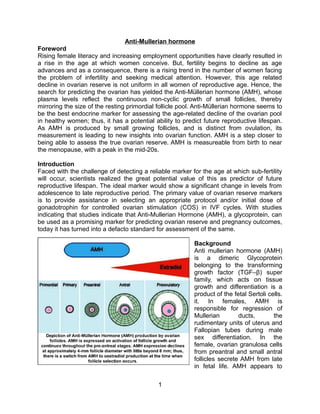
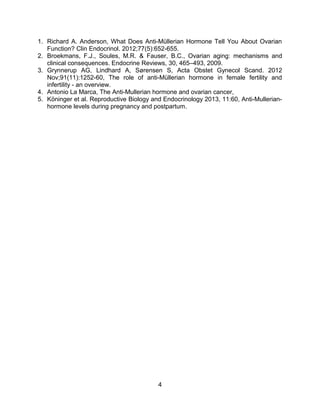
Anti-Mullerian hormone
Foreword
Rising female literacy and increasing employment opportunities have clearly resulted in
a rise in the age at which women conceive. But, fertility begins to decline as age
advances and as a consequence, there is a rising trend in the number of women facing
the problem of infertility and seeking medical attention. However, this age related
decline in ovarian reserve is not uniform in all women of reproductive age. Hence, the
search for predicting the ovarian has yielded the Anti-Müllerian hormone (AMH), whose
plasma levels reflect the continuous non-cyclic growth of small follicles, thereby
mirroring the size of the resting primordial follicle pool. Anti-Müllerian hormone seems to
be the best endocrine marker for assessing the age-related decline of the ovarian pool
in healthy women; thus, it has a potential ability to predict future reproductive lifespan.
As AMH is produced by small growing follicles, and is distinct from ovulation, its
measurement is leading to new insights into ovarian function. AMH is a step closer to
being able to assess the true ovarian reserve. AMH is measureable from birth to near
the menopause, with a peak in the mid-20s.
Introduction
Faced with the challenge of detecting a reliable marker for the age at which sub-fertility
will occur, scientists realized the great potential value of this as predictor of future
reproductive lifespan. The ideal marker would show a significant change in levels from
adolescence to late reproductive period. The primary value of ovarian reserve markers
is to provide assistance in selecting an appropriate protocol and/or initial dose of
gonadotrophin for controlled ovarian stimulation (COS) in IVF cycles. With studies
indicating that studies indicate that Anti-Mullerian Hormone (AMH), a glycoprotein, can
be used as a promising marker for predicting ovarian reserve and pregnancy outcomes,
today it has turned into a defacto standard for assessment of the same.
Background
Anti mullerian hormone (AMH)
is a dimeric Glycoprotein
belonging to the transforming
growth factor (TGF–β) super
family, which acts on tissue
growth and differentiation is a
product of the fetal Sertoli cells.
it. In females, AMH is
responsible for regression of
Mullerian
ducts,
the
rudimentary units of uterus and
Fallopian tubes during male
sex differentiation. In the
female, ovarian granulosa cells
from preantral and small antral
follicles secrete AMH from late
in fetal life. AMH appears to
1
have inhibitory effect upon the recruitment of primordial follicles and it may decrease the
sensitivity of large pre antral and small antral follicles to FSH. The patterns of
expression of AMH and its type II receptor in the post-natal ovary indicate that AMH may
play an important role in ovarian folliculogenesis. AMH is expressed in growing follicles
throughout the pre-antral and early antral stages, and then declines with little production
beyond 8 mm. Thus, AMH production has declined when follicles are selected for
dominance and when oestrogen production starts to escalate, and a contribution to this
switch may be one of the functions of AMH. Large follicles produce little AMH
(expression persists only in the cumulus cells surrounding the oocyte); thus, AMH does
not show clinically relevant changes across the menstrual cycle. This greatly adds to its
clinical utility, particularly compared to FSH, the historically established maker of ovarian
reserve. In harmony with the established relationship between age and declining
ovarian reserve, AMH falls linearly with increasing age. This occurs in conjuction with
reductions in the antral follicular count, which is strongly correlated, to plasma AMH
levels. AMH has consequently been explored as a predictor of ovarian response to FSH
and oocyte quality in cycles of ART. More over AMH levels have better cycle-to-cycle
reproducibility. But, AMH needs to be interpreted in the context of age.
The utility
Some recent studies have shown AMH as predictor of pregnancy outcome in assisted
reproductive techniques. There is considerable interest in its ability to predict remaining
reproductive lifespan, which clinically may be of value in the assessment of ovarian
reserve following damage, for
example postchemotherapy
or ovarian surgery. The
clearest potential of AMH is in
the diagnosis of polycystic
ovarian syndrome (PCOS).
Women with PCOS often
have very markedly increased
AMH concentrations. This
may sound more appealing
than just the assessment of
ultrasound findings, although
the two are functionally
related
as
high
AMH
production
reflects
the
increased numbers of small
growing follicles. The high
intra-ovarian
AMH
concentrations may also contribute to a reduced responsiveness to FSH.

2
Measurement of AMH may be of value in the differential diagnosis of oligomenorrhea,
and it is likely that it will be part of future revisions of the criteria for the diagnosis of
PCOS. Women with hyperprolactinaemia and hypothalamic amenorrhoea do not in
general show suppressed AMH concentrations. Prolonged gonadotrophin suppression
by GnRH analogue administration results in a progressive decline in AMH. AMH levels
decrease during pregnancy.
The decline in AMH levels
during pregnancy indicates
ovarian suppression. AMH
levels recover quickly after
delivery. Recent advances in
the study of AMH have
stimulated interest in the
significance of AMH as a
diagnostic
marker
for
monitoring of granulosa cell
tumours and maybe, ovarian
cancer, too. In children and
adolescents, AMH is likely to
be a useful indicator of
chemotherapy-induced
gonadotoxicity.
Conclusion
AMH measurement is rapidly becoming established in assisted conception. It is likely
that it will have a valuable role whenever the assessment of ovarian function is required,
and, despite the modest size of most studies on AMH in IVF, there is now sufficient
understanding to recognize its potential value in the assessment of ovarian reserve in
health and disease in both childhood and adult life. Over 80% of women show reduced
ovarian functional reserve, with a level of < 1 μg/l, and an inadequate ovarian response
to stimulation is seen in 90%. For this reason, patients with low AMH levels require
much higher rFSH doses for stimulation than women with normal or high levels. As with
any hormone, interpretation needs to be made in the clinical context and validated agespecific normal ranges, quality assurance and standardization of measurement are
required for confidence in interpretation. Anti-Müllerian hormone reflects a very different
aspect of ovarian function to the
historically available markers i.e. the
sex steroids, and this sea-change in our
ability to assess the submerged part of
the metaphorical iceberg is likely to lead
to considerable advances in our
understanding of ovarian function from
birth through puberty to the menopause.
Reference:

3
1. Richard A. Anderson, What Does Anti-Müllerian Hormone Tell You About Ovarian
Function? Clin Endocrinol. 2012;77(5):652-655.
2. Broekmans, F.J., Soules, M.R. & Fauser, B.C., Ovarian aging: mechanisms and
clinical consequences. Endocrine Reviews, 30, 465–493, 2009.
3. Grynnerup AG, Lindhard A, Sørensen S, Acta Obstet Gynecol Scand. 2012
Nov;91(11):1252-60, The role of anti-Müllerian hormone in female fertility and
infertility - an overview.
4. Antonio La Marca, The Anti-Mullerian hormone and ovarian cancer,
5. Köninger et al. Reproductive Biology and Endocrinology 2013, 11:60, Anti-Mullerianhormone levels during pregnancy and postpartum.

4

Amh

  • 1.
    Anti-Mullerian hormone Foreword Rising femaleliteracy and increasing employment opportunities have clearly resulted in a rise in the age at which women conceive. But, fertility begins to decline as age advances and as a consequence, there is a rising trend in the number of women facing the problem of infertility and seeking medical attention. However, this age related decline in ovarian reserve is not uniform in all women of reproductive age. Hence, the search for predicting the ovarian has yielded the Anti-Müllerian hormone (AMH), whose plasma levels reflect the continuous non-cyclic growth of small follicles, thereby mirroring the size of the resting primordial follicle pool. Anti-Müllerian hormone seems to be the best endocrine marker for assessing the age-related decline of the ovarian pool in healthy women; thus, it has a potential ability to predict future reproductive lifespan. As AMH is produced by small growing follicles, and is distinct from ovulation, its measurement is leading to new insights into ovarian function. AMH is a step closer to being able to assess the true ovarian reserve. AMH is measureable from birth to near the menopause, with a peak in the mid-20s. Introduction Faced with the challenge of detecting a reliable marker for the age at which sub-fertility will occur, scientists realized the great potential value of this as predictor of future reproductive lifespan. The ideal marker would show a significant change in levels from adolescence to late reproductive period. The primary value of ovarian reserve markers is to provide assistance in selecting an appropriate protocol and/or initial dose of gonadotrophin for controlled ovarian stimulation (COS) in IVF cycles. With studies indicating that studies indicate that Anti-Mullerian Hormone (AMH), a glycoprotein, can be used as a promising marker for predicting ovarian reserve and pregnancy outcomes, today it has turned into a defacto standard for assessment of the same. Background Anti mullerian hormone (AMH) is a dimeric Glycoprotein belonging to the transforming growth factor (TGF–β) super family, which acts on tissue growth and differentiation is a product of the fetal Sertoli cells. it. In females, AMH is responsible for regression of Mullerian ducts, the rudimentary units of uterus and Fallopian tubes during male sex differentiation. In the female, ovarian granulosa cells from preantral and small antral follicles secrete AMH from late in fetal life. AMH appears to 1
  • 2.
    have inhibitory effectupon the recruitment of primordial follicles and it may decrease the sensitivity of large pre antral and small antral follicles to FSH. The patterns of expression of AMH and its type II receptor in the post-natal ovary indicate that AMH may play an important role in ovarian folliculogenesis. AMH is expressed in growing follicles throughout the pre-antral and early antral stages, and then declines with little production beyond 8 mm. Thus, AMH production has declined when follicles are selected for dominance and when oestrogen production starts to escalate, and a contribution to this switch may be one of the functions of AMH. Large follicles produce little AMH (expression persists only in the cumulus cells surrounding the oocyte); thus, AMH does not show clinically relevant changes across the menstrual cycle. This greatly adds to its clinical utility, particularly compared to FSH, the historically established maker of ovarian reserve. In harmony with the established relationship between age and declining ovarian reserve, AMH falls linearly with increasing age. This occurs in conjuction with reductions in the antral follicular count, which is strongly correlated, to plasma AMH levels. AMH has consequently been explored as a predictor of ovarian response to FSH and oocyte quality in cycles of ART. More over AMH levels have better cycle-to-cycle reproducibility. But, AMH needs to be interpreted in the context of age. The utility Some recent studies have shown AMH as predictor of pregnancy outcome in assisted reproductive techniques. There is considerable interest in its ability to predict remaining reproductive lifespan, which clinically may be of value in the assessment of ovarian reserve following damage, for example postchemotherapy or ovarian surgery. The clearest potential of AMH is in the diagnosis of polycystic ovarian syndrome (PCOS). Women with PCOS often have very markedly increased AMH concentrations. This may sound more appealing than just the assessment of ultrasound findings, although the two are functionally related as high AMH production reflects the increased numbers of small growing follicles. The high intra-ovarian AMH concentrations may also contribute to a reduced responsiveness to FSH. 2
  • 3.
    Measurement of AMHmay be of value in the differential diagnosis of oligomenorrhea, and it is likely that it will be part of future revisions of the criteria for the diagnosis of PCOS. Women with hyperprolactinaemia and hypothalamic amenorrhoea do not in general show suppressed AMH concentrations. Prolonged gonadotrophin suppression by GnRH analogue administration results in a progressive decline in AMH. AMH levels decrease during pregnancy. The decline in AMH levels during pregnancy indicates ovarian suppression. AMH levels recover quickly after delivery. Recent advances in the study of AMH have stimulated interest in the significance of AMH as a diagnostic marker for monitoring of granulosa cell tumours and maybe, ovarian cancer, too. In children and adolescents, AMH is likely to be a useful indicator of chemotherapy-induced gonadotoxicity. Conclusion AMH measurement is rapidly becoming established in assisted conception. It is likely that it will have a valuable role whenever the assessment of ovarian function is required, and, despite the modest size of most studies on AMH in IVF, there is now sufficient understanding to recognize its potential value in the assessment of ovarian reserve in health and disease in both childhood and adult life. Over 80% of women show reduced ovarian functional reserve, with a level of < 1 μg/l, and an inadequate ovarian response to stimulation is seen in 90%. For this reason, patients with low AMH levels require much higher rFSH doses for stimulation than women with normal or high levels. As with any hormone, interpretation needs to be made in the clinical context and validated agespecific normal ranges, quality assurance and standardization of measurement are required for confidence in interpretation. Anti-Müllerian hormone reflects a very different aspect of ovarian function to the historically available markers i.e. the sex steroids, and this sea-change in our ability to assess the submerged part of the metaphorical iceberg is likely to lead to considerable advances in our understanding of ovarian function from birth through puberty to the menopause. Reference: 3
  • 4.
    1. Richard A.Anderson, What Does Anti-Müllerian Hormone Tell You About Ovarian Function? Clin Endocrinol. 2012;77(5):652-655. 2. Broekmans, F.J., Soules, M.R. & Fauser, B.C., Ovarian aging: mechanisms and clinical consequences. Endocrine Reviews, 30, 465–493, 2009. 3. Grynnerup AG, Lindhard A, Sørensen S, Acta Obstet Gynecol Scand. 2012 Nov;91(11):1252-60, The role of anti-Müllerian hormone in female fertility and infertility - an overview. 4. Antonio La Marca, The Anti-Mullerian hormone and ovarian cancer, 5. Köninger et al. Reproductive Biology and Endocrinology 2013, 11:60, Anti-Mullerianhormone levels during pregnancy and postpartum. 4