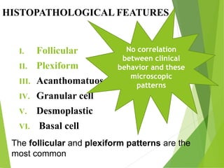
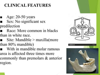
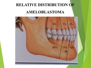
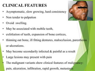
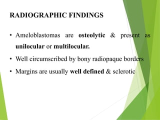
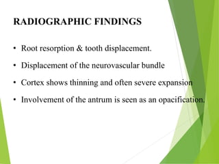
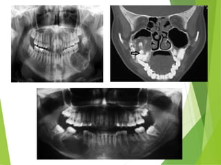
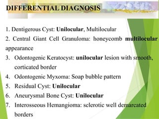
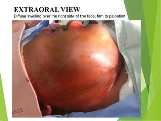
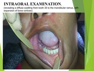
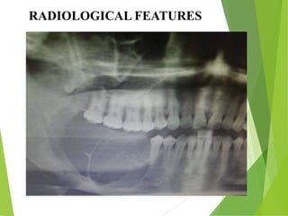
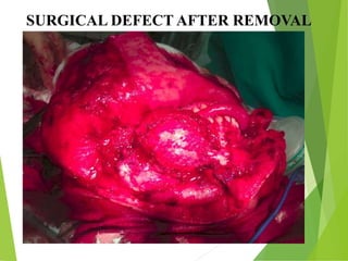
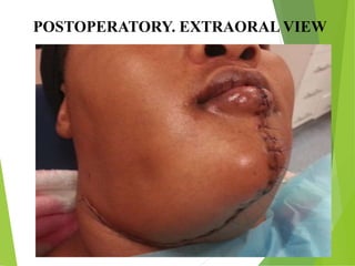
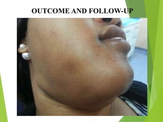
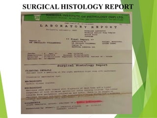
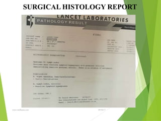
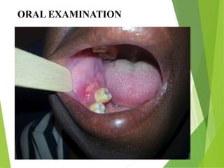
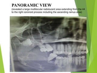
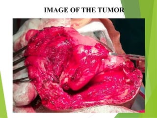
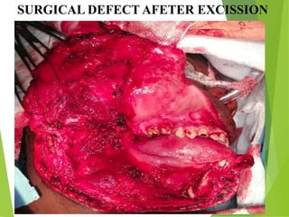
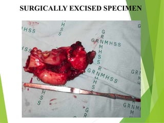
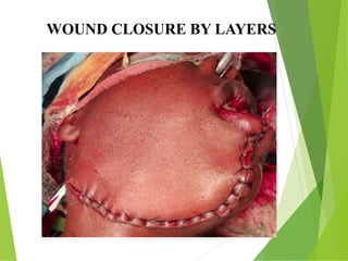
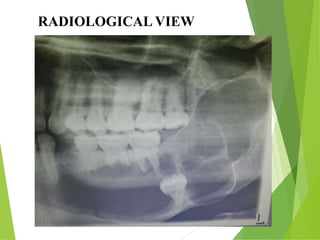
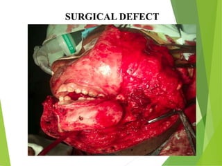
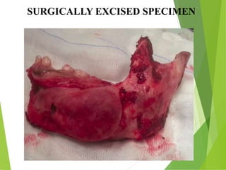
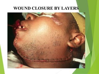
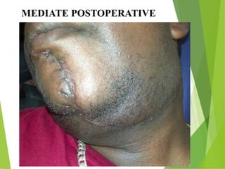
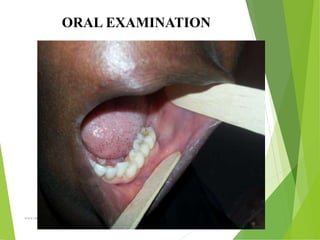
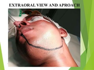
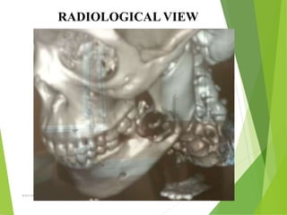
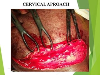
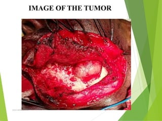
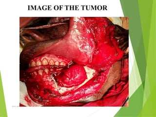
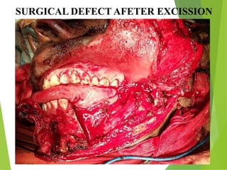
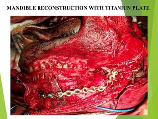
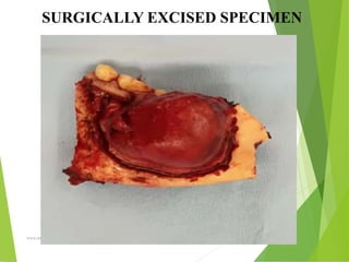
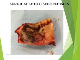
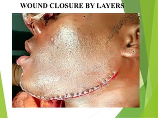
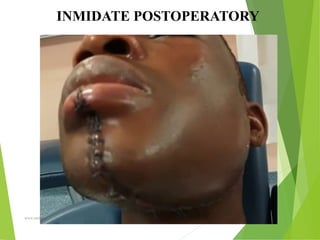
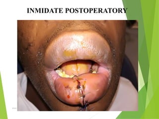
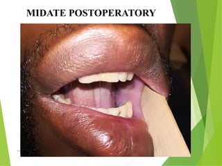
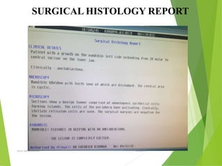
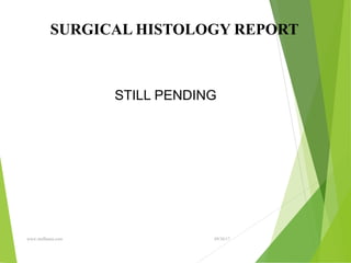
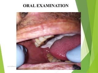
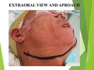
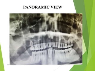
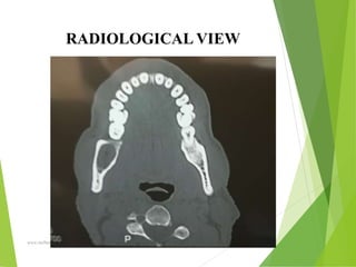
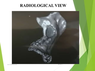
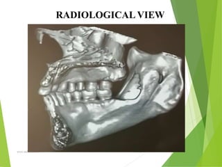
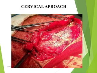
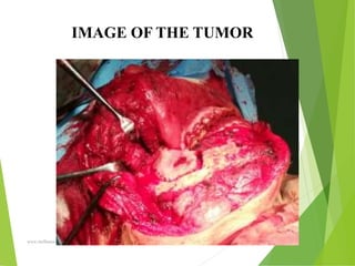
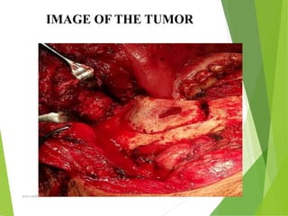
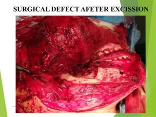
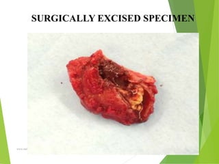
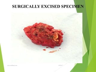
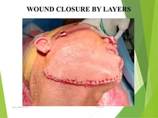
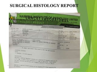
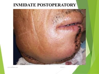
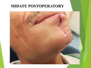
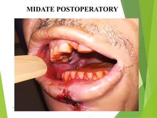
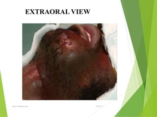
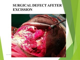
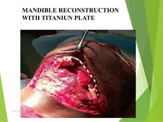
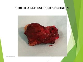
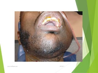
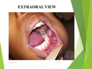
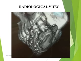
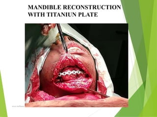
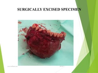
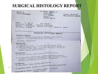
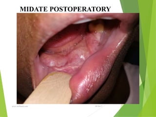
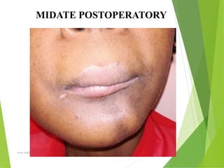
AMELOBLASTOMA. REVIEW OF THE
TUMOR AND CLINICAL CASE
PRESENTATIONS
Authors:
1. Dr. Ernesto Carmona Fernandez
First degree Specialist in Maxillofacial Surgery
Master in Dentistry Urgencies.
OSHAKATI STATE HOSPITAL. NAMIBIA
August 2019
OUTLINE
 Definition
 Epidemiology
 Classifications
 Radiological features
 Management
 Prognosis
 Differential diagnosis
 Conclusions
DEFINITION
• It is a benign tumor of odontogenic origin, derived
from odontogenic epithelium which is locally
invasive, intermittent in growth, clinically persistent
& with a strong tendency to recur.
EPIDERMIOLOGY
• It accounts for 1% of oral tumors & 18% of
odontogenic tumors.
• It is prevalent in Africans has no sex predilection
• Prevalent between 20-50 years
ETIOLOGY
♦♦♦Traumatic episodes: Extraction, cystectomy,
fractures
♦Chronic Infections
♦Dietary deficiency: vitamin D deficiency, Lack of
protein intake
♦Virus infection: HPV
ETIOLOGY
They may arise from:
Rests of dental lamina
A developing Enamel Organ
The epithelial lining of an Odontogenic
Cyst
The basal cells of the oral mucosa
The epithelial cell rests of
Malessez
CLINICAL CLASIFICATION
• Solid Ameloblastoma
• Cystic Ameloblastoma
• Peripheral Ameloblastoma
• Pituitary Ameloblastoma
• Malignant Ameloblastoma
WHO CLASIFICATION
• Solid / Multicystic
• Extraosseous or Peripheral
• Unicystic
• Desmoplastic
HISTOPATHOLOGICAL FEATURES
I. Follicular
II. Plexiform
III. Acanthomatuos
IV. Granular cell
V. Desmoplastic
VI. Basal cell
The follicular and plexiform patterns are the
most common
No correlation
between clinical
behavior and these
microscopic
patterns
■ Age: 20-50 years
■ Sex: No significant sex
predilection
■ Race: More common in blacks
than in white race.
■ Site: Mandible > maxilla(more
than 80% mandible)
■ With in mandible molar ramous
area is affected three times more
commonly than premolars & anterior
region.
CLINICAL FEATURES
RELATIVE DISTRIBUTION OF
AMELOBLASTOMA
CLINICAL FEATURES
• Asymptomatic, slow growing, hard consistency
• Non tender to palpation
• Ovoid swelling.
• May be associated with mobile teeth,
• exfoliation of teeth, expansion of bone cortices,
• thinning out bone, ill fitting dentures, malocclusion, paresthesia
or ulcerations.
• May become secondarily infected & painful as a result
• Large lesions may present with pain
• The malignant variants show clinical features of malignancy:
pain, ulceration, infiltration, rapid growth, metastasis
RADIOGRAPHIC FINDINGS
• Ameloblastomas are osteolytic & present as
unilocular or multilocular.
• Well circumscribed by bony radiopaque borders
• Margins are usually well defined & sclerotic
RADIOGRAPHIC FINDINGS
• Root resorption & tooth displacement.
• Displacement of the neurovascular bundle
• Cortex shows thinning and often severe expansion
• Involvement of the antrum is seen as an opacification.
1. Dentigerous Cyst: Unilocular, Multilocular
2. Central Giant Cell Granuloma: honeycomb multilocular
appearance
3. Odontogenic Keratocyst: unilocular lesion with smooth,
corticated border
4. Odontogenic Myxoma: Soap bubble pattern
5. Residual Cyst: Unilocular
6. Aneurysmal Bone Cyst: Unilocular
7. Interosseous Hemangioma: sclerotic well demarcated
borders
DIFFERENTIAL DIAGNOSIS
MANAGEMENT
• No single standard type of therapy advocated
• Depends on the age & general health of the patient,
the growth pattern of the tumor, the various physical
forms, whether cystic, solid, extraosseous or
malignant.
• Also depends on the duration, anatomical site of the
lesion, clinical size & extent, local behavior, virgin
or already treated before, therapy available and
working conditions.
MANAGEMENT
• Complete eradication of the lesion is required with
reconstruction of the resultant defect.
• Cystic ameloblastoma is treated less aggressively by
enucleation The solid lesions require at least excision
because recurrence follows in 50% to 90% of cases.
• Block excision or resection is reserved for larger
lesions
MANAGEMENT
 Resection could be marginal without continuity
defect, segmental with continuity defect,
hemimandibulectomy with or without
disarticulation, subtotal mandibulectomy & total
mandibulectomy.
 Peripheral ameloblastoma should be treated in a
conservative fashion while malignant lesions should
be managed as carcinomas
Marginal
Segmental
Subtotal Mandibulectomy
Hemimandibulectomy
MANAGEMENT
• The characteristic feature of this tumor is that it
microscopically infiltrates bone beyond the tumor-bone
interface seen in imaging so a safe margin of uninvolved
bone (1.5cm-2cm) should be removed when resecting.
• If complete excision of the tumor is ascertained by clinical
& radiographic examination then immediate
reconstruction can be carried out
MANAGEMENT
• Immediate reconstruction can be done by using autogenous
free bone grafts (Iliac or rib graft) or bank allogenic bone
crib and autogenous bone marrow with a reconstruction plate.
• Reconstruction plate with or without condylar prosthesis can
be used in cases where secondary reconstruction is planned,
where adequate soft tissue coverage is not available or in very
old patients
• In cases where there is insufficient soft tissue locally, a
vascularized composite pedicle graft of bone &
myocutaneous tissue can be used
• It may be extremely
disfiguring ,fungating and
ulcerative like carcinoma
• Invades surrounding tissues
• Bone destruction
• Root resorption
• Asymmetry
• Proptosis
• Severe pain
• Difficulty breathing
• Ill fit dentures
• Loss of teeth
IN THE ABSENCE OF TRETMENT
PROGNOSIS
• Prognosis of ameloblastoma is good as
long as management of the lesion is
carried out appropriately with follow-up
afterwards.
EXTRAORAL VIEW
Diffuse swelling over the right side of the face, firm to palpation
INTRAORAL EXAMINATION.
(revealing a diffuse swelling from tooth 35 to the mandibular ramus, with
expansion of bone cortices)
RADIOLOGICAL FEATURES
SURGICAL DEFECT AFTER REMOVAL
POSTOPERATORY. EXTRAORAL VIEW
OUTCOME AND FOLLOW-UP
SURGICAL HISTOLOGY REPORT
09/30/17
www.stuffamia.com
SURGICAL HISTOLOGY REPORT
09/30/17
www.stuffamia.com
EXTRAORAL VIEW
Diffuse swelling over the right side of the face, firm to palpation
ORAL EXAMINATION
PANORAMIC VIEW
(revealed a large multilocular radiolucent area extending from the 44
to the right coronoid process including the ascending ramus area)
CERVICALAPROACH
IMAGE OF THE TUMOR
SURGICAL DEFECT AFETER EXCISSION
SURGICALLY EXCISED SPECIMEN
WOUND CLOSURE BY LAYERS
SURGICAL HISTOLOGY REPORT
EXTRAORAL EXAMINATION
RADIOLOGICAL VIEW
SURGICAL DEFECT
SURGICALLY EXCISED SPECIMEN
WOUND CLOSURE BY LAYERS
SURGICAL HISTOLOGY REPORT
MEDIATE POSTOPERATIVE
09/30/17
www.stuffamia.com
ORAL EXAMINATION
09/30/17
www.stuffamia.com
EXTRAORAL VIEW AND APROACH
09/30/17
www.stuffamia.com
RADIOLOGICAL VIEW
09/30/17
www.stuffamia.com
CERVICALAPROACH
09/30/17
www.stuffamia.com
IMAGE OF THE TUMOR
09/30/17
www.stuffamia.com
IMAGE OF THE TUMOR
09/30/17
www.stuffamia.com
SURGICAL DEFECT AFETER EXCISSION
09/30/17
www.stuffamia.com
MANDIBLE RECONSTRUCTION WITH TITANIUN PLATE
09/30/17
www.stuffamia.com
SURGICALLY EXCISED SPECIMEN
09/30/17
www.stuffamia.com
SURGICALLY EXCISED SPECIMEN
09/30/17
www.stuffamia.com
WOUND CLOSURE BY LAYERS
09/30/17
www.stuffamia.com
INMIDATE POSTOPERATORY
09/30/17
www.stuffamia.com
INMIDATE POSTOPERATORY
09/30/17
www.stuffamia.com
MIDATE POSTOPERATORY
09/30/17
www.stuffamia.com
MIDATE POSTOPERATORY
09/30/17
www.stuffamia.com
SURGICAL HISTOLOGY REPORT
09/30/17
www.stuffamia.com
SURGICAL HISTOLOGY REPORT
STILL PENDING
09/30/17
www.stuffamia.com
ORAL EXAMINATION
09/30/17
www.stuffamia.com
EXTRAORAL VIEW AND APROACH
09/30/17
www.stuffamia.com
PANORAMIC VIEW
09/30/17
www.stuffamia.com
RADIOLOGICAL VIEW
09/30/17
www.stuffamia.com
RADIOLOGICAL VIEW
09/30/17
www.stuffamia.com
RADIOLOGICAL VIEW
09/30/17
www.stuffamia.com
CERVICALAPROACH
09/30/17
www.stuffamia.com
IMAGE OF THE TUMOR
09/30/17
www.stuffamia.com
IMAGE OF THE TUMOR
09/30/17
www.stuffamia.com
SURGICAL DEFECT AFETER EXCISSION
09/30/17
www.stuffamia.com
SURGICALLY EXCISED SPECIMEN
09/30/17
www.stuffamia.com
SURGICALLY EXCISED SPECIMEN
09/30/17
www.stuffamia.com
WOUND CLOSURE BY LAYERS
09/30/17
www.stuffamia.com
SURGICAL HISTOLOGY REPORT
09/30/17
www.stuffamia.com
INMIDATE POSTOPERATORY
09/30/17
www.stuffamia.com
MIDATE POSTOPERATORY
09/30/17
www.stuffamia.com
MIDATE POSTOPERATORY
09/30/17
www.stuffamia.com
EXTRAORAL VIEW
09/30/17
www.stuffamia.com
SURGICAL DEFECT AFETER
EXCISSION
09/30/17
www.stuffamia.com
MANDIBLE RECONSTRUCTION
WITH TITANIUN PLATE
09/30/17
www.stuffamia.com
SURGICALLY EXCISED SPECIMEN
09/30/17
www.stuffamia.com
SURGICALLY EXCISED SPECIMEN
09/30/17
www.stuffamia.com
09/30/17
www.stuffamia.com
EXTRAORAL VIEW
09/30/17
www.stuffamia.com
RADIOLOGICAL VIEW
09/30/17
www.stuffamia.com
MANDIBLE RECONSTRUCTION
WITH TITANIUN PLATE
09/30/17
www.stuffamia.com
SURGICALLY EXCISED SPECIMEN
09/30/17
www.stuffamia.com
SURGICAL HISTOLOGY REPORT
09/30/17
www.stuffamia.com
MIDATE POSTOPERATORY
09/30/17
www.stuffamia.com
MIDATE POSTOPERATORY
CONCLUSIONS
• Ameloblastoma has a recurrence rate of up to 50%
during the first five years postoperatively and as such
long term follow-up is a must.
• Treatment of these tumors require experience, training
and working conditions to restore proper function and
appearance of the patients.
REFERENCES
• Textbook of oral & maxillofacial surgery (3rd edition)by Neelima
Anil Malik
• Oral Pathology (5th edition) by Regezi, Scuibba & Jordan
• Textbook of oral pathology by Sanjay Saraf
• Burket’s oral medicine, diagnosis & treatment (12th edition)
09/30/17
www.stuffamia.com
““ Imagination is more important
than knowledge, for knowledge
is limited while imagination
embraces the entire world.”
Albert Einstein
THANKS

AMELOBLASTOMA JORNADA.ppt