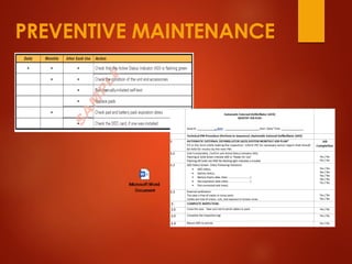
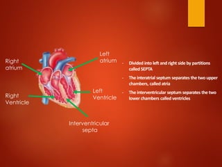
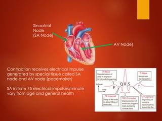
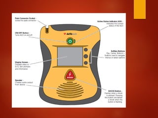
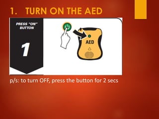
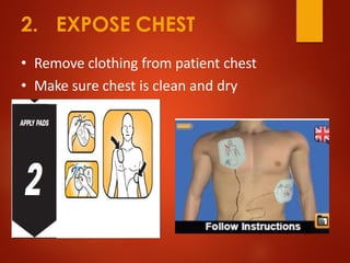
AUTOMATED EXTERNAL
DEFIBRILLATOR
(Defibtech)
AGENDA
01
02
03
04
Anatomy of Heart
Physiology of Heart
AED Instrumentation & PM
How to use AED
ANATOMY OF HEART
- Tricuspid valve located between right atrium and ventricle
- Pulmonary valve located between right ventricle and pulmonary artery (semilunar valve)
- Mitral valve aka Bicuspid valve located between left atrium and ventricle.
- Aortic valve located between left ventricle and aorta
Tricuspid
Pulmonary valve
Bicuspid
Aortic Valve
- Divided into left and right side by partitions
called SEPTA
- The interatrial septum separates the two upper
chambers, called atria
- The interventricular septum separates the two
lower chambers called ventricles
Right
atrium
Right
Ventricle
Interventricular
septa
Left
atrium
Left
Ventricle
PHYSIOLOGY OF HEART
BLOOD CIRCULATION
- Blood circulate via 2 pathways (Pulmonary & Systemic Circulation)
- They both carry DEOXYGENATED and OXYGENATED Blood
Pulmonary Artery Pulmonary Vein
Vena Cava Aorta
Systemic
Circulation
Pulmonary
Circulation
DEOXYGENATED (BLUE)
Pulmonary Artery Pulmonary Vein
OXYGENATED Blood (RED)
Lung
Oxygen from
Lungs RBC Oxygen to
Cells
Cell
- One round of heart contraction (beat) can be divided into;
❑ Relaxation phase – blood flow from atria into ventricles via open atrioventricular valves
❑ Atrial systole – contraction of atria. Pump the rest of blood into ventricles
❑ Ventricular systole – contraction of ventricle. Pump blood into systemic circulation
❑ Ventricular diastole – ventricles and atria relaxed
Sinoatrial
Node
(SA Node)
AV Node)
Contraction receives electrical impulse
generated by special tissue called SA
node and AV node (pacemaker)
SA initiate 75 electrical impulses/minute
vary from age and general health
- Due to medical condition & disease, heart contraction/beat become irregular, lowers in intensity or
even stop require troubleshoot
- The troubleshooting exercise require a device called Defibrillating Device or Automated Defibrillation
Device (AED)
INSTRUMENTATION
PREVENTIVE MAINTENANCE
HOW TO USE AED
1. TURN ON THE AED
p/s: to turn OFF, press the button for 2 secs
2. EXPOSE CHEST
• Remove clothing from patient chest
• Make sure chest is clean and dry
3. TOATTACH PADS
• Tear open the pads from packet
• Peel adhesive pads from blue liner
3A. ATTACH PADS
1. One pad goes on the upper right chest below
collarbone.
2. One pad goes on lower left side chest wall.
3. For Kid, put at front & back of the kid
3A.AED AND PACEMAKER
• Pacemakers are located under the collarbone
on right or left side of chest
• If located on right side place AED pad 1 inch
below pacemaker
• Continue with AED prompts
3A.AED AND BRAS
• Many bras contain metal (Clips, underwire)
• Remove / cut bra if AED
pads will touch the metal
3A.AED AND JEWELRY
• Ignore jewelry if it will not touch AED pad
• If in the way push jewelry to side of neck
away from AED pads.
• Do not waste time
removing jewelry
3A.AED AND WET PERSON
• Dry off victims chest (and back if a child under
age 8.)
• Attach pads following manufacturer directions
• The rest of the victim does not need to be dry.
• AEDs can be used on wet surfaces and snow
so long as patient is dry and not in a puddle of
water.
4. STAY CLEAR
• Make sure nobody , including yourself, is
touching the victim.
Say “Everyone stand clear!”
5. DELIVER SHOCK
If AED Prompts a Shock
• Make sure no one is
touching victim
• Push the “shock” button
6. AFTER SHOCK
• Do not turn off AED….
• It is now safe to touch Pads and patient
7. PERFORM CPR
• After shock, or if no shock is advised,
continue CPR.
• Every 2 minutes follow the AED prompts
• Stop CPR if you see signs of life
– (breathing, movement.)
THE END

AED Awareness.pdf

  • 1.
  • 2.
    AGENDA 01 02 03 04 Anatomy of Heart Physiologyof Heart AED Instrumentation & PM How to use AED
  • 3.
  • 4.
    - Tricuspid valvelocated between right atrium and ventricle - Pulmonary valve located between right ventricle and pulmonary artery (semilunar valve) - Mitral valve aka Bicuspid valve located between left atrium and ventricle. - Aortic valve located between left ventricle and aorta Tricuspid Pulmonary valve Bicuspid Aortic Valve
  • 5.
    - Divided intoleft and right side by partitions called SEPTA - The interatrial septum separates the two upper chambers, called atria - The interventricular septum separates the two lower chambers called ventricles Right atrium Right Ventricle Interventricular septa Left atrium Left Ventricle
  • 6.
  • 7.
    BLOOD CIRCULATION - Bloodcirculate via 2 pathways (Pulmonary & Systemic Circulation) - They both carry DEOXYGENATED and OXYGENATED Blood Pulmonary Artery Pulmonary Vein Vena Cava Aorta Systemic Circulation Pulmonary Circulation
  • 8.
    DEOXYGENATED (BLUE) Pulmonary ArteryPulmonary Vein OXYGENATED Blood (RED) Lung Oxygen from Lungs RBC Oxygen to Cells Cell
  • 9.
    - One roundof heart contraction (beat) can be divided into; ❑ Relaxation phase – blood flow from atria into ventricles via open atrioventricular valves ❑ Atrial systole – contraction of atria. Pump the rest of blood into ventricles ❑ Ventricular systole – contraction of ventricle. Pump blood into systemic circulation ❑ Ventricular diastole – ventricles and atria relaxed
  • 10.
    Sinoatrial Node (SA Node) AV Node) Contractionreceives electrical impulse generated by special tissue called SA node and AV node (pacemaker) SA initiate 75 electrical impulses/minute vary from age and general health
  • 11.
    - Due tomedical condition & disease, heart contraction/beat become irregular, lowers in intensity or even stop require troubleshoot - The troubleshooting exercise require a device called Defibrillating Device or Automated Defibrillation Device (AED)
  • 12.
  • 17.
  • 18.
  • 20.
    1. TURN ONTHE AED p/s: to turn OFF, press the button for 2 secs
  • 21.
    2. EXPOSE CHEST •Remove clothing from patient chest • Make sure chest is clean and dry
  • 22.
    3. TOATTACH PADS •Tear open the pads from packet • Peel adhesive pads from blue liner
  • 23.
    3A. ATTACH PADS 1.One pad goes on the upper right chest below collarbone. 2. One pad goes on lower left side chest wall. 3. For Kid, put at front & back of the kid
  • 24.
    3A.AED AND PACEMAKER •Pacemakers are located under the collarbone on right or left side of chest • If located on right side place AED pad 1 inch below pacemaker • Continue with AED prompts
  • 25.
    3A.AED AND BRAS •Many bras contain metal (Clips, underwire) • Remove / cut bra if AED pads will touch the metal
  • 26.
    3A.AED AND JEWELRY •Ignore jewelry if it will not touch AED pad • If in the way push jewelry to side of neck away from AED pads. • Do not waste time removing jewelry
  • 27.
    3A.AED AND WETPERSON • Dry off victims chest (and back if a child under age 8.) • Attach pads following manufacturer directions • The rest of the victim does not need to be dry. • AEDs can be used on wet surfaces and snow so long as patient is dry and not in a puddle of water.
  • 28.
    4. STAY CLEAR •Make sure nobody , including yourself, is touching the victim. Say “Everyone stand clear!”
  • 29.
    5. DELIVER SHOCK IfAED Prompts a Shock • Make sure no one is touching victim • Push the “shock” button
  • 30.
    6. AFTER SHOCK •Do not turn off AED…. • It is now safe to touch Pads and patient
  • 31.
    7. PERFORM CPR •After shock, or if no shock is advised, continue CPR. • Every 2 minutes follow the AED prompts • Stop CPR if you see signs of life – (breathing, movement.)
  • 32.