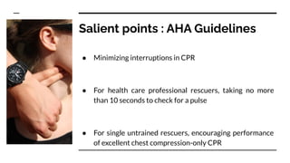
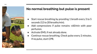
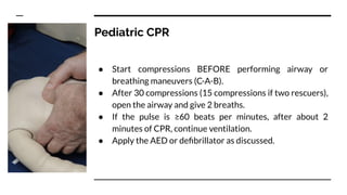
For medical students, pg residents, nursing students, and other medical professionals. Contains medical jargon and advice, not for use by general public. For use only under a trained instructor.
This document provides an overview of cardiac monitoring techniques including stethoscopy, electrocardiography, pulse rate monitoring, arterial blood pressure monitoring, central venous pressure monitoring, and pulmonary artery catheterization. Key points include: stethoscopy was introduced in 1818 but is not used for continuous monitoring; electrocardiography is the most common method to detect heart rate; differences exist between heart rate and pulse rate; noninvasive and invasive blood pressure monitoring methods are described along with their complications; central venous pressure monitoring provides information on right atrial pressure; and pulmonary artery catheters allow direct measurement of pressures and cardiac output in critically ill patients.
This document summarizes the 10th edition updates to the Advanced Trauma Life Support (ATLS) guidelines. It outlines the initial assessment process including the primary and secondary surveys. It provides updates to various body system-specific guidelines including new recommendations for airway management, shock classification and treatment, thoracic trauma management including tension pneumothorax, head trauma management including blood pressure targets, and spinal motion restriction. It also summarizes pediatric-specific guidelines including fluid resuscitation amounts and head CT criteria. Transfer communication is emphasized including avoiding unnecessary tests and using an ABC-SBAR template.
The initial assessment of a trauma patient involves a primary survey consisting of a rapid assessment of the airway, breathing, circulation, disability, and exposure (ABCDE). For a 34-year-old male brought to the emergency room after a road traffic accident with hoarseness, low blood pressure and rapid heart rate and breathing, the primary steps would be to open and secure the airway, assess breathing for tension pneumothorax, control bleeding, check neurological status, and fully expose the patient for further examination and resuscitation efforts. A secondary survey would then obtain a full history and examine all body regions for potential injuries.
CARDIOPULMONARY RESUSCITATION- BLS & ACLS-2020 AHA UPDATEAryaDasmahapatra
This document provides information about cardiopulmonary resuscitation (CPR) and the basic life support (BLS) and advanced cardiac life support (ACLS) protocols. It begins with definitions of CPR and its purposes to support life through circulation and prevent brain damage from lack of oxygen. The history of developments in CPR techniques from chest compressions to defibrillation are outlined. Adult and pediatric BLS protocols are described, including assessing responsiveness, calling for help, performing high-quality chest compressions, opening the airway, rescue breathing, and using an automated external defibrillator. Differences in CPR for adults, children and infants are also summarized.
This document discusses pulmonary artery pressure monitoring using a pulmonary artery catheter. It describes how Swan-Ganz catheters are inserted into the pulmonary artery to measure pressures. The document outlines the normal values of pressures in the heart and lungs. It also discusses how the catheter is used to monitor cardiac output through thermodilution and continuous cardiac output methods. Potential complications of the procedure are mentioned.
The document outlines the importance of post resuscitation care to stabilize patients after return of spontaneous circulation, including supporting oxygenation and circulation, treating injuries from CPR, monitoring for recurrent cardiac arrest, and promptly transferring patients to intensive care units for specialized monitoring and treatment. Proper post resuscitation care is critical to optimize outcomes in the hours after resuscitation.
Central venous catheterization and venous cut down techniques were presented. Central venous catheterization involves placing lines into large neck, chest, or groin veins and should only be done aseptically in operating rooms or high dependency units. It has indications for monitoring, infusing irritant drugs, pacing, dialysis, and emergencies. Sites include the subclavian, internal jugular, and femoral veins, each with advantages and disadvantages. Ultrasound guidance is becoming standard. Complications include infections, arterial puncture, and pneumothorax. Venous cut down is an open surgical technique to access veins and remains useful when other methods fail or are unavailable.
A presentation used to train medical professionals to perform BLS in emergency condition. it will provide a better understanding about the steps of BLS and the order in which it should be perfomed.
INITIAL ASSESSMENT OF TRAUMA PATIENTS....(INSPIRED FROM CTLS AND ATLS GUIDELI...Prerna Biswal
THIS PRESENTATION WAS MADE AT IMA HOUSE IN BHUBANESWAR,ODISHA, BY DR.NIBEDITA PANI,HOD ,DEPT. OF ANAESTHESIOLOGY AND DR.PRERNA BISWAL,PG,ANAESTHESIOLOGY,SCBMCH,CUTTACK,
Triage is the process of prioritizing patients according to the urgency of their need for care. It aims to ensure patients are treated in order of clinical urgency and receive timely care. There are three main types of triage - primary triage in the field, secondary triage in the emergency department, and tertiary triage by specialists. The START and SAVE methods are used for disaster triage in the field to categorize patients into immediate, delayed, or minimal care/expectant groups. In the ED, patients are assigned colors based on their condition - red for most urgent, yellow intermediate, green less urgent, and black for deceased. Documentation, equipment, and designated triage teams are needed to properly conduct triage
Initial assessment and management of traumaVASS Yukon
The document provides guidance on the initial assessment and management of trauma patients. It emphasizes that the golden hour refers to time to definitive treatment, not time in transport or the ED. For critical patients, EMS has just 10 minutes to provide lifesaving interventions. The primary survey involves assessing the patient's airway, breathing, circulation, disability and exposure to identify and treat life threats. Oxygenation and control of hemorrhage are top priorities. Only after life threats are addressed should providers begin the more detailed secondary exam and assessment. Rapid transport to the appropriate facility is critical for trauma patients.
Cardiopulmonary resuscitation (CPR) involves three key steps:
1) Assessment of the collapsed victim to determine unresponsiveness and activate emergency services.
2) Performance of chest compressions at a rate of 100-120 per minute and depth of 5-6 cm, allowing full chest recoil between compressions.
3) Use of an automated external defibrillator (AED) as soon as it is available to analyze the heart rhythm and deliver a shock if needed.
Comprehensive presentation on intra arterial blood pressure with a good insight into the the basic physics and brief look into the risks and complications.
Basic life support (BLS) involves procedures to restore oxygenated blood circulation after sudden cardiac or pulmonary arrest until full medical care can be provided. It includes chest compressions, rescue breathing, use of an automated external defibrillator, and establishing an open airway. BLS is essential for resuscitating someone and can double their chances of survival if performed immediately by bystanders before emergency services arrive. The key steps of BLS include assessing the scene and victim, calling for help, delivering chest compressions, giving rescue breaths, using an AED, and placing the victim in the recovery position if breathing returns.
This document provides information on assessing and managing critically ill patients using the ABCDE approach. It discusses:
- Common early signs of critical illness including hypoxia and hypotension.
- The ABCDE approach which prioritizes establishing a patient's airway, breathing, circulation, disability level and exposure for examination.
- Techniques for assessing and intervening on airway, breathing and circulation issues including providing oxygen, treating underlying causes, and starting IV fluids.
- The importance of continuous reassessment, calling for help early, and following basic life support protocols when indicated to stabilize critically ill patients.
Mechanical ventilation is used widely in patient care from initial injury through hospital transport, surgery, intensive care, and intermediate care. Various modes of ventilation have been developed to support patient breathing including controlled mandatory modes like CMV that do not allow spontaneous breathing and supported modes like PSV that augment patient effort. Key parameters monitored include pressures, volumes, and gas exchange. Complications can include barotrauma, decreased cardiac output, and pneumonia. Weaning protocols gradually reduce ventilator support as the underlying condition improves and respiratory function is adequate.
The document outlines an educational program on cardiac arrest for 4th year nursing students. The objectives are to review cardiac arrest, basic life support, advanced life support, and demonstrate skills like basic life support, airway insertion, and defibrillation. The program agenda includes topics on cardiac arrest, basic and advanced life support, defibrillation, drugs used in advanced life support, and demonstrations of skills. It provides details on cardiac arrest, basic life support procedures like chest compressions and rescue breathing, and advanced life support including defibrillation and drugs.
This document outlines the general approach and concepts for treating traumatic patients according to Advanced Trauma Life Support (ATLS) guidelines. It describes treating the greatest threats to life first using the ABCDE approach to assess the airway, breathing, circulation, disability, and exposure. The primary survey involves rapid assessment and interventions to stabilize the patient, while the secondary survey entails a full physical exam and diagnostic testing. Key interventions discussed include intubation, chest tube insertion, hemorrhage control, and use of the Focused Assessment with Sonography for Trauma (FAST) exam to evaluate for internal bleeding. Definitive care may involve transfer to the operating room or intensive care unit based on specialty consultations.
Basic life support (BLS) refers to emergency care provided to patients experiencing cardiac arrest, respiratory failure, or airway obstruction. It includes chest compressions, use of an automated external defibrillator, and relieving airway obstructions. The chain of survival emphasizes early CPR, early defibrillation, early advanced life support, and post-cardiac arrest care to maximize patient survival. BLS procedures include assessing the patient for responsiveness, activating emergency services, performing high-quality chest compressions, opening the airway, and providing rescue breaths. Defibrillation is key for shockable cardiac rhythms like ventricular fibrillation. BLS aims to provide oxygenated blood flow to vital organs until further medical help
This is a very simple presentation prepared for nurses. It will help nurses to understand the need of monitoring and the available methods. The presentation has been constructed on a clinical case base scenario and gradually different methods of monitoring has been introduced.
1. The document discusses the history and techniques of intracranial pressure (ICP) monitoring. It describes historical figures who contributed to the understanding of ICP and various monitoring methods that have been developed over time.
2. The current gold standard for ICP monitoring is an external ventricular drain, though fiberoptic and strain gauge monitors provide alternatives. Newer methods like optic nerve sheath ultrasound provide noninvasive options.
3. Careful analysis of ICP waveforms can provide insights into intracranial compliance and dynamics that help guide management of conditions with elevated ICP like traumatic brain injury.
The document provides information on basic life support (BLS) including definitions, the adult chain of survival, call or CPR first considerations, signs requiring CPR, approaching a victim, and high quality CPR techniques. It discusses refining the recognition of cardiac arrest and initiation of CPR or calling emergency services. Emphasis is placed on minimizing interruptions during chest compressions and avoiding excessive ventilation.
Fluid responsiveness in critically ill patientsUbaidur Rahaman
This document discusses fluid responsiveness in critically ill patients. It begins by defining fluid responsiveness as an increase in cardiac index after fluid infusion. It then describes three scenarios involving fluid resuscitation: patients with acute blood or fluid loss requiring immediate resuscitation, patients with suspected septic shock where early goal-directed therapy with fluids is important, and ICU patients who have already received fluids and their fluid responsiveness needs to be assessed. The document discusses various methods of assessing preload and preload dependence, including filling pressures, volumes, respiratory variations in inferior vena cava diameter, inspiratory drops in right atrial pressure, and predicting responsiveness through respiratory variations in parameters related to cardiac index. It emphasizes that preload alone
The document discusses sedation, analgesia, and paralysis in the ICU. It describes the goals of sedation as patient comfort while allowing interaction. The challenges include assessing sedation and altered drug pharmacology. An ideal sedation agent would have rapid onset and offset and lack respiratory depression. Monitoring scales like the Richmond Agitation Scale are used to standardize treatment. Dexmedetomidine, propofol, opioids and paralytics may be used. The optimal sedation approach balances adequate treatment while avoiding oversedation risks.
The document outlines guidelines for Basic Life Support (BLS) and Advanced Cardiovascular Life Support (ACLS) presented by interns at KIMS, BBSR. It discusses BLS guidelines including CPR technique and choking, and provides ACLS algorithms. Key aspects of BLS covered are assessing carotid pulse, initiating chest compressions if no pulse, and reassessing after 2 minutes of CPR. ACLS algorithms outlined include those for adult cardiac arrest, post-cardiac arrest care, tachycardia, and bradycardia. Identifying unstable patients using HASIA criteria is also summarized.
The Advanced Trauma Life Support (ATLS) system was created in the United States in 1976 after an orthopedic surgeon crashed his plane and found the emergency care for his critically injured children to be inadequate. ATLS provides a standardized approach to assessing and treating trauma patients, including maintaining the airway, breathing, and circulation during the primary survey to address life-threatening issues first before conducting a full secondary survey. Over 50 countries now provide the ATLS course to physicians to improve trauma care worldwide.
This document summarizes guidelines for cardiopulmonary resuscitation (CPR) from 2010. It discusses the history and development of CPR techniques from the 1960s onward. Key points covered include recommendations for chest compressions-only CPR by lay rescuers, a compression-to-ventilation ratio of 30:2, minimizing interruptions in chest compressions, and initial defibrillation energies of 150-200 joules for biphasic defibrillators. The guidelines provide evidence-based recommendations to optimize survival from cardiac arrest.
Cardiopulmonary Cerebral Resuscitation (CPCR) for Veterinary Techniciansupstatevet
Cardiopulmonary Cerebral Resuscitation (CPCR) involves providing artificial respiration and circulation to an animal that is not breathing and has no heartbeat. The document discusses CPCR outcomes, who is at risk, basic life support techniques like chest compressions and airway management, advanced techniques like drug administration and defibrillation, and the RECOVER initiative to improve resuscitation practices. Successful CPCR requires staff preparedness through training, having necessary equipment and supplies readily available, and effective teamwork during a resuscitation attempt.
Cardiopulmonary resuscitation (CPR) involves giving chest compressions and ventilations to patients whose breathing or heartbeat has stopped. CPR is commonly performed during cardiac arrest to circulate oxygenated blood to vital organs until spontaneous circulation can be restored. It is important to perform high-quality CPR with chest compressions that are fast, deep, and fully recoil between compressions. When possible, two rescuers should perform CPR to minimize interruptions in chest compressions. Drugs like epinephrine may be administered during CPR to increase heart rate and blood pressure.
This document summarizes a presentation on basic and advanced cardiac life support. It discusses key concepts in BLS including recognition of cardiac arrest, activating emergency services, performing chest compressions, minimizing interruptions, monitoring compression quality, ventilation, and use of an automated external defibrillator. It then covers ACLS, including treatment algorithms, airway management, defibrillation procedures, medications used during CPR, monitoring techniques, and management of specific arrhythmias like ventricular fibrillation, asystole, and pulseless electrical activity. The goal of BLS and ACLS is to provide immediate life-saving interventions for cardiac arrest patients until the underlying cause can be addressed.
BLS(basic life support) & ACLS with PALS by Dr. ShailendraShailendra Satpute
This document provides information on Basic Life Support (BLS) and Advanced Cardiac Life Support (ACLS). It defines cardiac arrest, outlines its causes and types including ventricular fibrillation, pulseless ventricular tachycardia, asystole, and pulseless electrical activity. It describes the signs and symptoms of cardiac arrest. It also summarizes the steps of BLS including chest compressions, airway management, rescue breathing, and defibrillation. Advanced techniques like intubation, use of laryngeal mask airways, endotracheal tubes, and automated external defibrillators are also outlined.
Cardio pulmonary resuscitation (CPR) involves basic life support techniques to maintain oxygenation to the heart, lungs and brain during cardiac or respiratory arrest. It includes chest compressions, opening the airway, and rescue breathing. The goals of CPR are to restore spontaneous circulation and breathing to prevent irreversible brain injury. It consists of early recognition of arrest, high quality chest compressions, airway management, rescue breathing, defibrillation if needed, and administration of drugs to support circulation.
The document discusses cardiopulmonary resuscitation (CPR) and emergency cardiac care procedures. It defines CPR, basic life support (BLS), advanced cardiac life support (ACLS), and the indications and contraindications for their use. Key aspects of BLS covered include chest compressions, airway management, breathing, and defibrillation. Commonly used medications in ACLS like epinephrine, atropine, and amiodarone are also outlined. The document provides details on the steps of BLS, complications to watch for, and a nurse's responsibilities in caring for a patient after resuscitation.
The document provides guidelines for performing basic life support, including checking for response, calling for help, opening the airway, checking for breathing, performing 30 chest compressions followed by 2 rescue breaths, and continuing cycles of compressions and breaths until emergency services arrive or the victim starts breathing on their own. Early CPR and defibrillation are critical for survival from cardiac arrest, with survival rates declining rapidly without intervention in the first few minutes. Bystander CPR can double or triple a victim's chance of survival.
This document provides information on cardiopulmonary resuscitation (CPR), including its definition, purpose, procedures, guidelines, and the roles of nurses. CPR is an emergency procedure performed when a person is unresponsive and not breathing or has abnormal breathing to manually maintain heart function until further measures can restore spontaneous circulation and breathing. It involves chest compressions, airway management, and rescue breathing in a cycle according to guidelines. Nurses play an important role in initiating and assisting with CPR, managing airways, monitoring the patient, and documenting the procedures. Knowledge of CPR techniques and guidelines is essential for nurses to be able to effectively resuscitate patients in cardiac or respiratory arrest.
This document discusses basic and advanced cardiovascular life support. It defines cardiac arrest as the sudden cessation of heart function and outlines the management of out-of-hospital and in-hospital cardiac arrest. For out-of-hospital cardiac arrest, it describes the steps of basic cardiopulmonary life support including calling for help, checking response, pulse and breathing, performing chest compressions and rescue breathing, using an automated external defibrillator, and positioning the victim for recovery. For in-hospital cardiac arrest, it outlines the steps of comprehensive cardiopulmonary life support including establishing intravenous access, airway management, medication administration, and treating reversible causes of arrest.
CPR involves basic techniques to manually support breathing and circulation until further medical help arrives. It aims to restore oxygenated blood flow to vital organs. The key steps are: [1] early recognition of cardiac or respiratory arrest; [2] beginning chest compressions immediately at a rate of 100 per minute; [3] giving rescue breaths if no signs of breathing. Advanced techniques use equipment like defibrillators, ventilators and drugs to further support breathing and circulation."
CPR is a process of oxygenating heart, lung through external cardiac massage and artificial respiration until the definite medical treatment can restore the normal functioning of heart, lung and brain.
Advanced cardiac life support (ACLS) involves techniques used to support or restore cardiac function and breathing in a person experiencing cardiac arrest, respiratory failure, or circulatory failure. The summary provides an overview of key aspects of ACLS including:
1. CPR techniques including chest compressions at a rate of 100-120 per minute and depth of at least 2 inches for adults. Early initiation of CPR and defibrillation are emphasized.
2. Advanced life support techniques including tracheal intubation, defibrillation, intravenous access, and medications like epinephrine, atropine, amiodarone to treat shockable and non-shockable cardiac rhythms.
3. Post-cardiac
Basic Life Support (BLS) refers to emergency care including CPR, use of an AED, and clearing obstructed airways. BLS aims to maintain circulation and breathing until emergency help arrives. It involves assessing for responsiveness, calling for help, providing chest compressions at 100-120 per minute and rescue breaths in a 30:2 ratio. An AED should be used as soon as available to analyze rhythms and deliver shocks if indicated. CPR techniques differ between adults and children, such as using two fingers to compress the chest of an infant or child. Foreign object airway obstructions are managed through back blows, chest thrusts and abdominal thrusts depending on responsiveness and age of the victim.
This document provides information on cardiac arrest, including objectives, case presentations, definitions of terms, and treatment algorithms. It discusses how to recognize cardiac arrest, perform CPR, differentiate shockable and non-shockable rhythms, understand key drugs, and review case examples. Advanced life support is described as well, with a focus on defibrillation, cardioversion, pacing, medications, intubation, and IV access to restore spontaneous circulation. The document emphasizes the importance of high-quality, continuous chest compressions in cardiac arrest resuscitation.
This document provides information on cardiac arrest, including objectives, case presentations, and details on cardiopulmonary resuscitation (CPR). It discusses recognizing cardiac arrest, performing chest compressions and rescue breathing, differentiating shockable and non-shockable rhythms, principal drugs used, and the ABCDE approach for assessing collapsed patients in a hospital setting. Case presentations provide scenarios to test understanding of sequential response and best interventions for pulseless patients.
Neonatal resuscitation also known as newborn resuscitation is an emergency procedure focused on supporting the approximately 10% of newborn children who do not readily begin breathing, putting them at risk of irreversible organ injury and death.
Similar to Advanced Trauma Life Support : Part 1 - Basic Life Support (20)
Basavarajeeyam is a Sreshta Sangraha grantha (Compiled book ), written by Neelkanta kotturu Basavaraja Virachita. It contains 25 Prakaranas, First 24 Chapters related to Rogas& 25th to Rasadravyas.
TEST BANK For Basic and Clinical Pharmacology, 14th Edition by Bertram G. Kat...rightmanforbloodline
TEST BANK For Basic and Clinical Pharmacology, 14th Edition by Bertram G. Katzung, Verified Chapters 1 - 66, Complete Newest Version.
Muktapishti is a traditional Ayurvedic preparation made from Shoditha Mukta (Purified Pearl), is believed to help regulate thyroid function and reduce symptoms of hyperthyroidism due to its cooling and balancing properties. Clinical evidence on its efficacy remains limited, necessitating further research to validate its therapeutic benefits.
Here is the updated list of Top Best Ayurvedic medicine for Gas and Indigestion and those are Gas-O-Go Syp for Dyspepsia | Lavizyme Syrup for Acidity | Yumzyme Hepatoprotective Capsules etc
2. Approach to Initial Assesment
● Preparation
● Triage
● Primary survey (ABCDEs) with immediate
● Adjuncts to the primary survey and resuscitation
● Consideration of the need for patient transfer
● Secondary survey (head-to-toe evaluation)
● Adjuncts to the secondary survey
● Continued postresuscitation monitoring/reevaluation
● Definitive care
6. Clinicians can quickly assess A, B, C, and D
in a trauma patient (10-second
assessment) by identifying themselves,
asking the patient for his or her name, and
asking what happened.
7. Primary Survey : Airway
● Establish a definitive airway if there is any doubt about
the patient’s ability to maintain airway integrity.
● While assessing and managing a patient’s airway, take
great care to prevent excessive movement of the cervical
spine. Based on the mechanism of trauma, assume that a
spinal injury exists.
Cervical spine motion restriction technique
8. Primary Survey : Airway
● To adequately assess jugular venous distention, position
of the trachea, and chest wall excursion, expose the
patient’s neck and chest.
● Perform auscultation to ensure gas flow in the lungs.
● Visual inspection and palpation can detect injuries to the
chest wall that may be compromising ventilation.
● Percussion of the thorax can also identify abnormalities,
but during a noisy resuscitation this evaluation may be
inaccurate.
9. A simple pneumothorax can be converted
to a tension pneumothorax when a patient
is intubated and positive pressure
ventilation is provided before
decompressing the pneumothorax with a
chest tube.
10. Primary Survey : Circulation
● The elements of clinical observation that yield important
information within seconds are
○ level of consciousness
○ skin perfusion
○ pulse
● Definitive bleeding control is essential, along with
appropriate replacement of intravascular volume
● Aggressive and continued volume resuscitation is not a
substitute for definitive control of hemorrhage.
11. Fluids are administered judiciously, as aggressive
resuscitation before control of bleeding has been
demonstrated to increase mortality and morbidity.
12. Hypothermia
Hypothermia can be present when the patient arrives, or it
may develop quickly in the ED if the patient is uncovered and
undergoes rapid administration of room-temperature fluids
or refrigerated blood. Because hypothermia is a potentially
lethal complication in injured patients, take aggressive
measures to prevent the loss of body heat and restore body
temperature.
14. Physiologic parameters such as pulse rate, blood
pressure, pulse pressure, ventilatory rate, ABG levels,
body temperature, and urinary output are assessable
measures that reflect the adequacy of resuscitation.
Values for these parameters should be obtained as
soon as is practical during or after completing the
primary survey, and reevaluated periodically
15. Adjuncts to Primary Survey
Chest and Aelvis AP radiography can provide valuable
information to aid resuscitation efforts.
FAST, eFAST, and DPL are useful tools for quick detection of
intraabdominal blood, pneumothorax, and hemothorax
16. Secondary Survey
The secondary survey does not begin until the primary survey
(ABCDE) is completed, resuscitative efforts are underway,
and improvement of the patient’s vital functions has been
demonstrated. It includes
● History
● Physical Examination
● Systemic evaluation
17. Adjuncts to Secondary Survey
Specialized diagnostic tests may be performed
● additional x-ray examinations of spine and extremities
● CT scans of the head,chest, abdomen, and spine;
● contrast urographyand angiography
● transesophageal ultrasound
● bronchoscopy
● esophagoscopy
21. 1957
External defibrillation
first described by
Kouwenhoven
1958
Elam and Safar
described the
technique of mouth to
mouth ventilation
1960
Kouwenhoven,
Knickerbocker, and
Jude described the
benefits of external
chest compressions
22. 1957
External defibrillation
first described by
Kouwenhoven
1958
Elam and Safar
described the
technique of mouth to
mouth ventilation
1960
Kouwenhoven,
Knickerbocker, and
Jude described the
benefits of external
chest compressions
1963
American Heart
Association formally
endorsed CPR
23. 1957
External defibrillation
first described by
Kouwenhoven
1958
Elam and Safar
described the
technique of mouth to
mouth ventilation
1960
Kouwenhoven,
Knickerbocker, and
Jude described the
benefits of external
chest compressions
1963
American Heart
Association formally
endorsed CPR
1966
Standardized AHA
guidelines for CPR to
lay rescuers
24. Random Fact
*Abella BS, Alvarado JP, Myklebust H, et al. Quality of
cardiopulmonary resuscitation during in-hospital cardiac arrest.
JAMA 2005; 293:305.
*Wik L, Kramer-Johansen J, Myklebust H, et al. Quality of
cardiopulmonary resuscitation during out-of-hospital cardiac arrest.
JAMA 2005; 293:299.
“Multiple studies assessing both
in-hospital and prehospital
performance of CPR have shown
that trained health care providers
consistently fail to meet basic life
support guidelines..”*
26. What is BLS ?
Basic life support consists of
cardiopulmonary resuscitation and,
when available, defibrillation using
automated external defibrillators
(AED)
30. Initially pulmonary alveoli are likely
to contain adequate levels of oxygen
and the pulmonary vessels and heart
likely contain sufficient oxygenated
blood to meet markedly reduced
demands, the importance of
compressions thus supersedes
ventilations.
Why C-A-B ?
(It’s Compression-Airway-Breathing….
But u already knew that)
31. Phases of Cardiac Arrest
Electrical
First 4 to 5 minutes
Immediate DC
cardioversion and
excellent chest
compressions are
required . Highest chance
of survival.
32. Phases of Cardiac Arrest
Electrical
First 4 to 5 minutes
Immediate DC
cardioversion and
excellent chest
compressions are
required . Highest chance
of survival.
Hemodynamic
From 4 to 10 minutes
Excellent chest
compressions continued
until just before
cardioversion. Resume
CPR immediately after
shock.
33. Phases of Cardiac Arrest
Electrical
First 4 to 5 minutes
Immediate DC
cardioversion and
excellent chest
compressions are
required . Highest chance
of survival.
Hemodynamic
From 4 to 10 minutes
Excellent chest
compressions continued
until just before
cardioversion. Resume
CPR immediately after
shock.
Metabolic
10 minutes without pulse
Post resuscitative
measures including
hypothermia therapy.
Do not usually survive if
not quickly converted
into a perfusing rhythm.
34. Is it beneficial during the hemodynamic
phase to delay defibrillation in order to
perform 2 to 3 minutes of CPR ?
35. Salient points : AHA Guidelines
● Immediate recognition of sudden cardiac arrest (SCA) by
noting unresponsiveness or absent/gasping breathing.
● Immediate initiation of excellent CPR – "push hard, push
fast" (but not too hard nor too fast) – with continuous
attention to the quality of chest compressions, and to the
frequency of ventilations.
36. Salient points : AHA Guidelines
● Minimizing interruptions in CPR
● For health care professional rescuers, taking no more
than 10 seconds to check for a pulse
● For single untrained rescuers, encouraging performance
of excellent chest compression-only CPR
37. Salient points : AHA Guidelines
● Using automated external defibrillators as soon as
available
● Activating emergency medical services as soon as
possible
38. Recognition of Cardiac Arrest
● Rescuer approaches the victim and confirms
unresponsiveness by tapping the person on the shoulder
and shouting: "are you all right?"
● Lay rescuers should not attempt to assess the victim's
pulse and, unless the patient has what appear to be
normal respirations, should assume the patient is apneic.
39. Recognition of Cardiac Arrest
● If the person does not respond, the rescuer calls for help,
activates the emergency response system, and initiates
excellent chest compressions.
● A knowledgeable clinician may check for a carotid pulse;
however, no more than 10 seconds should be spent
assessing pulselessness alongside apnea.
40. Excellent Chest
Compressions
Coronary perfusion pressure and
return of spontaneous circulation
(ROSC) are maximized when
excellent chest compressions are
performed.
Abella BS, Sandbo N, Vassilatos P, et al. Chest compression rates
during cardiopulmonary resuscitation are suboptimal: a prospective
study during in-hospital cardiac arrest. Circulation 2005; 111:428.
Guidelines 2000 for cardiopulmonary care: international consensus
on science. Circulation 2000; 102(suppl):384.
43. Excellent Chest Compressions
● Maintain the rate of chest compression at 100 to 120
compressions per minute.
● Compress the chest at least 5 cm (2 inches) but no more
than 6 cm (2.5 inches) with each down-stroke
44. Excellent Chest Compressions
● Allow the chest to recoil completely after each
down-stroke
● Minimize the frequency and duration of any
interruptions
46. What is proper recoil ?
It should be easy to pull a piece of paper
from between the rescuer's hand and the
patient's chest just before the next
down-stroke.
47. Minimizing Interruptions
● Interruptions in chest compressions are reduced by
changing the rescuer performing compressions at the
2-minute interval when the rhythm is assessed, and the
patient is defibrillated if needed.
● Pulse checks and rhythm analysis without compressions
should only be performed at preplanned intervals (every
2 minutes)
48. Minimizing Interruptions
● Such interruptions should not exceed 10 seconds, except
for specific interventions, such as defibrillation.
● No more than three to five seconds should elapse
between stopping chest compressions and shock
delivery.
49. Airway
Management
Inadequate ventilation can result
from low respiratory effort and
airway obstruction. While the latter
is more prominent due to abnormal
sounds, professionals often fail to
determine adequacy of respiratory
efforts.
50. lemon assessment for difficult
intubation
L = Look Externally
E = Evaluate the 3-3-2 Rule
M = Mallampati class
O = Obstruction
N = Neck Mobility
51. lemon assessment for difficult
intubation
The 3 -3 -2 Rule
• The distance between the patient’s incisor teeth should be
at least 3 finger breadths (3)
• The distance between the hyoid bone and chin should be at
least 3 finger breadths (3)
• The distance between the thyroid notch and floor of the
mouth should be at least 2 finger breadths (2)
52.
53. Airway Maneuvers
● Head-tilt chin-lift - This technique has been shown in
multiple studies to improve airway patency.
● The clinician uses two hands to extend the patient's neck
and open the airway. While one hand applies downward
pressure to the patient's forehead, the tips of the index
and middle finger of the second hand lift the mandible at
the mentum, which lifts the tongue from the posterior
pharynx.
54. Airway Maneuvers
● The jaw-thrust maneuver - is used to relieve upper
airway obstruction by moving the tongue anteriorly with
the mandible.
● Performed by placing the heels of both hands on the
parieto-occipital areas on each side of the patient's head,
then grasping the angles of the mandible with the index
and long fingers, and displacing the jaw anteriorly.
55. Which one to be performed in a patient
with suspected cervical spine injury ?
56. Which one to be performed in a patient
with suspected cervical spine injury ?
The jaw-thrust maneuver
57. Compression Ventilation Ratio
● A ratio of 30 excellent compressions to 2 ventilations
until an advanced airway has been placed.
● Following placement of an advanced airway, excellent
compressions are continuous, and asynchronous
ventilations are delivered approximately 10 times per
minute.
60. Proper Ventilation
● Not exceeding one second per breath
● Avoid excessive ventilation. Positive pressure ventilation
raises intrathoracic pressure which causes a decrease in
venous return, pulmonary perfusion, cardiac output, and
cerebral and coronary perfusion pressures.
62. How much volume per ventilation ?
Provide only enough tidal volume to see
the chest rise (approximately 500 to 600
mL, or 6 to 7 mL/kg)
63. Defibrillation
● Defibrillate using the highest available energy (generally
200 J with a biphasic defibrillator and 360 J with a
monophasic defibrillator)
● Compressions should not be stopped until the
defibrillator has been fully charged.
64. Compression
only CPR
(CO-CPR)
Lay rescuers should not interrupt
excellent chest compressions to
palpate for pulses or check for the
return of spontaneous circulation,
and should continue CPR until an
AED is ready to defibrillate, EMS
personnel assume care, or the
patient wakes up.
65. Compression
only CPR
(CO-CPR)
If a sole lay rescuer is present or
multiple lay rescuers are reluctant to
perform mouth to mouth ventilation,
the AHA Guidelines encourage the
performance of CPR using excellent
chest compressions alone.
Note that CO-CPR is not
recommended for children or arrest
of noncardiac origin (eg, near
drowning)
66. Findings of a
retrospective
observational study
The first group was treated between 2001 and 2003
according to the 2000 AHA Guidelines (standard
compressions and ventilations), while the second group
was treated between 2004 and 2007 according to the
2005 AHA Guidelines (compression-only CPR without
ventilations). Among 92 patients in the first group, 18
survived of whom 14 (15 percent) were neurologically
intact. Of the 89 patients in the second group, 42
survived of whom 35 (39 percent) were neurologically
intact.
Kellum MJ, Kennedy KW, Barney R, et al. Cardiocerebral
resuscitation improves neurologically intact survival of
patients with out-of-hospital cardiac arrest. Ann Emerg
Med 2008; 52:244.
67. For patients receiving high-quality CPR
from trained emergency medical
personnel, the use of continuous chest
compressions (ie, ventilations are
performed without interrupting CPR) may
not improve outcomes significantly*
*Nichol G, Leroux B, Wang H, et al. Trial of Continuous or
Interrupted Chest Compressions during CPR. N Engl J Med
2015; 373:2203.
69. Cardiopulmonary arrest among infants
and children is typically caused by
progressive tissue hypoxia and acidosis as
the result of respiratory failure and/or
shock, in contrast to adults, for whom the
most common cause of cardiac arrest is
ischemic cardiovascular disease.
70. 78%
Rate of acute resuscitation survival among children with in-hospital
cardiopulmonary arrest.
71. Conventional CPR may increase
one-month survival with favorable
neurologic outcomes compared with
CO-CPR for children 1 to 17 years of age
whose arrest is due to noncardiac causes.
72. No normal breathing but pulse is present
● Start rescue breathing by providing 1 breath every 3 to 5
seconds (12 to 20 breaths/min).
● Add compressions if pulse remains ≤60/min with poor
perfusion.
● Activate EMS, if not already done.
● Continue rescue breathing. Check pulse every 2 minutes.
If no pulse, start CPR.
73. No breathing or only gasping and no
definite pulse after 10 seconds
● If this is not a witnessed sudden collapse then the
provider should immediately start cardiopulmonary
resuscitation for 2 minutes and then activate EMS.
● If this is a witnessed sudden collapse, then the provider
should activate EMS and use an AED available and then
initiate CPR.
74. Pediatric CPR
● Start compressions BEFORE performing airway or
breathing maneuvers (C-A-B).
● After 30 compressions (15 compressions if two rescuers),
open the airway and give 2 breaths.
● If the pulse is ≥60 beats per minutes, after about 2
minutes of CPR, continue ventilation.
● Apply the AED or defibrillator as discussed.
75. Proceed based upon AED analysis
● Shockable rhythm – Give 1 shock and resume CPR
immediately for about 2 minutes or until prompted by the
AED.
● No shockable rhythm – Resume CPR immediately for
about 2 minutes or until prompted by the AED.
76. Compressions
Chest compressions should be
performed over the lower half of the
sternum. Compression of the xiphoid
process can cause trauma to the
liver, spleen, or stomach, and must
be avoided.
77. Effective Chest Compressions
● The chest should be depressed at least one-third of its
anterior-posterior diameter with each compression.
● The optimum rate of compressions is approximately 100
to 120 per minute. Each compression and decompression
phase should be of equal duration.
78. Compression
in Infants
Chest compressions for infants
(younger than one year) may be
performed with either two fingers or
with the two thumb-encircling hands
technique.
79. Two fingers Technique-
Compressions are performed with
index and middle fingers, placed on
the sternum just below the nipples.
Because of the infant's large occiput,
slight neck extension and the
placement of a hand or rolled towel
beneath the upper thorax and
shoulders may be necessary to
ensure that the work of compression
is focused on the heart.
80. Two thumb-encircling hands —
The two thumb-encircling hands
technique is suggested when there
are two rescuers. The thorax is
encircled with both hands and
cardiac compressions are performed
with the thumbs. The thumbs
compress over the lower half of the
sternum, avoiding the xiphoid
process, while the fingers are spread
around the thorax.
81. Older Children
From one year until the start of
puberty, compressions should be
performed over the lower half of the
sternum with either the heel of one
hand or with two hands.
84. Ventilation in Children
● A child with a pulse ≥60 bpm who is not breathing should
receive 1 breath every 3 to 5 seconds.
● Each rescue breath should be delivered over 1 second,
just enough to see the chest wall rise.
85. Compression Ventilation Ratio
● For lone rescuers, two ventilations should be delivered
during a short pause at the end of every 30th
compression.
● For two rescuers, two ventilations should be delivered at
the end of every 15th compression.
86. Compression Ventilation Ratio
● Once the trachea is intubated, ventilation and
compression can be performed independently.
● Ventilations are given at a rate of 8 to 10 per minute after
intubation.
87. For infants and children <8 years of age, an
AED with a pediatric dose attenuating
system should be used whenever possible.
However, if it is not available, then use of
an AED without a dose attenuator is
advised.