This document summarizes an experiment investigating the shape memory properties of microscale polymer particles. The researchers were able to deform polystyrene microparticles into flattened, temporary shapes using nanoimprinting above the glass transition temperature, then lock in the shapes by cooling below the transition temperature. Upon reheating above the transition temperature, the particles recovered their original shapes while constrained by the substrate. Removing the substrate allowed for full, unconstrained recovery of the original particle shapes, demonstrating shape memory behavior at the microscale for the first time. Atomic force microscopy and scanning electron microscopy were used to characterize the particles throughout the shape memory cycling process.
Ekeeda Provides Online Engineering Subjects Video Lectures and Tutorials of Mumbai University (MU) Courses. Visit us: https://ekeeda.com/streamdetails/University/Mumbai-University
Ekeeda Provides Online Engineering Subjects Video Lectures and Tutorials of Mumbai University (MU) Courses. Visit us: https://ekeeda.com/streamdetails/University/Mumbai-University
170male reproductive systemmale reproductive system
xtestis is covered by three layers (from
outside to inwards):
̞visceral layer of tunica vaginalis:
̎it is lined by flat mesothelial cells.ons of seminiferous tubules lined by spermatogonia, primary and secondary
xThese tight junctions form the blood–testis
barrier.
xThe tight junction divides the intercellular
compartment between the Sertoli cells
into basal and luminal compartment.
xBasal compartment contains spermato
gonia and primary spermatocytes.
xLuminal compartment contains secondary
spermatocytes and spermatids (Fig. 19.5).
Functions of Sertoli Cells
xSertoli cells provide support and nutrition
to spermatogenic cells.
xThe bloodtestis barrier protects the
spermatogenic cells from the harmful
substances (antigens) of blood.
xThey phagocytose the residual bodies.
xSertoli cells secrete androgen-binding
protein (ABP), which concentrates the
testosterone.
xIn fetal testis, Sertoli cells produce anti
mullerian hormone, which inhibits the
development of mullerian duct.
xSertoli cells are nondividing cells, highly
resistant to infection, malnutrition, and
radiation.
xThese produce inhibin, which inhibits the
secretion of follicle-stimulating hormone
(FSH).
Interstitial Cells of Leydig
xThese are large polyhedral cells lying in
the connective tissue between seminif
erous tubules.
xThese are pale staining cells with eccen
tric nucleus and cytoplasm shows unique
needleshaped crystalline inclusion
(Reinke’s crystal).
spermatocytes, spermatids, and sperms are seen.
2.Sertoli cells are seen in between the spermatogenic cells.
3.Interstitial ces of Leydig are seen in between the seminiferous tubules.
xThey secrete testoster
̞tunica albuginea:
̎it is a thin layer of connective tissue
containing collagen, blood vessels,
and lymphatics.
̎along the posterior border, tunica
albuginea is thickened to form medi
astinum testis.
̎septa arising from the mediastinum
testis divide the substance of the
testis into 200 to 300 lobules.
̎each lobule contains one to four
seminiferous tubules.
̎seminiferous tubules contain coiled
part in the front and straight part
behind.
̎straight part enters the medi
astinum testis where it joins and
forms a network called as rete testis.
̎from the upper end of rete testis
12 to 14 efferent ductules arise and
enter the epididymis.
̞tunica vasculosa:
̎highly vascularized connective
tissue which covers the individual
lobule.
microscopic structure
oftestis
seminiferous tubule
xthere are 400 to 600 seminiferous tubules
in each testis.
xeach tubule is surrounded by a basal
lamina supported by connective tissue
which contains muscle-like myoid cells.
xcontraction of myoid cells helps to move
the spermatozoa along the tubule.
xeach seminiferous tubule is lined by
stratified seminiferous epithelium which
contains spermatogenic cells and sertoli
cells(figs. 19.2and19.3).
fig. 19.2diagram of testis (h&e pencil). h&e, hematoxylin and eosin. 3.Interstitia
Electrospinning, a broadly used technology for electrostatic fiber formation which utilizes electrical forces to produce polymer fiber with diameters ranging from 2 nm to several micrometers using polymer solutions of both natural and synthetic polymers.
This presentation dives into the deep realms of nano-chemistry starting from the very basics to a sufficient advanced level. Nano-chemistry has always been a very intriguing topic for most of us as we see it in movies more than frequently. If not, we at least hear some explanation about a curious event that relates directly to nano-chemistry.
Diving into the depths of those explanations related to nano-chemistry and revealing the actual facts about nano-chemistry and its related topics. We have formulated this presentation to become a crucial source of information regarding nano-chemistry and its other related terms.
It is also a study material for Basics of Chemistry subject taught during the 1st or 2nd semesters during B.E. / B.Tech degree courses.
In this paper, the analysis of optically responsive microfibers with uniaxially ordered liquid crystal (LC) molecules at their cores is discussed. LC microfibers were electrospun from a solution of poly(vinyl pyrrolidone) (PVP) and N-(4-methoxybenzylidene)-4$-butylaniline (MBBA) using absolute alcohol as a solvent. Two parallel copper (Cu) collectors were used to obtain ordered fibers. The microfibers with oriented LC molecules were well fabricated at a voltage of 5 kV. A thermal-optical analysis revealed that the fibers were responsive to temperature. The rise of temperature from nematic to isotropic phase of LC decreased the LC intensity under a polarized optical microscope (POM).
Similar to AdoraYabutShapeMemoryMicroparticles (20)
Thermal-optical analysis of polymer–liquid crystal microfibers
AdoraYabutShapeMemoryMicroparticles
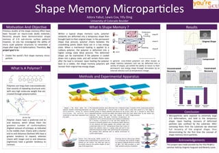
1. Results
What
Is
Shape
Memory
?
Mo4va4on
And
Objec4ve
Previous
studies
of
the
shape
memory
effect
have
been
focused
on
macro-‐scale
(bulk)
materials.
Recently
people
have
demonstrated
shape
memory
of
2-‐D
sub-‐micron
surface
paHerns
however,
no
one
has
inves4gated
the
ability
of
micro
scale
polymer
structures
to
remember
a
shape
aLer
large
3-‐D
deforma4ons.
Therefore,
the
project
goal
is
to:
• Create
the
world’s
first
shape
memory
micro-‐
par4cle
Within
a
typical
shape
memory
cycle,
polymer
networks
are
deformed
into
a
temporary
shape
then
brought
back
to
their
original
shape.
In
the
permanent
shape
(top
picture),
polymer
chains
between
the
crosslinking
points
(black
dots)
are
in
a
low
energy
state.
When
a
mechanical
loading
is
applied
to
a
rubbery
polymer,
the
polymer
is
deformed
into
a
higher
energy
state
(blue
picture).
This
deformed
shape
can
be
maintained
if
the
polymer
is
cooled
down
into
a
glassy
state,
and
will
remain
there
even
aLer
the
load
is
removed.
Upon
hea4ng
the
polymer
back
to
a
rubber,
the
shape
memory
polymers
will
recover
their
original
low
energy
shape.
In
general,
cross-‐linked
polymers
are
oLen
known
as
shape
memory
polymers
and
can
be
deformed
into
a
variety
of
shapes;
yet
exhibit
the
ability
to
return
to
their
permanent,
low
energy
shape
through
s4mula4on
by
an
external
s4mulus
such
as
temperature
change.
0
0.5
1
1.5
2
2.5
3
3.5
4
Original
Shape
Compressed
Constrained
Recovery
Unconstrained
Recovery
Micrometers
Shape
memory
was
aCained!
Original
Shape
Compressed
Unconstrained
Recovery
Constrained
Recovery
What
Is
A
Polymer?
End
to
End
Distance
Probability
Polymers
are
long-‐chain
macromolecules
that
consists
of
repea4ng
structural
units
with
very
high
molecular
weight
that
are
created
through
polymeriza4on.
ΔG=ΔH-‐TΔS
Polymer
chains
have
a
preferred
end
to
end
distance,
which
allows
them
the
greatest
number
of
conforma4ons
(highest
amount
of
entropy),
as
indicated
in
the
middle
chain.
Chains
with
a
shorter
end
to
end
distances
(farthest
leL)
have
a
greater
tendency
to
expand,
whereas
chains
with
longer
end
to
end
distances
(rightmost)
have
a
greater
tendency
to
contract.
Shape
Memory
Micropar4cles
Adora
Yabut,
Lewis
Cox,
Yifu
Ding
University
of
Colorado
Boulder
Conclusion
• Micropar4cles
were
exposed
to
extremely
large
3-‐D
deforma4ons,
and
held
in
the
temporary
shape.
Upon
hea4ng,
recovery
of
deformed
par4cles
was
confined
by
the
substrate.
ALer
removing
them
from
the
substrate
we
observed
full
recovery
of
the
original
shape,
thus
demonstra4ng
for
the
first
4me
the
concept
of
shape
memory
micro-‐par4cles.
Acknowledgements
This
project
was
made
possible
by
the
YOU’RE@CU
seminar
held
by
Virginia
Ferguson
and
Beverly
Louie.
Methods
and
Experimental
Apparatus
Dipped
a
flat
silicon
wafer
into
a
aqueous
solu4on
containing
polystyrene
micro-‐par4cles.
Using
an
op4cal
microscope,
the
par4cles
were
confirmed
to
have
been
deposited
onto
the
wafer.
The
deposited
par4cles
were
deformed
into
a
flaHened
shape
by
using
a
nanoimprinter.
A
second
piece
of
silicon
with
a
treated
surface
to
reduce
adhesion
was
placed
on
top
of
the
deposited
par4cles,
and
the
two
plates
were
placed
within
the
imprinter.
The
environment
was
then
heated
to
120°C
(significantly
above
the
glass
transi4on
temperature
of
polystyrene:
95°C)
and
the
par4cles
were
allowed
to
equilibrate
for
3
minutes.
A
pressure
of
15
bar
was
then
applied
for
5
minutes
to
mold
the
par4cles
into
a
temporary
shape.
With
the
15
bar
pressure
s4ll
being
applied,
the
par4cles
were
then
cooled
back
down
to
35°C
(temperature
below
Tg)
in
order
to
freeze
the
polymer
chains
in
a
glassy
state
and
lock
in
the
deformed
temporary
shape.
ALer
performing
the
compression,
the
par4cles
were
observed
with
an
op4cal
microscope
to
confirm
deforma4on.
A
por4on
of
the
compressed
par4cles
were
then
placed
on
a
hot
stage
at
120°C
for
2
minutes
to
heat
them
back
above
their
Tg
and
induce
the
shape
recovery.
Atomic
Force
Microscopy
(AFM)
consists
of
a
can4lever
with
a
sharp
4p
that
is
used
to
scan
the
par4cle
on
the
surface.
The
AFM
was
used
to
accurately
measure
the
par4cle
heights
at
each
step
of
the
experiment.
Scanning
Electron
Microscope
(SEM))
is
a
microscope
that
produces
images
by
capture
scaHered
electrons
instead
of
light.
The
SEM
provided
us
with
high
resolu4on
pictures
of
par4cles.