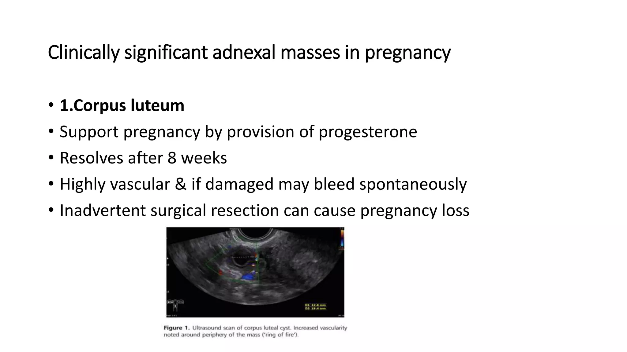
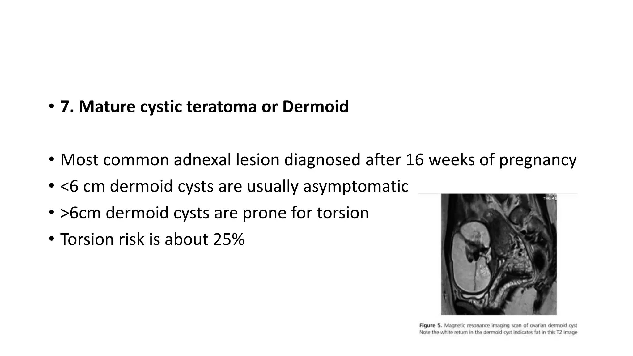
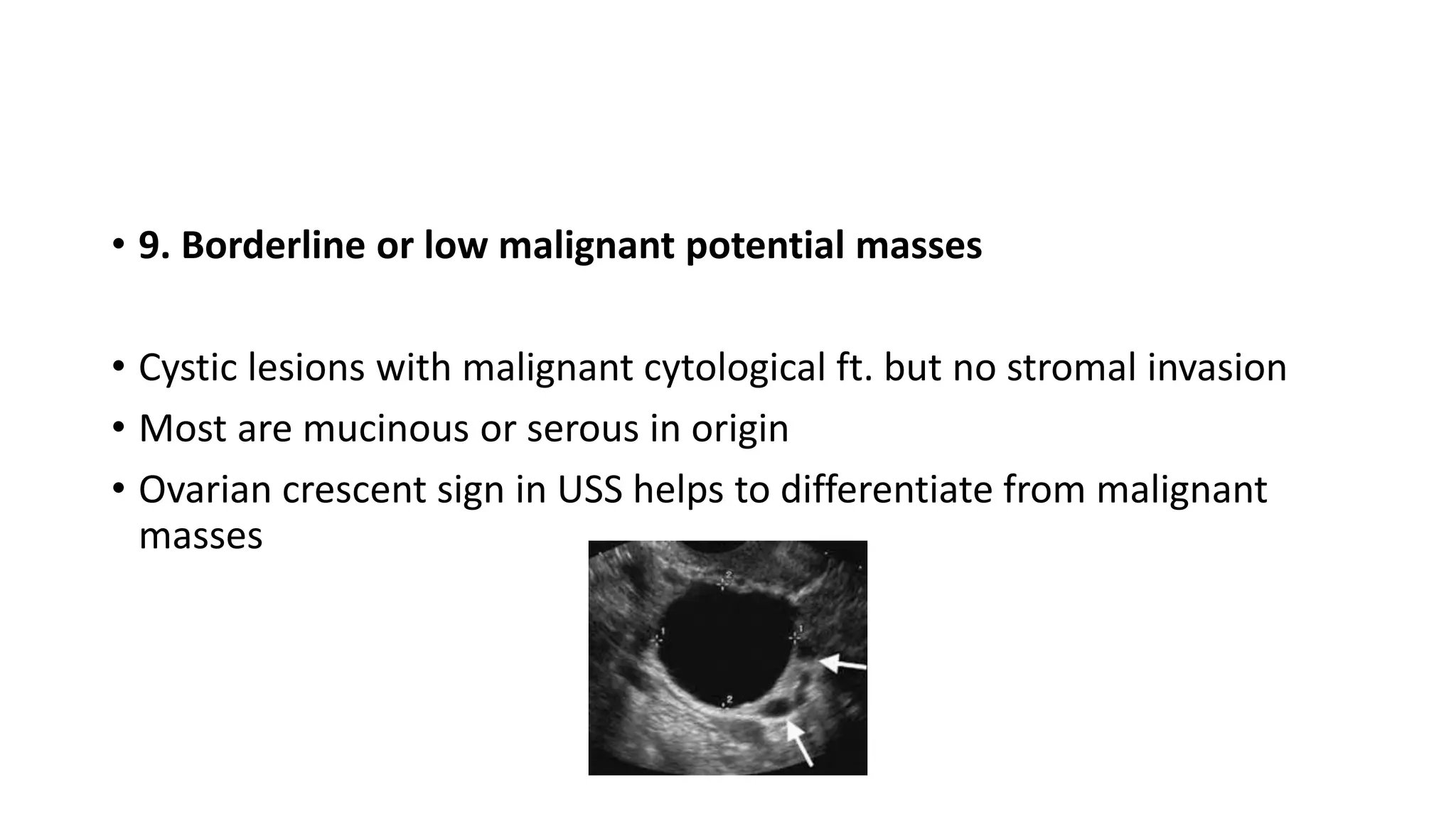
How to manage
adnexal mass in
pregnancy?
Dr.KG Hewawitharana
• This is common
• 0.1% - 8.8% incidence
• Ovarian malignancy is found in 1 / 1500-32000 pregnancies
• Management depend on benign or malignant nature of mass
• Most are benign & can mx with conservative measures
Clinically significant adnexal masses in pregnancy
• 1.Corpus luteum
• Support pregnancy by provision of progesterone
• Resolves after 8 weeks
• Highly vascular & if damaged may bleed spontaneously
• Inadvertent surgical resection can cause pregnancy loss
• 2.Follicular cyst
• Physiological cyst is about 2 cm
• If fails to rupture or ovulate can enlarge up to 6cm
• Typically resolves before 16 weeks
3.Haemorrhagic cysts
Present with acute pain and if clinically stable, best mx conservatively
There is no color flow in solid looking clot in contrast to true solid area
• 3.Ovarian Hyperstimulation
• In conventional IVF, incidence of OHSS is 33%
• 3%-8% cases are composed of moderate to severe OHSS
• Severe cases may have ovarian size of 12 cm which are likely to
rupture or undergo torsion
• Surgery is only indicated in such conditions
• During handling ovaries may damage & bleed profusely
• 4. Hyperreactio luteinalis
• USS ft are like OHSS
• Beta HCG endogenous production causes this in the absence of
ovulation induction
• Maternal virilization may occur in up to 25% pts.
• 5.Luteoma of pregnancy
• Benign & rare
• Luteinized stroma replaces ovarian parenchyma
• Excess androgen production cause maternal virilization in 25% cases
• If maternal virilization occurs, 50% risk of female fetal virilization
exists
• 6.Heterotropic pregnancy
• Rare
• 1-2% risk if ARTs done
• 7. Mature cystic teratoma or Dermoid
• Most common adnexal lesion diagnosed after 16 weeks of pregnancy
• <6 cm dermoid cysts are usually asymptomatic
• >6cm dermoid cysts are prone for torsion
• Torsion risk is about 25%
• 8.Malignant germ cell tumors
• 40% 0f invasive ovarian neoplasm includes this
• Dysgerminomas are commonest with good prognosis-sensitive to
chemo- radiation
• 9. Borderline or low malignant potential masses
• Cystic lesions with malignant cytological ft. but no stromal invasion
• Most are mucinous or serous in origin
• Ovarian crescent sign in USS helps to differentiate from malignant
masses
• 10. TOAs/PID
• Rare in pregnancy as thick cervical mucus is protective
• TOAs can happen in ARTs
• Tetracyclines are not used in mx until 15 weeks since dental
discoloration may occur. But they are not teratogenic.
• 11.Appendicular mass
• Acute appendicitis occurs in 1/1500 pregnancies
• Masses seen in 5%
• Appendicectomy is the most common non-obstetric surgery in
pregnancy
• After T1 , appendix moves upwards to pelvic brim at T2 & reaches
lower Right upper quadrant in T3
Assessment of adnexal mass
• USS - sensitivity 85-95% and specificity 70-90%
• Factors suggestive of malignancy include
• 1.solid components in situ
• 2. papillary projection >6mm
• 3.high color flow in papillary projections
• 4.size increment >20% in subsequent USS
• 5.presence of septations
• 6.free fluid extend beyond POD
• Color doppler- assess tumor vascularity and help to differentiate
benign from malignant
• IOTA suggests
• If no flow- benign
• If high flow -malignant
• If other flow rates – undetermined
• But in pregnancy as with increasing vascularity, this is a debatable
fact
• Pooled sensitivity 85% And specificity 90%
IOTA tool – sensitivity 95% AND specificity 91%, if any M rule, refer to gynae-
oncologist & RMI /ROMA score – not useful in pregnancy
Tumor Markers
• 1. AFP & HCG- both altered in pregnancy and less valuable.
• 2. LDH-levels rise with Dysgerminoma and pregnancy does not
affect. useful in pregnancy.
• 3.CA 125- Level become peak in T1 and then decline with POG. High
cut off>112 can be used in 11-14 weeks. This can still be use as a
baseline in pregnancy. If levels grossly elevated that highlight for
malignancy
Cont.
• 4-HE 4
• glycoprotein increase in ovarian CA.
• Also increase in Mesothelioma, lung, breast & endometrial CA.
• HE4 do not increase with endometriosis.
• has less false positive results compare with CA125 for benign
conditions
• better in sensitivity & specificity in differentiating benign form
malignant
• Serum level drops in pregnancy.
Management
1.Conservative management
• 75% of adnexal masses in pregnancy are simple & < 5cm, they often functional
cysts and resolve spontaneously by 16 weeks. Therefore, no further follow-ups
required for such cysts.
• Follow up scans should offer for those larger or complex cysts at around 14-16
weeks & interventions should delay until 16 weeks over, thereby allow
spontaneous resolution and avoid damage to luteal cysts.
• Persistent ovarian or para-ovarian cysts have low risk of CA thus can be mx
conservatively
• Complex masses which are ultrasonically benign (eg-Dermoid) left untreated
unless symptomatic. But educate patients over possibility of cyst accidents.
2.USS guided aspiration
• useful in acute pain relief and reduces cyst rupture/torsion risk
• well tolerated procedure with fewer side effect
• recurrence rate is high as 40%
• only suitable for simple cysts
3.Role of surgery
Surgical Approach
• Laparoscopy is safe in pregnancy
• RCT shows- less blood loss, good visualization, less uterine irritation
• Pneumoperitoneum does not modify uteroplacental flow in studies
• No difference in FGR/IUD rates
• No adverse fetal outcome in the pressure range of 12-15 mmHg
• Possible uterine injury can be prevented with Open Hassen technique
or Palmers point entry
• Laparoscopy become difficult after 12 week and impossible in T3
• If in T3-do the delivery with LSCS same time
• Incision site & Type change with POG
Adnexal torsion
• 1-5/10000 pregnancies
• High as 16% if OHSS exists
• Can occur in anytime of pregnancy but more common in T1/T2
• Can occur even without adnexal mass
• even normal ovary may undergo torsion during pregnancy present
with acute abdomen
• N/V present in about 85% cases.
• Recurrent torsion risk in pregnancy is about 2X high.
• USS features- tender mass with thick edematous capsule, Avascular
center with bland
• Mx-immediate SX, ideally laparoscopy with detorsion, aspiration &
cystectomy or Salphingo-oophorectomy
• Aim of sx-detorsion to revascularize & preserve ovary, reduce size of
ovary to avoid recurrence
• Ischemic, friable, edematous ovaries may mx with simple puncture &
drainage with minimal risk for pregnancy.
• In case if obvious necrosis with no revascularization after detorsion,
proceed with U/L salphingo-oophorectomy.
Suspected malignant mass
• Majority of these are borderline or early stage Dx with good
prognosis.
• Requires MDT
• MRI require with left lateral tilt of pregnant lady( if it takes time )
• Think about-Woman’s age , parity , POG ,future fertility desires &
Stage of disease
• T1-discuss about risk of miscarriage, termination of pregnancy with
advanced disease
• Late T2/T3 expedite delivery with CS & continue with gynae
oncological mx.
Further more
• Primary surgery can be salphingo-oophorectomy or surgical staging
laparotomy (cytology, omentectomy, peritoneal biopsy +
appendicectomy if abnormal)
• Re-Staging requires after pregnancy together with
imaging/biomarkers especially with invasive epithelial tumors as
extra-ovarian disease may not sufficiently addressed during
pregnancy surgery.
Reference
• TOG-2017/0ct

Adnexal masses in pregnancy

  • 1.
    How to manage adnexalmass in pregnancy? Dr.KG Hewawitharana
  • 2.
    • This iscommon • 0.1% - 8.8% incidence • Ovarian malignancy is found in 1 / 1500-32000 pregnancies • Management depend on benign or malignant nature of mass • Most are benign & can mx with conservative measures
  • 4.
    Clinically significant adnexalmasses in pregnancy • 1.Corpus luteum • Support pregnancy by provision of progesterone • Resolves after 8 weeks • Highly vascular & if damaged may bleed spontaneously • Inadvertent surgical resection can cause pregnancy loss
  • 5.
    • 2.Follicular cyst •Physiological cyst is about 2 cm • If fails to rupture or ovulate can enlarge up to 6cm • Typically resolves before 16 weeks
  • 6.
    3.Haemorrhagic cysts Present withacute pain and if clinically stable, best mx conservatively There is no color flow in solid looking clot in contrast to true solid area
  • 7.
    • 3.Ovarian Hyperstimulation •In conventional IVF, incidence of OHSS is 33% • 3%-8% cases are composed of moderate to severe OHSS • Severe cases may have ovarian size of 12 cm which are likely to rupture or undergo torsion • Surgery is only indicated in such conditions • During handling ovaries may damage & bleed profusely
  • 8.
    • 4. Hyperreactioluteinalis • USS ft are like OHSS • Beta HCG endogenous production causes this in the absence of ovulation induction • Maternal virilization may occur in up to 25% pts.
  • 9.
    • 5.Luteoma ofpregnancy • Benign & rare • Luteinized stroma replaces ovarian parenchyma • Excess androgen production cause maternal virilization in 25% cases • If maternal virilization occurs, 50% risk of female fetal virilization exists
  • 10.
    • 6.Heterotropic pregnancy •Rare • 1-2% risk if ARTs done
  • 11.
    • 7. Maturecystic teratoma or Dermoid • Most common adnexal lesion diagnosed after 16 weeks of pregnancy • <6 cm dermoid cysts are usually asymptomatic • >6cm dermoid cysts are prone for torsion • Torsion risk is about 25%
  • 12.
    • 8.Malignant germcell tumors • 40% 0f invasive ovarian neoplasm includes this • Dysgerminomas are commonest with good prognosis-sensitive to chemo- radiation
  • 13.
    • 9. Borderlineor low malignant potential masses • Cystic lesions with malignant cytological ft. but no stromal invasion • Most are mucinous or serous in origin • Ovarian crescent sign in USS helps to differentiate from malignant masses
  • 14.
    • 10. TOAs/PID •Rare in pregnancy as thick cervical mucus is protective • TOAs can happen in ARTs • Tetracyclines are not used in mx until 15 weeks since dental discoloration may occur. But they are not teratogenic.
  • 15.
    • 11.Appendicular mass •Acute appendicitis occurs in 1/1500 pregnancies • Masses seen in 5% • Appendicectomy is the most common non-obstetric surgery in pregnancy • After T1 , appendix moves upwards to pelvic brim at T2 & reaches lower Right upper quadrant in T3
  • 17.
    Assessment of adnexalmass • USS - sensitivity 85-95% and specificity 70-90% • Factors suggestive of malignancy include • 1.solid components in situ • 2. papillary projection >6mm • 3.high color flow in papillary projections • 4.size increment >20% in subsequent USS • 5.presence of septations • 6.free fluid extend beyond POD
  • 19.
    • Color doppler-assess tumor vascularity and help to differentiate benign from malignant • IOTA suggests • If no flow- benign • If high flow -malignant • If other flow rates – undetermined • But in pregnancy as with increasing vascularity, this is a debatable fact • Pooled sensitivity 85% And specificity 90%
  • 20.
    IOTA tool –sensitivity 95% AND specificity 91%, if any M rule, refer to gynae- oncologist & RMI /ROMA score – not useful in pregnancy
  • 21.
    Tumor Markers • 1.AFP & HCG- both altered in pregnancy and less valuable. • 2. LDH-levels rise with Dysgerminoma and pregnancy does not affect. useful in pregnancy. • 3.CA 125- Level become peak in T1 and then decline with POG. High cut off>112 can be used in 11-14 weeks. This can still be use as a baseline in pregnancy. If levels grossly elevated that highlight for malignancy
  • 22.
    Cont. • 4-HE 4 •glycoprotein increase in ovarian CA. • Also increase in Mesothelioma, lung, breast & endometrial CA. • HE4 do not increase with endometriosis. • has less false positive results compare with CA125 for benign conditions • better in sensitivity & specificity in differentiating benign form malignant • Serum level drops in pregnancy.
  • 23.
  • 24.
    1.Conservative management • 75%of adnexal masses in pregnancy are simple & < 5cm, they often functional cysts and resolve spontaneously by 16 weeks. Therefore, no further follow-ups required for such cysts. • Follow up scans should offer for those larger or complex cysts at around 14-16 weeks & interventions should delay until 16 weeks over, thereby allow spontaneous resolution and avoid damage to luteal cysts. • Persistent ovarian or para-ovarian cysts have low risk of CA thus can be mx conservatively • Complex masses which are ultrasonically benign (eg-Dermoid) left untreated unless symptomatic. But educate patients over possibility of cyst accidents.
  • 26.
    2.USS guided aspiration •useful in acute pain relief and reduces cyst rupture/torsion risk • well tolerated procedure with fewer side effect • recurrence rate is high as 40% • only suitable for simple cysts
  • 27.
  • 28.
    Surgical Approach • Laparoscopyis safe in pregnancy • RCT shows- less blood loss, good visualization, less uterine irritation • Pneumoperitoneum does not modify uteroplacental flow in studies • No difference in FGR/IUD rates • No adverse fetal outcome in the pressure range of 12-15 mmHg • Possible uterine injury can be prevented with Open Hassen technique or Palmers point entry • Laparoscopy become difficult after 12 week and impossible in T3 • If in T3-do the delivery with LSCS same time • Incision site & Type change with POG
  • 29.
    Adnexal torsion • 1-5/10000pregnancies • High as 16% if OHSS exists • Can occur in anytime of pregnancy but more common in T1/T2 • Can occur even without adnexal mass • even normal ovary may undergo torsion during pregnancy present with acute abdomen • N/V present in about 85% cases. • Recurrent torsion risk in pregnancy is about 2X high.
  • 30.
    • USS features-tender mass with thick edematous capsule, Avascular center with bland • Mx-immediate SX, ideally laparoscopy with detorsion, aspiration & cystectomy or Salphingo-oophorectomy • Aim of sx-detorsion to revascularize & preserve ovary, reduce size of ovary to avoid recurrence • Ischemic, friable, edematous ovaries may mx with simple puncture & drainage with minimal risk for pregnancy. • In case if obvious necrosis with no revascularization after detorsion, proceed with U/L salphingo-oophorectomy.
  • 31.
    Suspected malignant mass •Majority of these are borderline or early stage Dx with good prognosis. • Requires MDT • MRI require with left lateral tilt of pregnant lady( if it takes time ) • Think about-Woman’s age , parity , POG ,future fertility desires & Stage of disease • T1-discuss about risk of miscarriage, termination of pregnancy with advanced disease • Late T2/T3 expedite delivery with CS & continue with gynae oncological mx.
  • 32.
    Further more • Primarysurgery can be salphingo-oophorectomy or surgical staging laparotomy (cytology, omentectomy, peritoneal biopsy + appendicectomy if abnormal) • Re-Staging requires after pregnancy together with imaging/biomarkers especially with invasive epithelial tumors as extra-ovarian disease may not sufficiently addressed during pregnancy surgery.
  • 33.